What does the cecum do in pigs? The main functions of the cecum are to absorb fluids and salts that remain after completion of intestinal digestion and absorption and to mix its contents with a lubricating substance, mucus. The internal wall of the cecum is composed of a thick mucous membrane, through which water and salts are absorbed.
Why is the fetal pig a good specimen to dissect?
Why is the fetal pig good for dissection? A fetal pig is a great choice for dissection because the size of the organs make them easy to find and identify. It is also a very exciting dissection because, like sheep and their organs, the internal anatomy is similar to humans! It is fascinating to see how all the organs fit and work together.
What tissue covers the lungs in a fetal pig?
What kind of tissue covers and protects the lungs? Pleura- connective tissue. Why do the lungs appear collapsed in the fetal pig? Because the fetal pig's lungs have not been filled with air, the fetal pig does not need to carry out gas exchange through breathing yet because the mother does that for it, so the lungs are deflated ...
What cavity is the pancreas located in the fetal pig?
Where is the pancreas located in a fetal pig? The pancreas is located dorsal to the spleen and within a fold of the duodenum and stomach . It is a gland organ in the digestive and endocrine system.
What does the pancreas do in a fetal pig?
what is the function of the pancreas in a fetal pig? The pancreas is involved with both exocrine and endocrine excretions. This means the pancreas is responsible for secretion of insulin and glucagon in response to high or low glucose levels in the body. In addition, it has exocrine functions of secreting digestive enzymes and sodium bicarbonate.

What is the function of the cecum?
It is separated from the ileum (the final portion of the small intestine) by the ileocecal valve (also called Bauhin valve), which limits the rate of food passage into the cecum and may help prevent material from returning to the small intestine . The main functions of the cecum are to absorb fluids and salts that remain after completion ...
How many cecums are there in a rock hyrax?
Cecum number can also vary; for example, the rock hyrax ( Procavia capensis) has two ceca, whereas certain insectivores (such as hedgehogs, moles, and shrews) lack a cecum. The Editors of Encyclopaedia Britannica This article was most recently revised and updated by Adam Augustyn, Managing Editor, Reference Content.
What is the cecum of the intestine?
The cecum, the first part of the large intestine, is a sac with a closed end that occupies the right iliac fossa, the hollow of the inner side of the ilium ( the upper part of the hipbone). Guarding the opening of the ileum (the terminal portion…
What is the internal wall of the cecum?
The internal wall of the cecum is composed of a thick mucous membrane, through which water and salts are absorbed. Beneath that lining is a deep layer of muscle tissue that produces churning and kneading motions. Variations in cecum size and structure occur among animals.
What is an encyclopedia editor?
Encyclopaedia Britannica's editors oversee subject areas in which they have extensive knowledge, whether from years of experience gained by working on that content or via study for an advanced degree. ...
What is the name of the pouch that houses bacteria used to digest plant materials such as cellulose?
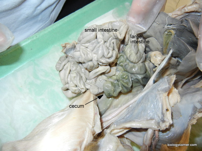
The cecum is a blind pouch where the small intestine joins the large intestine. It houses bacteria used to digest plant materials such as cellulose. The cecum is large in herbivores but much of it has been lost during evolution in humans. The appendix in humans is the evolutionary remains of a larger cecum in human ancestors.
Why is the heart and blood vessels of the neck region removed?
In the photograph below, the heart and blood vessels of the neck region have been removed so that the trachea can be seen more clearly . You should not remove these structures yet because you will need to identify the blood vessels later in the dissection.
What is Figure 14?
Figure 14. The surrounding tissues have been separated to reveal the thymus and thyroid gland.
How to cut the bottom of your jaw?
Use a scalpel to cut the sides of the mouth so that the bottom jaw can be opened for easier viewing (see the photograph below). You will need to cut through the musculature and the joint that holds the lower jaw to the skull.
How far from the chin do you cut a pig?
Extend a single cut along the midline of the ventral surface of the animal to about 2 cm from the chin. Cut completely through the body wall in the abdominal area but keep the cut shallow in the neck region.
What is the dorsal side of a pig?
Dorsal refers to the back side . The pig in the first photograph below is laying on its dorsal side.
How to cut the umbilical cord?
Insert one blade of scissors through the body wall on one side of the umbilical cord and cut posteriorly to the base of the leg as shown in the first photograph below. Continue cutting from the anterior end of this cut so that it resembles an upside-down U. Your finished cut will be anterior to the navel and along each side of the navel. The flap of body wall that contains the navel can be folded posteriorly to reveal the internal organs of the abdomen.