Can the lateral collateral ligament heal itself?
Your LCL (lateral collateral ligament) is a vital band of tissue on the outside of your knee. Athletes are more likely to tear it, causing a lot of pain and other symptoms. LCL tears usually heal after three to 12 weeks, depending on severity. You have to take care of yourself, though.
What is collateral and what does it mean to me?
Strictly speaking, collateral is the asset or assets pledged by a borrower to back up a request for a loan. If the borrower gets the loan and fails to repay it, the lender has the right to seize the asset (i.e. collateral) to make up for the lost income. In the real world, collateral works like this:
What are the types of collateral?
Types of Collateral
- Consumer Goods. These are primarily items bought by any consumer. ...
- Property on Paper. This type of collateral involves stocks, bonds, and funds in a savings account. ...
- Farm Products. These products may involve farm animals and crops. ...
- Inventory. This type of collateral includes contents of a building, a list of properties, and goods in stock. ...
- Equipment. ...
What does it feel like to tear your LCL?
What does it feel like to tear your LCL? The symptoms of an LCL injury are similar to other ligament injuries. You may experience pain and tenderness along the outside of the knee, along with swelling. Some people also describe a feeling of instability in their knee when walking, as if the knee may give out, lock or catch.
What are the collateral ligaments?
The lateral collateral ligament (LCL) is on the outer side of your knee and runs from the top part of the fibula (the bone on the outside of the lower leg) to the outside part of the lower thigh bone. The ligament helps keep the outer side of your knee joint stable.
What is the function of the medial and lateral collateral ligaments?
Condition: The medial and lateral collateral ligaments (MCL and LCL) are bands of tissue that connect the thigh bone to lower leg bones at the knee and help stabilize the knee.
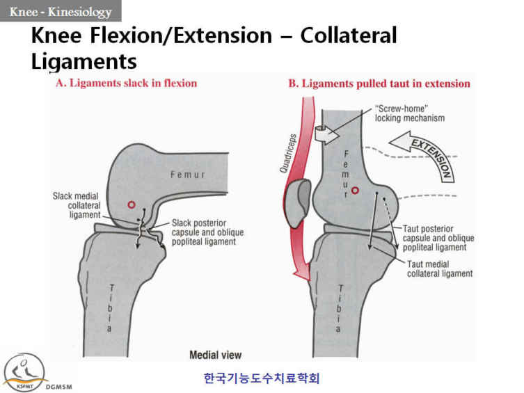
What movements do the collateral ligaments prevent?
The function of the collateral ligaments is to keep the femoral and tibial condyles together,and thus to prevent the knee joint from bending from side to side like this, or like this.
What is the difference between the lateral and medial collateral ligaments?
The collateral ligaments of the knee are located on the outside part of your knee joint. They help connect the bones of your upper and lower leg, around your knee joint. The lateral collateral ligament (LCL) runs on the outer side of your knee. The medial collateral ligament (MCL) runs along the inside of your knee.
Where does the collateral ligament attach?
The medial collateral ligament (MCL) is on the inside. It connects the femur to the tibia. The lateral collateral ligament (LCL) is on the outside. It connects the femur to the fibula (the smaller bone in the lower leg).
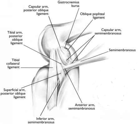
What ligament keeps the knee from hyperextending?
Conclusion: The oblique popliteal ligament was found to be the primary ligamentous restraint to knee hyperextension.
How do you check for collateral ligaments?
0:392:06Varus Stress Test of the Knee Lateral Collateral Ligament - YouTubeYouTubeStart of suggested clipEnd of suggested clipYour patient to relax as much as possible grab on to the lower leg. With one hand just above theMoreYour patient to relax as much as possible grab on to the lower leg. With one hand just above the ankle joint and fixate. With the other hands on the medial side of the femur.
What are the 3 ligaments in the knee?
They are:Anterior cruciate ligament (ACL). This ligament is in the center of the knee. ... Posterior cruciate ligament (PCL). This ligament is in the back of the knee. ... Medial collateral ligament (MCL). This ligament gives stability to the inner knee.Lateral collateral ligament (LCL).
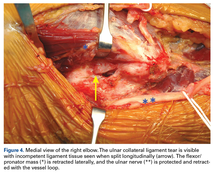
Which ligaments stabilize the medial and lateral surfaces of the elbow joint?
The important ligaments of the elbow are the medial collateral ligament (on the inside of the elbow) and the lateral collateral ligament (on the outside of the elbow.) Together these ligaments provide the main source of stability for the elbow, holding the humerus and the ulna tightly together.
What is the lateral collateral ligament of the ankle?
The lateral collateral ligament (complex) of the ankle is a set of three ligaments that resist inversion of the ankle joint. They are more commonly injured than the medial collateral (deltoid) ligament of the ankle. They run from the lateral malleolus of the fibula to the talus and calcaneus.
Is the radial collateral ligament medial or lateral?
The medial (ulnar) collateral ligament (MCL) supports the ulnohumeral and radiohumeral joints medially, and is a fan-shaped structure. The lateral (radial) collateral ligament (LCL) also supports the ulnohumeral and radiohumeral joints, but laterally. It is more of a cord-like structure.
What does medial collateral ligament pain feel like?
MCL injuries hurt. Most people feel pain along the inside edge of the knee, and they also have swelling. You might hear a pop when the damage to the knee takes place, and your knee may lurch to the side. You may find it hard to walk, or feel like you can't put pressure on the leg with the hurt knee.
What is the fibular collateral ligament?
The fibular collateral ligament is one of the ligaments that make up the knee joint. Ligaments are bands of fibrous, durable tissue that connect and strengthen joints. They can be likened to rubber bands.
Which ligament is attached to the femur?
The fibular collateral ligament is attached to the femur (thigh bone) on one end, goes through the biceps femoris muscle, and attaches to the fibula (calf bone) on the other end. It collaborates with the tibial collateral ligament to form the system of bone, ligament, and tendon that is known as the knee joint.
What is the anterior part of the humerus?
The anterior is attached to the front part of the medial epicondyle of the humerus. The posterior section is attached to the lower and back section of the medial epicondyle . An injury of the ulnar collateral ligament of the elbow can be either due to a slow deterioration or an acute rupture. Stress on the ulna will cause slow deterioration, ...
What ligament runs beside the metacarpophalangeal joint?
In the thumb, the ulnar collateral ligament runs beside the metacarpophalangeal joint. If injured, the thumb may be immobilized for treatment. This ligament is not the same as the ulnar collateral ligament of the wrist. The ulnar collateral ligament of the wrist joint is a rounded cord. It attaches to the end of the ulna’s styloid process.
Where is the Ulnar Collateral ligament located?
Ulnar collateral ligament. Ulnar collateral ligaments are found in the thumb, wrist, and elbow regions. In the thumb, the ulnar collateral ligament runs beside the metacarpophalangeal joint. If injured, the thumb may be immobilized for treatment.
What is the role of the DMCL in knee rotation?
The dMCL helps stabilize internal rotation of the knee from full extension through 90-degree flexion (assists the knee in rotational stability primarily in extension moving through into early flexion).. Despite the relationship of the dMCL with the medial meniscus, there is no influence of the MCL on the stability of the medial meniscus.
What is the most common ligamentous injury in the knee?
MCL injuries often occur in sports, being the most common ligamentous injury of the knee, and 60% of skiing knee injuries involve the MCL) . NB The MCL is also known as the tibial collateral ligament (see image) Provides valgus stability to the knee joint .
What is the MCL?
Description. The medial collateral ligament (MCL) is a flat band of connective tissue that runs from the medial epicondyle of the femur to the medial condyle of the tibia and is one of four major ligaments that supports the knee. MCL injuries often occur in sports, being the most common ligamentous injury of the knee, ...
How many degrees of flexion is required for MCL testing?
Perform with the knee in approximately 30 degrees flexion rather than extension, ensuring isolated testing of the MCL (flexion helps to relax surrounding structures including the posterior capsule).
What is the most common injury to the MCL?
The MCL is one of the most commonly injured ligaments of the knee. Valgus stress is the most common mechanism of injury. Injuries can be contact (a direct blow to the outer aspect of the lower thigh or upper leg) or non-contact (common in skiing). Contact injuries are usually more severe.
What is the valgus force on the knee?
A valgus force is then applied, a positive result of the knee in this position would be an increase in joint space medially.
What is the VST of MCL?
The VST assesses laxity of the MCL compared to the contralateral knee as a control. An increase in laxity and joint space usually distinguishes damage to the medical collateral ligament.
What are the two groups of ligaments that connect the thigh to the lower leg?
Knee ligaments are bands of tissue that connect your thigh bone to your lower leg bones. They can be classified into two main groups: collateral ligaments and cruciate ligaments . Sprained and torn knee ligaments are common, especially among athletes. They may be mild, requiring rest and simple treatment, to severe, requiring surgery.
What are the two ligaments that connect the femur to the tibia?
Cruciate ligaments: The two cruciate ligaments are inside your knee joint and connect your femur to your tibia. They cross each other to create an X. The anterior cruciate ligament (ACL) is located toward the front of the knee. The posterior cruciate ligament (PCL) is behind the ACL. The cruciate ligaments control the way your knee moves front to back.
What is the ligament that connects the thigh bone to the lower leg?
The knee ligaments are bands of tissue that connect your thigh bone in your upper leg (femur) to your lower leg bones (tibia and fibula).
What are the collateral ligaments?
Collateral ligaments: The two collateral ligaments are like straps on each side of your knee. The medial collateral ligament (MCL) is on the inner side of your knee. It attaches the thigh bone (femur) to the shin bone (tibia). The lateral collateral ligament (LCL) is on the outer side of your knee. It connects your femur to your calf bone (fibula). The collateral ligaments prevent the knee from moving side to side too much.
What are the ligaments in the knee?
Knee ligaments are bands of tissue that connect the thigh bone in the upper leg to the lower leg bones. There are four major ligaments in the knee: ACL, PCL, MCL and LCL. Injuries to the knee ligaments are common, especially in athletes. A sprained knee can range from mild to severe. Talk to a healthcare provider if you have a severe knee injury or repeat injuries. Proper diagnosis and treatment can help prevent pain and future injuries.
What is a grade 1 knee injury?
Grade 1: A grade 1 injury to a knee ligament is a minor sprain. The ligament is overstretched or just slightly torn. With a grade 1 knee strain, you’ll have minimal pain, swelling or bruising. You’ll still be able to put weight on the affected leg and bend the knee.
What tests are needed to check knee ligaments?
Order imaging tests if necessary, such as MRI, to take pictures of the knee ligaments.
What is the collateral ligament?
The collateral knee ligaments are found on either side of the knee joint. They are responsible for providing sideways stability by holding the femur and tibia bones together. There are two collateral ligaments, medial (MCL) and lateral (LCL) 1. Medial Collateral Ligament (MCL)
Why does my knee feel unstable?
This results in pain and swelling on the inner side of the knee joint and the knee may feel unstable depending on the severity of the injury.
How many ligaments are there in the knee?
There are two pairs of ligaments in the knee, These ligaments are frequently damaged by sudden twisting movements e.g. changing direction quickly when running, or a force through the knee e.g. a fall or tackle. After a ligament injury, the knee can feel painful, weak and unstable.
How thick is the cruciate ligament?
The cruciate knee ligaments are each about as thick as a pencil and are extremely strong, with a breaking strain of about 60kg.
How long does it take to recover from an ACL injury?
It can take up to a year to recover from an ACL injury so prevention has become an important factor in sports training.
What are the components of knee stability?
Knee Ligaments. The knee ligaments are one of the vital components of knee stability and control. Ligaments are thick fibrous bands, like ropes, and their job is to provide stability and control movement. The knee ligaments connect the thigh and shin bones (femur & tibia) and work together to control how the knee moves to keep it stable ...
Which ligament is the primary structure for proprioception?
1. Anterior Cruciate Ligament (ACL) The anterior cruciate liga ment sits deep in the middle of the knee joint. It attaches to the front of the tibia and the back of the femur. The ACL stops the tibia sliding too far forward in relation to the femur and is the primary structure for proprioception.
What is a joint held together by collagen?
Synarthrosis - a joint held together by collagen.
Which side of the foot is the tibia bind to?
To bind the tibia to the fibula on the medial side. To bind the tibia to the foot on the medial side. To bind the tibia to the foot on the lateral side. To bind the fibula to the foot on the lateral side.
Is the knee more complicated than the hip?
True or false: In terms of structure, the knee is no more complicated than either the hip or ankle joints.
Is there a joint between the maxilla and the mandible?
There is not a joint between the maxilla and the mandible. The mandible forms the "jaw joint" with the temporal bone.
Description
Attachments
- The superficial medial collateral ligament (sMCL) has one femoral and two tibial attachments.
- The femoral attachment is situated on the medial epicondyle.
- The proximal attachment 1. blends into semimembranosustendon and 2. distal attachment is at the posteromedial crest of the tibia.
Function
- The medial collateral ligament is recognised as being a primary static stabiliser of the kneeand assists in passively stabilising the joint. The superficial and deep ligaments each have a unique function, making the MCL the primary responder to valgus stress and a secondary restraint to rotational forces. 1. The sMCL, specifically the proximal divi...
Blood Supply
- Branches of the superior and inferior genicular arteries supply the MCL. The area near the bony insertions is more richly vascularized.
Nerves
- The MCL is innervated by the medial articular nerve, a branch of the saphenous nerve. 1. Innervation is greatest in the epiligament and near the insertions. 2. The ligament can perceive pain and process proprioception through specialized sensory mechanoreceptors like Ruffini endings, Pacinian corpuscles, Golgi receptors, and bare nerve endings. 3. Complete MCL tears w…
Clinical Relevance
- The MCL is one of the most commonly injured ligaments of the knee. 1. Valgus stress is the most common mechanism of injury. 2. Injuries can be contact (a direct blow to the outer aspect of the lower thigh or upper leg) or non-contact (common in skiing). Contact injuries are usually more severe. Injuries to the MCL can have detrimental effects to surrounding structures. 1. It is recog…
Assessment
- Assessment of the MCL is best within 20 to 30 minutes of injury before pain, swelling, and muscle spasms make examination difficult. The assessment includes palpation and a special test, the valgus stress test VST 1. Palpation The anterior aspect of the ligament can be palpated moving vertically, roughly midway along the medial joint line. Focal tenderness indicates an MCL injury. …
See Also