What is the function of the fascia lata?
The fascia lata wraps the large muscles of the thigh and forms the outer limit of the fascial compartments. In doing so, it limits the outward expansion of contracting muscles, making muscular contraction more efficient in compressing veins to push blood towards the heart .
Where does the tensor fasciae latae attach to the thigh?
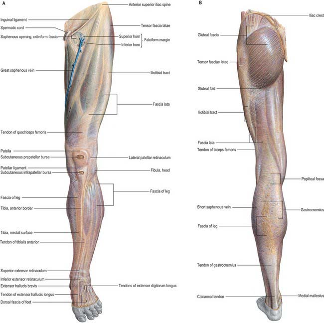
Tensor fasciae latae muscle - ventral view. The muscle originates from the iliac crest and the anterior superior iliac spine on the iliac bone. Distally its fibers attach to the fascia lata - a deep fascia surrounding the entire thigh musculature.
What is the difference between ligaments and fasciae Lata?
Fascia Lata. Fasciae are very similar to ligaments, aponeuroses, and tendons as they are all made up of collagen fibers; however, ligaments join bones together, tendons join muscles to bone, but fasciae wrap around muscles or other structures like fat – superficial fasciae.
What is the fascia of the leg called?
It is continuous with what is renamed the deep fascia of the leg (also known as the crural fascia). The depth of the fascia lata varies considerably across the thigh. It is thickest along the superolateral aspect of the thigh, where it arises from the fascial condensations of gluteus maximus and medius.
What is the fascia lata?
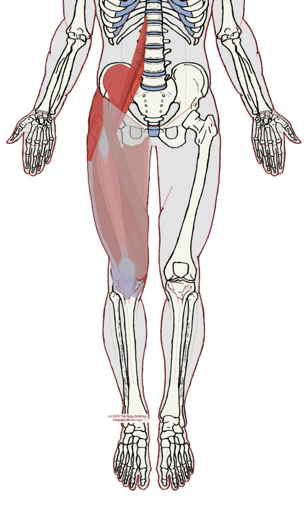
The fascia lata (FL) is a fascial plane that surrounds the deep tissues of the thigh. It varies in thickness throughout its course from the hip to the leg. It receives fibers from gluteus maximus and tensor fascia lata (TFL) laterally.
Where does the fascia lata insert?
It inserts distally to the IT track/band, which is comprised of the fascial aponeurosis of the gluteus maximus and the tensor fascia latae. The IT band then runs along the lateral aspect of the thigh, where it attaches to the lateral condyle of the tibia, specifically the Gerdy tubercle.
Is fascia lata a muscle?
The tensor fasciae latae (or tensor fasciæ latæ or, formerly, tensor vaginae femoris) is a muscle of the thigh. Together with the gluteus maximus, it acts on the iliotibial band and is continuous with the iliotibial tract, which attaches to the tibia.
Which muscle is covered by fascia lata?
tensor fasciae latae muscleThe fascia lata surrounds the tensor fasciae latae muscle. It is a fibrous sheath that encircles the thigh subcutaneously. This encircling of the muscle allows the muscles to be bound together tightly.
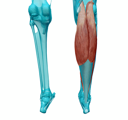
What is the deep fascia of the leg?
The crural fascia or deep fascia of the lower leg is a thick connective tissue fascia that invests the muscles of the lower leg and divides them into the four compartments of the lower leg 1,2: anterior compartment. lateral or peroneal compartment. deep posterior compartment.
Does fascia have blood supply?
Fascia may be innervated by nerves and may send pain signals to your brain. Microcapillaries supply blood and nutrients to fascia. It is easy to understand fascia by comparing it to a sausage casing around tendons, muscles, bones, organs, and joints.
What causes tensor fascia lata pain?
Activities that place excess strain on the TFL include climbing up or downhill without proper support, walking or running on sloped surfaces, excessive pronation, and sports like running or soccer that involve a lot of hip flexion and medial rotation.
Can you tear your tensor fasciae latae?
An injury to the TFL is due to a tear or strain in the muscle. TFL tear or strain has been experienced by many runners, this is because the TFL is used greatly as it provides pelvic stability with the dominant one-sided bearing of weight.
What passes through the fascia lata?
The fascia lata contains an oval opening or hiatus known as the saphenous opening or the fossa ovalis through which the great saphenous vein and efferent lymphatic vessels of the superficial inguinal lymph nodes pass through.
Where does the IT band originate and insert?
It originates at the anterolateral iliac tubercle portion of the external lip of the iliac crest and inserts at the lateral condyle of the tibia at Gerdy's tubercle.
What passes through saphenous opening?
the great saphenous veinThe saphenous opening is an oval aperture located in the fascia lata to allow the passage of the great saphenous vein.
Where is Scarpa's fascia?
Scarpa's fascia lies below the Camper's fascia and above the external oblique muscle. It is connected laterally to the aponeurosis of the external oblique muscle. Medially it fades into the linea alba and pubic symphysis. In the upper thigh just below the inguinal ligament, it blends in with the fascia lata.
What is the fascia lata?
Anatomical Structure. The fascia lata is a deep fascial investment of the musculature of the thigh, and is analogous to a strong, extensible, and elasticated stocking. It begins proximally around the iliac crest and inguinal ligament and ends distal to the bony prominences of the tibia. It is continuous with what is renamed the deep fascia ...
Which part of the fascia lata attaches to the femur?
The deep aspect of fascia lata produces three intermuscular septa which attach centrally to the femur. The lateral septum joins to the lateral lip of the linea aspera and the medial and anterior septa attach to the medial lip. These attachments then continue along the whole length of the femur to include the supracondylar lines.
What is the iliotibial tract?
The iliotibial tract (sometimes known as the iliotibial band or IT band) is a longitudinal thickening of the fascia lata, which is strengthened superoposteriorly by fibres from the gluteus maximus.
What are the three compartments of the fascia lata?
The septa divide the thigh musculature into three compartments; anterior, medial, and lateral . The lateral intermuscular septum is the strongest of the three due to reinforcement from the iliotibial tract (see later), whereas the other two septa are proportionately weaker.
How does a femoral hernia develop?
A femoral hernia develops when an out-pouching of abdominal viscera protrudes through the femoral canal. The protrusion becomes noticeable when it exits superficially through the saphenous opening within the fascia lata – producing a swelling inferior to the inguinal ligament.
Why do you need fascia lata grafts?
Dermatofasciotomy and debridement can leave large wound sites that require post-operative grafts to facilitate tissue regeneration and healing. The fascia lata graft is a popular choice as the iliotibial tract provides a particularly high concentration of connective tissue fibres, and can be surgically harvested whilst leaving the majority of fibres intact.
Where does the tensor fascia originate?
The muscle originates from the iliac crest and descends inferiorly to the superolateral thigh. At the junction of the middle and upper thirds of the thigh, it inserts into the anterior aspect of the iliotibial tract. When stimulated, the tensor fasciae lata tautens the iliotibial band and braces the knee, especially when the opposite foot is lifted.
What is the function of the Tensor Fasciae Latae?
The main task of the tensor fasciae latae is to sustain tension of the iliotibial tract. As the femoral shaft meets the pelvis, an angled pressure from above imposes a high bending strain to the femur. Both the hip abductors and the tensor fasciae latae counteract the pressure on the opposite side and help stabilize the bone (tension banding effect). Furthermore, the activation of the muscle leads to an abduction, flexion and internal rotation of the hip joint.
Where is the Tensor Fasciae Latae located?
In its superior aspect, tensor fasciae latae is found between the sartorius and gluteus medius muscle, where it overlays the gluteus minimus muscle.
What muscle causes external rotation of the hip?
Furthermore, the activation of the muscle leads to an abduction, flexion and internal rotation of the hip joint. Contraction of the tensor fasciae latae muscle also causes external rotation ...
How long does it take to read Tensor Fasciae Latae?
Reading time: 4 minutes. Tensor fasciae latae muscle (Musculus tensor fasciae latae) Tensor fasciae latae is a fusiform muscle located in the lateral aspect of the thigh. It belongs to the muscles of the gluteal region, along with the gluteus maximus, gluteus medius and gluteus minimus muscles. Tensor fasciae latae is found superficial in ...
What is the iliotibial tract?
The tensor fasciae latae, the fibers of the gluteus maximus and the aponeurosis of the gluteus medius form a horizontal reinforcement known as the iliotibial tract. This band of connective tissue runs laterally over the knee joint and inserts at the lateral condyle of tibia and lateral patellar retinaculum. Learn the anatomy of the body muscles ...
Where does the tensor fascia originate?
The muscle originates from the outer lip of the anterior iliac crest and the anterior superior iliac spine. Distally its fibers attach to the fascia lata - a deep fascia surrounding the entire thigh musculature. The tensor fasciae latae, the fibers of the gluteus maximus and the aponeurosis of the gluteus medius form a horizontal reinforcement ...
Where is the descending muscle located?
While descending down the thigh, the muscle is found between the two layers of fascia lata. Due to its superficial position, this muscle is easily palpated. This is usually performed at the area located halfway between the anterior superior iliac spine and the greater trochanter of femur.
Where is the fascia located?
It is located on the bottom of your foot and stretches from your heel bone to your toes. This thick band of fascia supports your medial arch and gives shape to the bottom of your foot.
What is fascia tissue?
Injury. Rehabilitation. Fascia is a system of connective tissue that encases our body parts and binds them together. Fascia, made primarily of collagen, can be thought of as a sausage casing for your body's tissues. It surrounds muscles, nerves, tendons, and ligaments and gives them shape.
How does the fascia connect to the brain?
Fascia may be innervated by nerves and may send pain signals to your brain. Microcapillaries supply blood and nutrients to fascia. It is easy to understand fascia by comparing it to a sausage casing around tendons, muscles, bones, organs, and joints. Fascia also helps support proper movement and function in your body.
What is the collagen pattern in the fascia?
The collagen that makes up fascia is organized in a wavy pattern. When pulled, these lines of tissue resist tensile and shear loads, helping to keep your body parts together.
Why is my fascia tight?
Sometimes fascia compartments can become tight and not allow for normal movement of blood into and out of the compartment. A condition called compartment syndrome occurs when muscles fill with blood during activity, but the fascia covering around the muscles is tight and does not allow the blood to easily exit the muscle compartment.
What are the different types of fascia?
Fascia is located all over your body, and while it surrounds all tissues, it can be divided into three distinct types based on location. Types of fascia include: 1 Superficial fascia: This type of fascia is associated with your skin. 2 Deep fascia: Deep fascia surrounds your bones, nerves, muscles, and arteries and veins. 3 Visceral fascia: This fascia surrounds your internal organs.
Why is gentle motion important for fascia?
Fascia is like any other collagen type tissue in the body. When it becomes torn or injured, it needs appropriate time to heal properly. As it is healing , gentle motion can be started to ensure that the collagen cells are properly aligned. This is thought to eliminate the build up of scar tissue in the body.
Where does the tensor fascia latae muscle attach?
The tensor fasciae latae originates just behind (posterior) or to the outside of the anterior superior iliac spine or ASIS.
What does tensor fascia latae mean?
The muscle name describes what the muscle does and where it’s located. The word “tensor” comes from the Latin word “tendere” which means “to tense”. “Fascia” comes from the Latin word for “band”. “Latae” comes from the Latin word “lata” which means “side or lateral”. You could combine those three words to get a muscle that tenses a band on the side. Tensor fasciae latae is often shortened to the TFL.
What does iliotibial band mean?
Band refers to the fact that the iliotibial band is a narrow tendinous strip. Iliotibial band is often shortened to IT band.
What actions does the iliotibial band do?
The iliotibial band acts to stabilize the knee, especially in walking and running.
What about contracting or lengthening the IT band in yoga?
The tension in the iliotibial band is really a function of the tension in the muscles that it originates from, the tensor fascia latae and the gluteus maximus. We wouldn’t likely have the intention of contracting or lengthening the IT band itself in yoga. But, if we’re aware of tension there, we could have the intention of lengthening either or both of the tensor fascia latae and the gluteus maximus to release some tension there.
Which muscle inserts onto the iliotibial band?
Tensor fascia latae is one of two muscles that insert onto the iliotibial band. The other muscle that inserts onto the iliotibial band is the gluteus maximus.
Where does the iliotibial band originate?
The iliotibial band is really a continuation of the tissue of the tensor fascia latae that originates on the ilium and becomes a tendinous band.
What tract aids lateral rotation of the leg?
As part of the iliotibial tract it aids lateral rotation of the leg.
Which nerve innervates the tensor fascia?
The Tensor fasciae latae is innervated by superior gluteal nerve, originating from lumbar nerve 4, 5, and first sacral nerve (L4-S1) roots. It also innervates gluteus minimus and medius muscles before terminating with innervation of tensor fasciae latae muscle.
Why is stretching important for hip and knee pain?
The Tensor fascia latae Tightness is most common seen in adult and stretching exercise is very important for keep your posture correct and also helps in reducing pain in Hip and Knee pain.
Which tract attaches to the lateral condyle of the tibia?
TFL together with the gluteus maximus joins to form the Iliotibial tract, which attaches to lateral condyle of tibia
What is a TFL?
TFL is prime mover in hip medial rotation and a weak hip abductor
How far below the horizontal is the thigh?
With the knee straight and the pelvis in a neutral position, the thigh drops about 10 degrees below the horizontal. It is suggestive of normal length.
Who described the test for tightness of TFL and ITB?
Frank Ober described the test for tightness of TFL and ITB in an article entitled “Back Strain and Sciatica”, wherein he discussed the relationship of a contracted TFL and ITB to low backache .
Classification
Structure
- In this article, we shall look at the anatomy of the deep fascia of the thigh the fascia lata. A detailed account of its structure, anatomical relations, function, attachments, and clinical relevance will be outlined. The fascia lata is a deep fascial investment of the musculature of the thigh, and is analogous to a strong, extensible, and elasticated stocking. It begins proximally aro…
Variations
- The deepest aspect of the fascia lata gives rise to three intermuscular septa that attach centrally to the femur. The septa divide the thigh musculature into three compartments; anterior, medial, and lateral. The lateral intermuscular septum is the strongest of the three due to reinforcement from the iliotibial tract (see later), whereas the other two septa are proportionately weaker.
Clinical significance
- An ovoid hiatus known as the saphenous opening is present in the fascia lata just inferior to the inguinal ligament. The opening serves as an entry point for efferent lymphatic vessels and the great saphenous vein, draining into superficial inguinal lymph nodes and the femoral vein respectively. The iliotibial tract (sometimes known as the iliotibial band or IT band) is a longitudi…
Function
- The tensor fascia lata is a gluteal muscle that acts as a flexor, abductor, and internal rotator of the hip. Its name, however, is derived from its additional role in tensing the fascia lata. It is innervated by the superior gluteal nerve, like gluteus medius and minimus, but is located more anterolaterally than the other gluteal muscles. The prope...
Pathophysiology
- The lateral thickening of fascia lata forms the iliotibial tract and receives tendon insertions superiorly from gluteus maximus and tensor fascia lata. The widened band of fibres descends the lateral thigh and attaches to the lateral tibial condyle on the anterolateral (Gerdy) tubercle.