What is the function of the pisiform?
Function The pisiform serves as an attachment for tendons and ligaments. As it is a sesamoid bone, it acts as a pulley that provides a smooth surface for the flexor carpi ulnaris tendon to glide over. The pisiform also forms part of the ulnar canal or as otherwise called the Guyon canal.
What is the function of the pisiform bone in the wrist?
The pisiform bone may provide mechanical stability to the ulnar column of the wrist by preventing triquetral subluxation. Thus, surgical excision of the pisiform might cause loss of function to the wrist. We performed a functional evaluation of 20 hands in 20 patients who had undergone pisiformectom …
What is the structure of the pisiform bone?
Structure. The pisiform bone has four surfaces: The dorsal surface is smooth and oval, and articulates with the triquetral: this facet approaches the superior, but not the inferior border of the bone. The palmar surface is rounded and rough, and gives attachment to the transverse carpal ligament, the flexor carpi ulnaris and...
When does the pisiform bone ossify?
Development and Ossification The pisiform bone develops within the flexor carpi ulnaris (FCU) tendon, which makes it a sesamoid bone. It is the last wrist bone to ossify, becoming visible on an x-ray only when a child is about 8 to 12 years old, often later in boys than girls.
Do you need pisiform bone?
The pisiform bone is a sesamoid bone which lies embedded within the flexor carpi ulnaris tendon, providing a smooth surface for it to glide over. It acts as an important attachment site for both the flexor carpi ulnaris and abductor digiti minimi muscles.
Can the pisiform bone be removed?
Conclusions Pisiformectomy is a surgery used sparingly in cases with refractory pain associated with arthrosis of the pisotriquetral joint or enthesopathy of the flexor carpi ulnaris/pisiform interface.
What happens if you break your pisiform?
Pisiform fractures may be associated with triquetrum, hamate, or dorsal radius fractures. Clinical presentation includes pain, swelling, and tenderness of the hypothenar eminence. Ulnar nerve irritation may occur, because the pisiform makes up the ulnar wall of Guyon's canal.
What other carpal bone does the pisiform function with?
The pisiform is the only carpal bone with insertions and attachments for the abductor digiti minimi and the flexor carpi ulnaris.
What causes pain in the pisiform bone?
Pain in the area of the pisiform can be because of a wide variety of pathologies including tendinitis at the insertion FCU, arthritis of the pisotriquetral joint, subluxation of the pisiform with associated synovitis, fracture of the triquetrum or pisiform, rheumatism, or osteonecrosis.
How long does it take for a pisiform fracture to heal?
Pisiform fractures are most commonly acute injuries and can be treated non-operatively with symptomatic immobilization in a wrist brace, padding and activity modification for a period of 4–6 weeks.
How hard is it to break your pisiform?
Pisiform fractures are an uncommon injury accounting for only 0.2% of all carpal fractures. They are managed by immobilisation in either a plaster cast or a wrist splint. This fracture can be easily missed on first presentation due the superimposition of adjacent carpal bones.
What does a broken pisiform feel like?
This injury presents as chronic wrist pain, grip weakness, and/or restriction of wrist movements. Pisiform fractures may also be associated with tenderness in the affected area. Most pisiform fractures are a result of falling onto an outstretched hand (FOOSH injuries).
What is the most common bone to break in your wrist?
The most commonly injured carpal bone is the scaphoid bone, located near the base of your thumb.
Can you dislocate your pisiform?
Dislocation of the pisiform bone is a relatively rare injury associated with hyperextension traction of the flexor carpi ulnaris (FCU), tearing the pisohamate and/or pisometacarpal ligament.
What is the bone on wrist that sticks out?
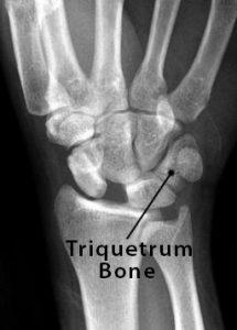
Triquetrum. The triquetrum is a bone on the small finger side of the wrist in the first row of wrist bones.
What type of bone is pisiform name other bones of the same type?
The pisiform (os pisiforme) is a small carpal bone on the medial side of the proximal carpal bones row. It is considered a sesamoid bone within the tendon flexor carpi ulnaris.
What is a pisiform excision?
Pisiform excision is a relatively safe procedure for patients with chronic ulnar-sided wrist pain due to pisotriquetral osteoarthritis, FCU tendinitis, or ulnar neuropathy when a conservative treatment is insufficient. Mixed diagnoses are often encountered in clinical practice.
Why does the bone on my wrist stick out?
With osteoarthritis, the cartilage starts to wear away over time. In extreme cases, the cartilage can completely wear away, leaving nothing to protect the bones in a joint, causing bone-on-bone contact. Bones may also bulge, or stick out at the end of a joint, called a bone spur.
How big is the pisiform bone?
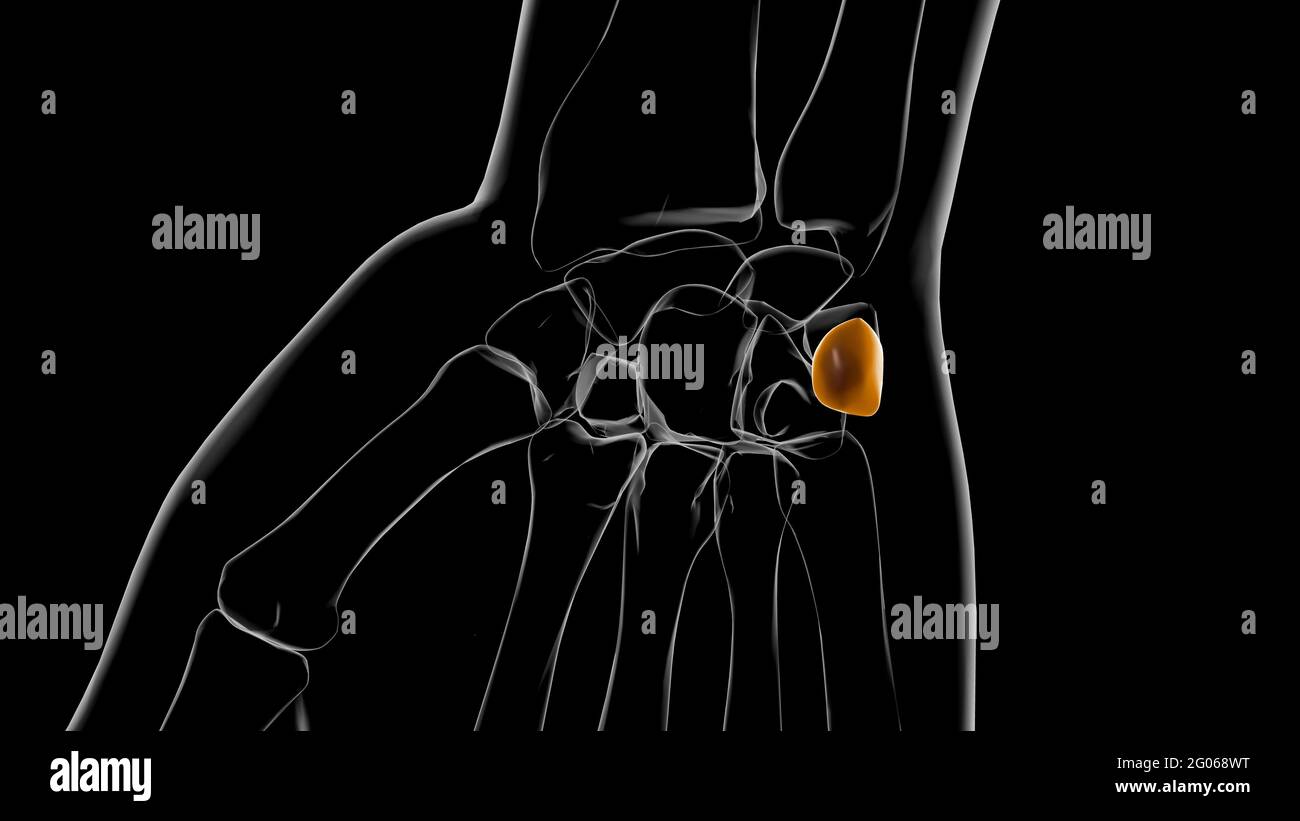
The pisiform is one of eight and smallest carpal bones that forms part of the wrist joint. It is a small pea-shaped bone. it develops in a tendon and is a sesamoid bone The name pisiform is derived from the Latin word pisum which means "pea". It can be felt on the anteromedial side of the wrist.
What type of bone is the pisiform?
sesamoid bone10.1. The pisiform is the smallest of the carpals. Because it develops within a tendon, it is actually a sesamoid bone. There are other, much smaller sesamoid bones found embedded in flexor tendons, for example, at some metacarpophalangeal and interphalangeal joints.
What is the function of the pisiform bone?
Along with the other seven carpal bones, pisiform helps in maintaining the structure of the human wrist, its mobility, and functioning. This small bone also protects the FCU tendon by allowing it to connect with the triquetral bone through itself, so it can support the tendon’s movements during flexing ...
Where is the pisiform located?
Situated on the outer side of the proximal row, it is bordered by the hamate bone on its inner side. The pisiform can be felt on the palmar side of the wrist, below the little finger.
What is the last wrist bone to ossify?
The pisiform bone develops within the flexor carpi ulnaris (FCU) tendon, which makes it a sesamoid bone [1]. It is the last wrist bone to ossify, becoming visible on an x-ray only when a child is about 8 to 12 years old [6], often later in boys than girls [3].
What bone forms an attachment with the carpal ligament?
The pea-shaped bone also forms an attachment with the carpal ligament on its lateral, medial, and palmar surfaces [5].
Which side of the triquetral bone is the flat dorsal surface of the pisiform articulate?
The flat dorsal surface of pisiform articulates with the palmar side of the triquetral bone [7].
What is the smallest bone in the human wrist?
What is the Pisiform Bone . The pisiform (Latin: os pisiforme) is a pea-shaped knobbly bone in the human wrist and the smallest of the eight carpal bones [1]. The name is actually derived from ‘pisum’, the Latin word for ‘pea’, referring to the characteristic shape of the bone [2].
What is the pisiform bone?
Fluid collection and the thickening of soft tissue are common in the pisiform bone. The pisiform is sphere-shaped, like a pea. In fact, its name means ‘pea-shaped.’. The pisiform has four types of surfaces: dorsal, palmar, lateral, and medial. The latter three surfaces are rough, allowing the pisiform to attach to the carpal ligament.
Why does my pisiform hurt?
Chronic or acute pain is common in the pisiform because it is where tendinopathy of the FCU occurs at insertion. Osteoarthritis, mechanical overuse, and bony fractures can also affect the pisiform. Pain in the pisiform is usually examined by a sonographic evaluation.
What is the flexor carpi ulnaris?
Pisiform. The pisiform is a sesamoid bone. It is located in the flexor carpi ulnaris ( FCU) wrist tendon. It protects this tendon by supporting and bearing its forces as it moves across the triquetrum during wrist movement. The triquetrum is a proximal carpal bone located between the pisiform and lunate bones.
Which surface of the carpal ligament is smooth?
The latter three surfaces are rough, allowing the pisiform to attach to the carpal ligament. However, the dorsal surface is smooth, allowing for the bone’s articulation with the triquetrum. Last medically reviewed on January 19, 2018.
Where is the triquetrum located?
The triquetrum is a proximal carpal bone located between the pisiform and lunate bones. The pisiform is located opposite the wrist’s carpal base plate and communicates with the abductor digiti minimi of the hand. Specifically, it is located where the carpus joins the ulna, which is the inner forearm bone. Chronic or acute pain is common in the ...
What is the pisiform bone?
The pisiform bone is most recognizable as an unassuming palmar projection forming the heel of your hand. The pisiform bone, along with the hamulus of the hamate, defines the medial boundary of the carpal tunnel because the pisiform body acts as one of the four attachments points of the flexor retinaculum. It also acts as an attachment site ...
Where is the pisiform bone located?
It is the last carpal bone to ossify. The pisiform bone is a small bone found in the proximal row of the wrist ( carpus ). It is situated where the ulna joins the wrist, within the tendon of the flexor carpi ulnaris muscle. It only has one side that acts as a joint, articulating with the triquetral bone.
What is the effect of Hox knockouts on the formation of the pisiform?
Studies looking at the effect of Hox gene knockouts on the formation of the pisiform in mice have suggested that the modification of Hoxa11 or Hoxd11 genes, or the downstream targets they affect , could have acted as the mechanism for the reduction we see in the human pisiform condition.
What is the name of the bone that is a small knobbly, sessile, and shaped?
1254. FMA. 23718. Anatomical terms of bone. The pisiform bone ( / ˈpaɪsɪfɔːrm / or / ˈpɪzɪfɔːrm / ), also spelled pisiforme (from the Latin pisifomis, pea-shaped), is a small knobbly, sesamoid bone that is found in the wrist. It forms the ulnar border of the carpal tunnel .
What is the name of the bone in the left hand?
Pisiform bone. Left hand anterior view (palmar view). Pisiform bone shown in red. The pisiform bone ( / ˈpaɪsɪfɔːrm / or / ˈpɪzɪfɔːrm / ), also spelled pisiforme (from the Latin pisifomis, pea-shaped), is a small knobbly, sesamoid bone that is found in the wrist. It forms the ulnar border of the carpal tunnel .
Why did we see pisiform reduction during the course of Hominin evolution?
Some suggest that the reduction of the pisiform allowed for ulnar deviation and that allowed for greater extension in the human wrist which increased our capacity for throwing .
Why is the pisiform removed?
In clinical studies, the pisiform has been removed as treatment for osteoarthritis in the pisotriquetral joint. While some studies came to the conclusion that the pisiform "contributes to the stability of the ulnar column of the wrist", others suggested that while excision slightly impairs the range of motion of the wrist (especially wrist extension), the forces generated within the wrist are not significantly impacted. Subjects in the latter study did report impaired function after excision when performing heavy lifting and weightbearing activities, but this is suggested to be subjective considering that they did not have to change occupation or their level of activity as a result of the excision.
What is the function of the pisiform?
Function. The pisiform serves as an attachment for tendons and ligaments. As it is a sesamoid bone, it acts as a pulley that provides a smooth surface for the flexor carpi ulnaris tendon to glide over. The pisiform also forms part of the ulnar canal or as otherwise called the Guyon canal.
What is the pisiform part of?
The pisiform also forms part of the ulnar canal or as otherwise called the Guyon canal. It is a fibro-osseous structure that forms a groove between the pisiform and the hook of the hamate. The ulnar nerve and ulnar artery pass through this canal from the distal forearm into the hand.
Where is the pisiform located?
Structure. The pisiform can be found on the anteromedial side of the wrist in the proximal row of carpal bones. It is a small sesamoid bone, enveloped in the flexor carpi ulnaris tendon and can be easily palpated from the exterior.
What is the name of the bone in the left hand?
Pisiform bone (Left Hand) The pisiform is one of eight and smallest carpal bones that forms part of the wrist joint. It is a small pea-shaped bone. it develops in a tendon and is a sesamoid bone The name pisiform is derived from the Latin word pisum which means "pea".
Where is the pisiform bone?
Possible but rare: The pisiform bone is one of the eight carpal (wrist) bones. It is a small pea-shaped sesamoid bone located where the ulna (one of the lower arm bones ... Read More
How is a fractured pisiform bone in wrist treated?
Usually by cast: For few weeks, then splint if you have no more problems. Good result usually.
Where is the sesamoid bone located?
It is a small pea-shaped sesamoid bone located where the ulna (one of the lower arm bones) joins the wrist (on the pinky side). Dislocation of the pisiform is possible, but rare. Fracture of the pisiform is also uncommon. 2.3k views Answered >2 years ago.
Is it possible to dislocate the pisiform bone?
Possible but rare: The pisiform bone is one of the eight carpal (wrist) bones. It is a small pea-shaped sesamoid bone located where the ulna (one of the lower arm bones... Read More
What is pisotriquetral trauma?
Trauma- Where the pisotriquetral joint suffers acute or chronic trauma.
What is the condition that attacks bones by destroying cartilage?
Arthritis is an inflammatory health condition that attacks bones by destroying the cartilage. This makes them to rub against each other to cause pain, particularly, within a joint. There is a wide variety of arthritic conditions, especially because, the condition may affect any joint of the body: large joints like the knee and small joints like the fingers.
What is the name of the joint on the ulnar side of the wrist?
Pisotriquetral arthritis is arthritis of the pisotriquetral joint. At times, this condition can be referred to as osteoarthritis of the pisotriquetral joint. This is the small joint on the ulnar side of the wrist. With this condition, you experience pain over your wrist (pisiform). The pisiform bone is a small and pea-shaped sesamoid bone that is part of the eight carpal or wrist bones. It is situated where the ulna (a forearm bone) connects with the wrist (the side of the little finger).
Can pisotriquetral joints be injected?
Steroid Injections – The pisotriquetral joint can be injected with steroid injections for pain relief. Steroids are used when medication does not control inflammation. They can weaken ligaments and joints and therefore, they are repeated minimally.
Overview
Function
The pisiform bone is most recognizable as an unassuming palmar projection forming the heel of your hand.
The pisiform bone, along with the hamulus of the hamate, defines the medial boundary of the carpal tunnel because the pisiform body acts as one of the four attachments points of the flexor retinaculum. It also acts as an attachment site for tendons of the abductor digiti minimi and for the flexor …
Structure
The pisiform is a sesamoid bone, with no covering membrane of periosteum. It is the last carpal bone to ossify. The pisiform bone is a small bone found in the proximal row of the wrist (carpus). It is situated where the ulna joins the wrist, within the tendon of the flexor carpi ulnaris muscle.
It only has one side that acts as a joint, articulating with the triquetral bone. It is on a plane anterior to the other carpal bones and is spheroidal in form.
Etymology
The etymology derives from the Latin pīsum which means "pea" ultimately derived from the Greek "pison" (pea).
Development
Compared with other non-human primates, humans have a short pisiform bone. This dramatic size difference is suggested to be the outcome of a lost growth plate in hominins some time between Australopithecus afarensis, who has been shown to have an elongated and ape-like pisiform, and Homo neanderthalensis, who is suggested to have a pisiform resembling the modern human condition.
Evolution
There are several hypotheses that seek to explain why we see pisiform reduction during the course of hominin evolution. Some suggest that the reduction of the pisiform allowed for ulnar deviation and that allowed for greater extension in the human wrist which increased our capacity for throwing. Scholars with this point of view would believe that these anatomical changes would improve the action of clubbing in our hominin ancestors.
Other animals
All other tetrapods have a pisiform, being the most common sesamoid. In mammals and non-human primates, the pisiform is an enlarged and elongated bone that articulates with the distal ulna. In some taxa, the pisiform even articulates with the hammate or radius. In these non-human taxa, the pisiform develops from two ossification centers that are divided by a palmar epiphyseal plate. Because in other mammals, the bone does not follow a typical sesamoid development pattern an…
See also
• Carpal bone
• Intercarpal articulations