
What is the function of the sternohyoid?
The hyoid is a bone near the top of the throat. The sternum is the bone at the front of the rib cage. Due to its location, the sternohyoid is useful for several functions, including depression (lowering) of the hyoid bone, head and neck movement, and speech.
What is the difference between the sternohyoid and sternothyroid?
The sternothyroid muscle is shorter and wider than the sternohyoid muscle. The sternothyroid muscle lies underneath the sternohyoid muscle. Nerves from the upper cervical nerve travel through the cervical ansa, or cervical loop, and supply the sternothyroid muscle. The main function of the sternothyroid is to depress the larynx.
What is the blood supply to the sternohyoid muscle?
Vascular supply to sternohyoid comes from the superior thyroid artery (a branch of the external carotid artery ), while the venous blood is conveyed by the superior thyroid vein. The action of the sternohyoid muscle is to depress the hyoid bone after it has been elevated by the suprahyoid muscles.
What is the function of the infrahyoid muscle?
The infrahyoid muscles, together with the suprahyoid muscles, are responsible for the placing of the hyoid bone & the larynx. Specifically, the infrahyoid muscles, & omohyoid, depress the hyoid bone following its raising during the work of swallowing.
What is the sternohyoid function?
Function. The action of the sternohyoid muscle is to depress the hyoid bone after it has been elevated by the suprahyoid muscles. The elevation of the hyoid bone and thus the larynx, happens during swallowing. This action closes the airways, preventing the food from being inhaled.
What is the action of the sternothyroid muscle?
Action. The sternothyroid muscles primarily depress and fix the hyoid bone and underlying larynx.
What is the function of the omohyoid and sternohyoid muscles?
It belongs to a group of muscles called infrahyoid muscles, along with three others: sternohyoid, sternothyroid and thyrohyoid. The function of this muscle is to depress the hyoid bone and larynx and reestablish breathing following the act of swallowing.
Where is sternohyoid muscle located?
The sternohyoid muscle is a long, thin muscle located along the entire length of the front of the neck. This muscle is connected by tendons — strong, flexible tissue that usually connects muscle to bone — to the hyoid bone at its top end, and connected to the sternum at its lower end.
What does sternothyroid mean in anatomy?
Medical Definition of sternothyroid : an infrahyoid muscle on each side of the body below the sternohyoid that arises from the sternum and from the cartilage of the first and sometimes of the second ribs, inserts into the thyroid cartilage, and acts to draw the larynx downward by depressing the thyroid cartilage.
Can you feel the sternohyoid muscle?
The sternohyoid is superficial so it is easy to palpate. However, because it is so small, it requires a fine touch to feel and discern it from adjacent musculature. To palpate the sternohyoid, place your palpating fingers between the sternum and hyoid bone and then gently resist the client from depressing the mandible.
Why does my sternohyoid hurt?
Some causes of sternocleidomastoid pain include: carrying a heavy object, such as a child or backpack, in an awkward position. poor posture, for example, when a person spends long days hunched over a computer or straining their neck to reach things in the garden.
How do I stretch my sternohyoid?
Slowly move the ear toward the shoulder while the hands stay behind the back. Keep the shoulders down and the hands behind the back. Do not lift the shoulders up when tilting the head to the side. Hold the stretch for at least 20 seconds.
What is the insertion of the sternohyoid muscle?
Sternohyoid muscleOriginmanubrium of sternumInsertionhyoid boneArterysuperior thyroid arteryNerveC1-C3 by a branch of ansa cervicalis10 more rows
What is the shape of the sternohyoid muscle?
butterfly-shapedIt is a reddish-brown, butterfly-shaped organ with right and left lobes joined by a thin isthmus, which forms a bridge across the ventral and anterior aspect of the trachea, respectively. It typically extends from the posterior larynx along the first three or four tracheal cartilage rings in mice (Figs.
Does sternohyoid muscle attach to the clavicle?
The sternohyoid muscle has fibers originating inferomedially along the posterior surface of the clavicle in addition to the manubrium and posterior sternoclavicular ligament.
Does the sternohyoid depress the larynx?
Infrahyoid muscles: Together, the infrahyoid muscles play an active role in swallowing through the movement of the larynx. The omohyoid, sternohyoid, and thyrohyoid act to depress the hyoid bone. The thyrohyoid elevates the larynx whereas the sternothyroid depress the larynx.
What muscle depresses the thyroid cartilage?
Thyrohyoid muscleArterysuperior thyroid arteryNervehypoglossal nerve, first cervical nerve (C1) via hypoglossal nerveActionselevates thyroid and depresses the hyoid boneIdentifiers10 more rows
What is the stylohyoid muscle?
The stylohyoid muscle connects the hyoid bone to the base of the skull, and it pulls the hyoid bone upward and backward, resulting in elevation of the base of the tongue and elongation of the floor of the mouth. This movement helps in deglutition and this muscle functions in association with other suprahyoid muscles.
What is the origin of sternothyroid muscle?
Sternothyroid muscleOriginManubriumInsertionThyroid cartilageArterySuperior thyroid arteryNerveAnsa cervicalis10 more rows
What is origin and insertion of the sternothyroid muscle?
The sternothyroid muscle originates from the dorsal surface of the manubrium. It has its insertion on the oblique line of thyroid cartilage. That is why the sternothyroid is the only hyoid muscle that does not directly attach to the hyoid bone.
Where is the sternohyoid muscle located?
The sternohyoid muscle is a long, thin muscle located along the entire length of the front of the neck.
What is the function of the sternum?
The sternum is the bone at the front of the rib cage. Due to its location, the sternohyoid is useful for several functions, including depression (lowering) of the hyoid bone, head and neck movement, and speech. The sternohyoid muscle’s main function is the depression of the hyoid bone.
Which bone is connected to the sternum?
This muscle is connected by tendons — strong, flexible tissue that usually connects muscle to bone — to the hyoid bone at its top end, and connected to the sternum at its lower end. The hyoid is a bone near the top of the throat. The sternum is the bone at the front of the rib cage.
Which bone is responsible for swallowing?
The hyoid bone is located below the mandible, or lower jaw, and is a ‘U’ shaped bone that is partially responsible for tongue movement and the action of swallowing. The sternohyoid is one of a pair of muscles responsible for this action.
What is the sternohyoid muscle?
FMA. 13341. Anatomical terms of muscle. The sternohyoid muscle is a thin, narrow muscle attaching the hyoid bone to the sternum. It is one of the paired strap muscles of the infrahyoid muscles, serving to depress the hyoid bone. It is innervated by the ansa cervicalis.
Where is the sternohyoid muscle located?
The sternohyoid muscle arises from the posterior border of the medial end of the clavicle, the posterior sternoclavicular ligament, and the upper and posterior part of the manubrium of the sternum. Passing upward and medially, it is inserted by short tendinous fibers into the lower border of the body of the hyoid bone.
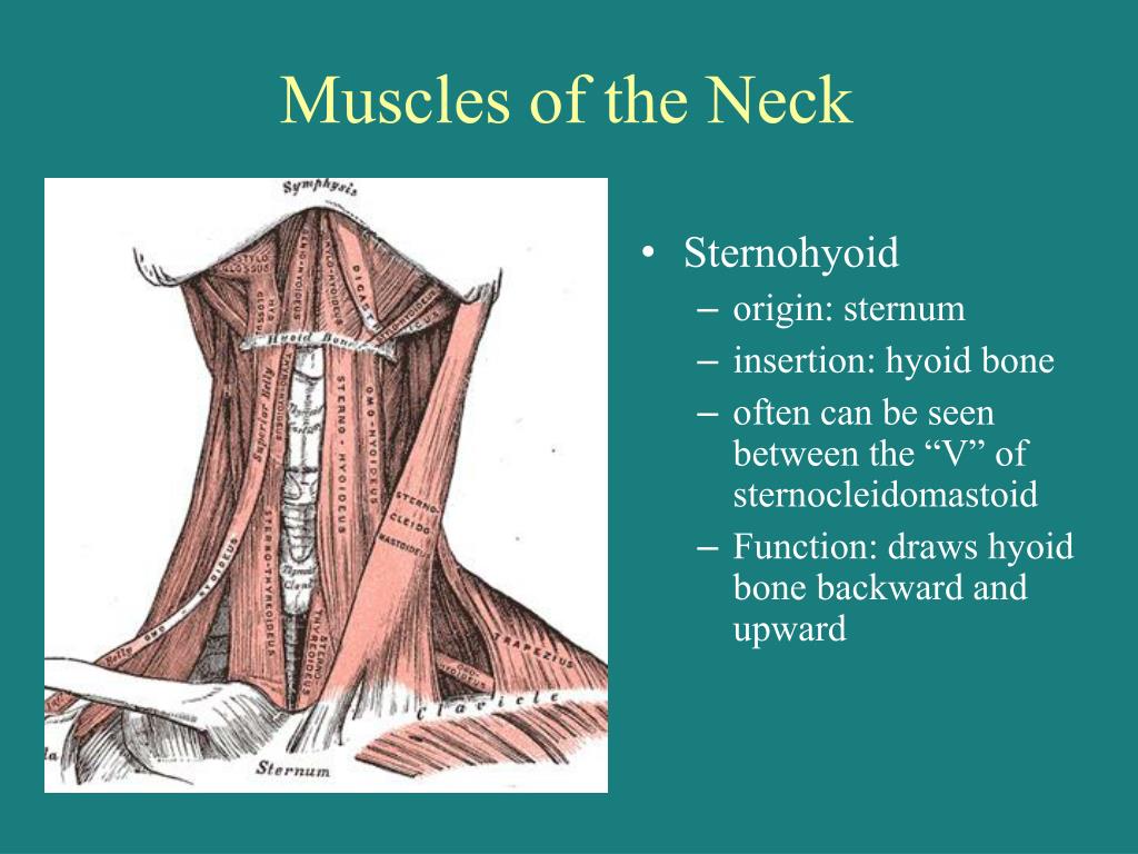
What is the muscle that attaches to the hyoid bone?
Muscles of the neck. Lateral view. Sternohyoid muscle labeled. The sternohyoid muscle is a thin, narrow muscle attaching the hyoid bone to the sternum. It is one of the paired strap muscles of the infrahyoid muscles, serving to depress the hyoid bone. It is innervated by the ansa cervicalis.
Which muscle is supplied by a branch of the ansa cervicalis?
The sternohyoid muscle is supplied by a branch of the ansa cervicalis.
Which surface of the sternum and costal cartilages shows transversus thoracis?
Posterior surface of sternum and costal cartilages, showing Transversus thoracis.
Which muscle is crossed anteriorly by the oblique fibers of the sternohyoid?
Sternothyroid has numerous important anatomical relationships to neurovascular and glandular structures of the neck. It is crossed anteriorly by the oblique fibers of the sternohyoid and the superior belly of omohyoid. On either side of the neck, the muscle is anterior to the lateral lobe of the thyroid gland.
Where does the Sternothyroid originate?
It originates from the posterior edge of the costal cartilage of the first rib as well as the posterior surface of the manubrium of the sternum.
What muscle pulls the lamina of the thyroid cartilage away from the hyoid bone,?
When acting alone, the sternothyroid muscle pulls the lamina of the thyroid cartilage away from the hyoid bone, thus opening the laryngeal inlet. This is particularly beneficial during forced inspiration so that air enters the lower airway.
What is the deep layer of the infrahyoid muscle?
The superficial layer includes sternohyoid and omohyoid, while the deep layer is made up of sternothyroid and thyrohyoid.
Which lobe of the thyroid is anterior to the thyroid?
On either side of the neck, the muscle is anterior to the lateral lobe of the thyroid gland. The muscle lies anterior to the brachiocephalic trunk on the right and the common carotid artery and brachiocephalic vein on the left. It is also anterior to the external laryngeal nerve and the superior thyroid artery.
Where is blood supplied to sternothyroid?
Blood is supplied to sternothyroid by the branches of the lingual and superior thyroid arteries.
Can sternothyroid muscle be divided?
The sternothyroid muscle may be divided during thyroidectomy (removal of the diseased thyroid gland ). This allows the surgeon to gain access to the superior pedicle of the gland. To date, there is no evidence to support any relationship between the division of this muscle and adverse functional outcomes postoperatively.
Which muscle is shorter, sternohyoid or sternothyroid?
The sternothyroid muscle is shorter and wider than the sternohyoid muscle. The sternothyroid muscle lies underneath the sternohyoid muscle. Nerves from the upper cervical nerve travel through the cervical ansa, or cervical loop, and supply the sternothyroid muscle. The main function of the sternothyroid is to depress the larynx.
What is the function of the sternothyroid gland?
The main function of the sternothyroid is to depress the larynx. This is important for mastication, or chewing, as well as swallowing. This raising and lowering of the larynx can also affect vocal range, the ability to control pitch, and volume.
Where is the sternothyroid muscle located?
The sternothyroid muscle, also called the sternothyroideus, is in the neck area. The muscle goes from the sternum, or breastbone, to the edge of the thyroid cartilage.
