What are suboccipital muscles?
Suboccipital muscles are located just below the occipital bone, right where the base of your skull meets your neck. This cluster of muscles is mainly responsible for posture and movements between your skull and top vertebrae.
Why is it important to strengthen your suboccipital muscles?
Specifically, strengthening your suboccipital muscles will not only prevent tension but also improve posture. Suboccipital muscles are located just below the occipital bone, right where the base of your skull meets your neck. This cluster of muscles is mainly responsible for posture and movements between your skull and top vertebrae.
What is the function of the suboccipital nerve?
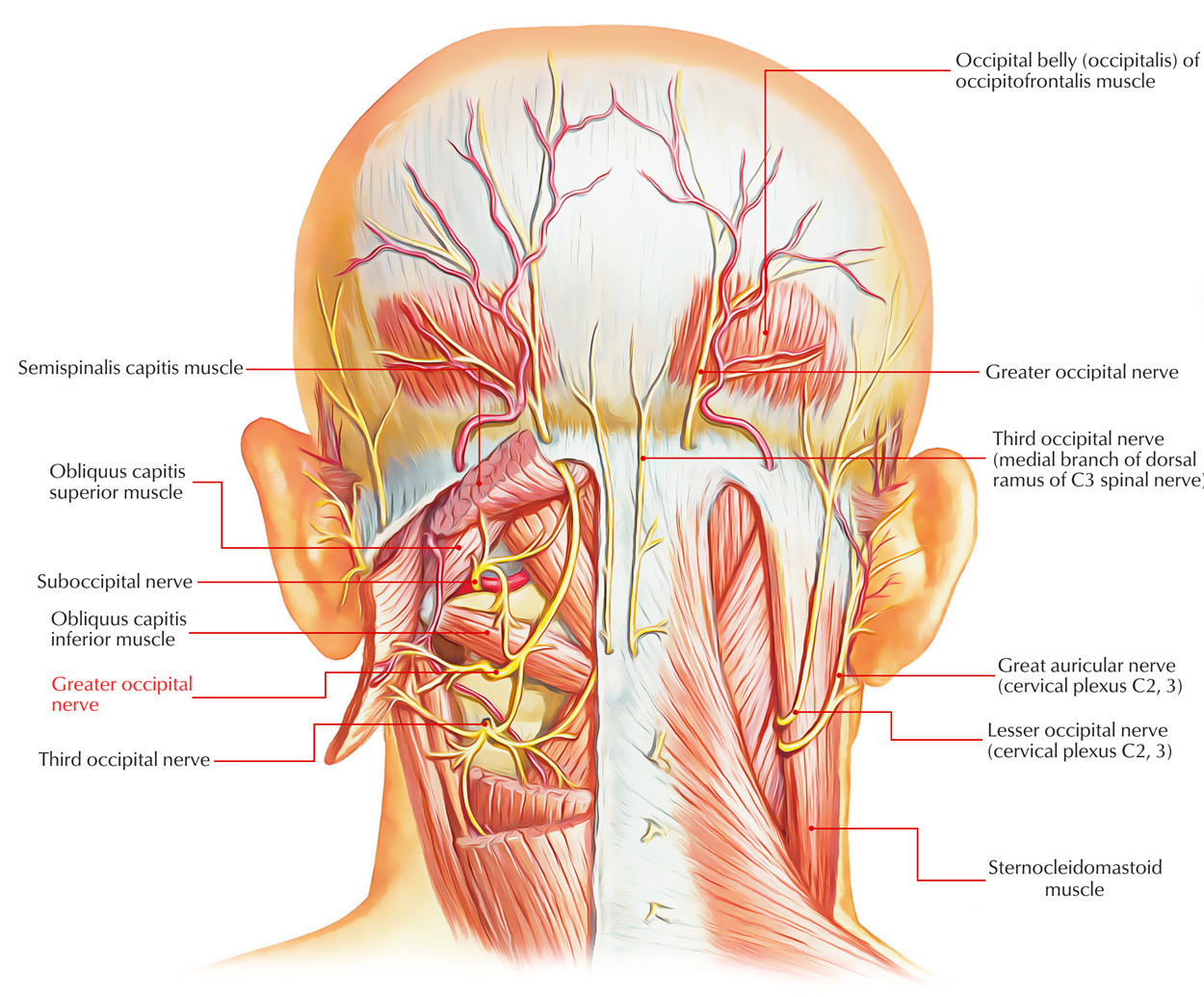
Neuroanatomy, Suboccipital Nerve - StatPearls - NCBI Bookshelf The suboccipital nerve, also known as the dorsal ramus of the first cervical nerve, arises from the posterior ramus of the C1 nerve. The primary function of the suboccipital nerve is the innervation of the suboccipital muscles.
What is the function of the suboccipital triangle?
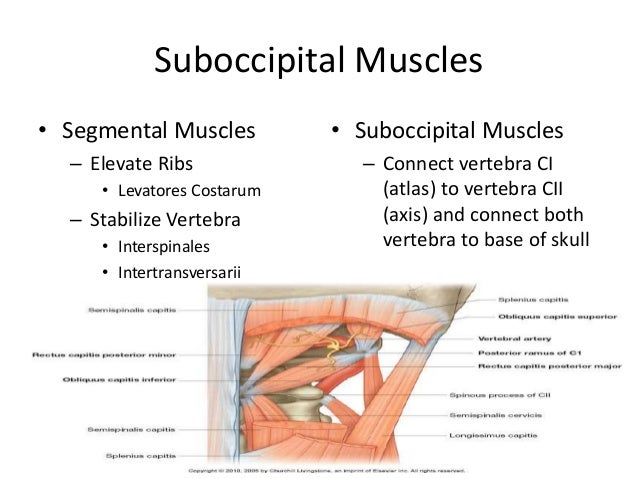
The suboccipital triangle is made up of the rectus capitis posterior major and both of the obliquus capitis muscles. This trio provides fine motor function in movements of the head. Each muscle in the suboccipital triangle is responsible for its respective region and direction of movement.
What are the actions of suboccipital muscles?
The suboccipital muscles are a group of four paired deep muscles located at the base of the occipital bone. Their main actions are to extend and rotate the head. They consist of the rectus capitis posterior major, rectus capitis posterior minor, obliquus capitis superior and obliquus capitis inferior.
What are the 4 suboccipital muscles?
The suboccipital muscles are a group of four muscles located inferior to the occipital bone. These four muscles include the rectus capitis posterior major, rectus capitis posterior minor, obliquus capitis superior, and obliquus capitis inferior.
How do you release tension in the suboccipital muscle?
0:532:22Suboccipital Tension Release - YouTubeYouTubeStart of suggested clipEnd of suggested clipAsk your patient to relax their head into your hands create a fulcrum by extending your fingersMoreAsk your patient to relax their head into your hands create a fulcrum by extending your fingers anteriorly into the suboccipital.
What causes tight suboccipital muscles?
The suboccipital muscles commonly become tense and tender due to factors such as eye strain, wearing new eyeglasses, poor ergonomics at a computer workstation, grinding the teeth, slouching posture, and trauma (such as a whiplash injury).
How do you engage suboccipital muscles?
0:060:46Suboccipital Stretch for Neck Pain and Headaches - YouTubeYouTubeStart of suggested clipEnd of suggested clipSo in order to get a good stretch on these muscles you have to maximally retract your cervical spineMoreSo in order to get a good stretch on these muscles you have to maximally retract your cervical spine and chin all the way down.
What causes suboccipital trigger points?
2:1911:28Trigger Point Therapy | Tension Headache | Suboccipital MusclesYouTubeStart of suggested clipEnd of suggested clipAre a whiplash type injury. Or sub-optimal posture the very common situation where people slumpMoreAre a whiplash type injury. Or sub-optimal posture the very common situation where people slump forward their chin juts forward and and they shorten their neck.
Can you do a Suboccipital release on yourself?
Self release is a great way to release the tension that builds up in the suboccipital muscles. These are a group of muscles just behind and below the skull that are infamous headache generators.
How do I get a knot out of the base of my skull?
1:059:26How to Fix Muscle Knots in Your Neck and Shoulder in 30 SECONDSYouTubeStart of suggested clipEnd of suggested clipAnd let's go over the base of the skull. Or over the spot that feels very tender. Put a little bitMoreAnd let's go over the base of the skull. Or over the spot that feels very tender. Put a little bit of pressure in there and start to do nice circular motions over those muscles.
How do you get rid of a Suboccipital headache?
You can try to:Apply heat to your neck.Rest in a quiet room.Massage tight and painful neck muscles.Take over-the-counter anti-inflammatory drugs, like naproxen or ibuprofen.
How do you release the occipital nerve?
Surgical options include occipital release surgery. In this outpatient procedure, your doctor makes an incision in the back of the neck to expose your occipital nerves and release them from the surrounding connective tissue and muscles that may be compressing them.
Can suboccipital muscles cause headaches?
At the base of the skull, there is a group of muscles called the suboccipital muscles. They can cause headache pain for many people. These four pairs of muscles are responsible for subtle movements between the skull and first and second vertebrae in the neck.
Why do I have Suboccipital pain?
Over time, the upper cervical ligaments stretch too far, recruiting the suboccipital muscles to tighten. Once fatigued, they also become a source of significant pain. Suboccipital muscle tension or headache is one of the major symptoms of someone having upper cervical instability.
What is the suboccipital muscle?
Already a member? Log In. The suboccipital muscles are a group of four muscles situated underneath the occipital bone. All the muscles in this group are innervated by the suboccipital nerve.
Which suboccipital muscle is most medial?
The rectus capitis posterior minor is the most medial of the suboccipital muscles. There is a connective tissue bridge between this muscle and the dura mater (outer membrane of the meninges) – which may play a role in cervicogenic headaches.
Where is the rectus capitis posterior major located?
The rectus capitis posterior major is the larger of the rectus capitis muscles. It is located laterally to the rectus capitis posterior minor. Attachments: Originates from the spinous process of the C2 vertebrae (axis), and inserts into the lateral part of the inferior nuchal line of the occipital bone.
Which muscle has no attachment to the cranium?
As its name suggests, the obliquus capitis inferior is the most inferiorly positioned of the suboccipital muscles. Additionally, it is the only capitis muscle that has no attachment to the cranium. Attachments: Originates from the spinous process of the C2 vertebra, and attaches into the transverse process of C1.
Where are the splenic muscles located?
They are located within the suboccipital compartment of the neck; deep to the sternocleidomastoid, trapezius, splenius and semispinalis muscles. They collectively act to extend and rotate the head.
Which muscle is inferior to the cranium?
As its name suggests, the obliquus capitis inferior is the most inferiorly positioned of the suboccipital muscles. Additionally, it is the only capitis muscle that has no attachment to the cranium.
Which nerve innervates all of the suboccipital muscles?
The suboccipital nerve innervates all of the suboccipital muscles. From which nerve root does it arise?
Where are the suboccipital muscles located?
Suboccipital muscles are located just below the occipital bone, right where the base of your skull meets your neck. This cluster of muscles is mainly responsible for posture and movements between your skull and top vertebrae.
What is the suboccipital triangle?
The suboccipital triangle is made up of the rectus capitis posterior major and both of the obliquus capitis muscles. This trio provides fine motor function in movements of the head.
What is the superior obliquus?
OBLIQUUS CAPITIS SUPERIOR. This muscle is explicitly in charge of flexing your head sideways. It arises from the atlas bone and, like the rectus capitis muscles, inserts into the occipital bone. The obliquus capitis superior actually looks like two muscles, located on each side of the occipital region.
What is the Latin name for the posterior minor rectus capitis?
RECTUS CAPITIS POSTERIOR MINOR. You may have guessed it already - rectus capitis posterior minor is Latin for “lesser posterior straight muscle of the head.”. This muscle begins at the atlas, which is the spine’s first vertebra, located above the axis.
What muscles extend the head forward?
While they all contribute to the suboccipital muscles’ overall functions, each has its own distinctions. RECTUS CAPITIS. Made up of the rectus capitis posterior major and the rectus capitis posterior minor, the rectus capitis works to extend your head forward. RECTUS CAPITIS POSTERIOR MAJOR.
Which muscle is responsible for the direction of the head?
Each muscle in the suboccipital triangle is responsible for its respective region and direction of movement. The rectus capitis posterior major assists with moving the head medially or rotating it inward. The obliquus capitis muscles move the head laterally, which describes outward rotation of the head.
Where does the posterior straight muscle of the head start?
Latin for “larger posterior straight muscle of the head,” this muscle begins at the spinous process (the protrusion from the vertebra) of the axis (the second vertebra of your spine). The end inserts into the occipital bone at the base of your skull.
What are the two muscles on the occipital bone?
These are four paired muscles on the underside of the occipital bone; the two straight muscles ( rectus) and the two oblique muscles ( obliquus ). The muscles are named. Rectus capitis posterior major goes from the spinous process of the axis (C2) to the occipital bone.
Where does the obliquus capitis go?
Obliquus capitis superior goes from the transverse process of the atlas to the occiput. Obliquus capitis inferior goes from the spine of the axis vertebra to the transverse process of the atlas. They are innervated by the suboccipital nerve .
What are the suboccipital muscles?
Key points. The suboccipital muscles are a group of four paired deep muscles located at the base of the occipital bone. Their main actions are to extend and rotate the head. They consist of the rectus capitis posterior major, rectus capitis posterior minor, obliquus capitis superior and obliquus capitis inferior.
Which muscle forms the medial border of the suboccipital triangle?
Three of the suboccipital muscles form the boundaries of the suboccipital triangle: The medial border of the triangle is formed by the rectus capitis posterior major muscle. The lateral border of the triangle is formed by the obliquus capitis superior muscle.
What muscles are in the neck?
The suboccipital muscles are a set of four paired muscles in the back of the neck. The suboccipital muscles attach to the atlas (C1), axis (C2) and occipital bone, connecting the atlas to the axis and the two vertebrae to the base of the skull. They are deep to the trapezius, splenius and semispinalis muscles and superficial to ...
What muscle forms the inferior border of the triangle?
The inferior border of the triangle is formed by the obliquus capitis inferior muscle.
Which muscles are involved in the head movement?
They are deep to the trapezius, splenius and semispinalis muscles and superficial to the atlanto-occipital and atlanto-axial joints. These small muscles contribute to the movements of the head. When the suboccipital muscles contract, they extend the head at the atlanto-occipital joint and rotate the head at the atlanto-axial joints. Figure 1.
Which muscles extend the head at the axial joints?
These small muscles contribute to the movements of the head. When the suboccipital muscles contract, they extend the head at the atlanto-occipital joint and rotate the head at the atlanto-axial joints.
What is the purpose of Part One of our two part special on diabetes?
Part one of our two part special on diabetes focuses on the firsthand experience of a patient living with diabetes. We discuss what it is like to be diagnosed with Type 1 diabetes and to live with and manage the condition day-to-day. We hope that this episode will be useful for students, medical professionals, and anyone who wants to understand more about the challenges of managing this condition. Guest: Ashwin Bali