Why is the circle of Willis so important?
The circle of Willis plays an important role, as it allows for proper blood flow from the arteries to both the front and back hemispheres of the brain. The arteries that stem off from the circle of Willis supply much of the blood to the brain.
What is the circle of Willis and what does it do?
Overview. The Circle of Willis is the joining area of several arteries at the bottom (inferior) side of the brain. At the Circle of Willis, the internal carotid arteries branch into smaller arteries that supply oxygenated blood to over 80% of the cerebrum.
What is the circle of Willis and why is it important quizlet?
The circle of willis is an important means of collateral circulation in the event of gradual obstruction of one of the major arteries forming the circle. Sudden occlusion, even in only partial, results in neurological deficits.
How does blood flow in the circle of Willis?
The main cerebral distribution center for blood flow is the circle of Willis (see [15, 37]), a ring-like network of collateral vessels; see Figure 1(left). Blood is delivered to the brain through the two internal carotid arteries and the two vertebral arteries that join intracranially to form the basilar artery.
How do you remember the circle of Willis?
0:003:08MNEMONIC Brain's Blood Supply: MEMORIZE in 3 Minutes - YouTubeYouTubeStart of suggested clipEnd of suggested clipOther blood vessels hence the Circle of Willis is the spirit bowl appearing on top of the meditatingMoreOther blood vessels hence the Circle of Willis is the spirit bowl appearing on top of the meditating cow's head as well as the meditating cows horns.
What arteries are involved in the circle of Willis?
The circle of Willis begins to form when the right and left internal carotid artery (ICA) enters the cranial cavity and each one divides into two main branches: the anterior cerebral artery (ACA) and middle cerebral artery (MCA).
What does the circle of Willis surround?
The circle of Willis surrounds the optic tracts, pituitary stalk, and basal hypothalamus. It includes the three sets of paired cerebral arteries plus the anterior communicating artery, interconnecting the ACAs, and the posterior communicating arteries, interconnecting the MCAs and PCAs.
What are the three main vessels that compose the circle of Willis?
Internal carotid artery (left and right) Posterior cerebral artery (left and right) Posterior communicating artery (left and right)
What is the most common anomaly of the circle of Willis?
hypoplasiaThe most common anomaly of the circle of Willis in normal brains was hypoplasia of one or other components of the circle. Arteries of less than 1 mm in external diameter were considered hypoplastic, except for the communicating arteries, where less than 0.5 mm was considered hypoplastic.
What arteries are part of the circle of Willis?
The anterior communicating, anterior cerebral, internal carotid, posterior communicating, posterior cerebral, and basilar arteries are all part of the circle of Willis (see Fig. 3-13).
What are the 4 arteries that make up the circle of Willis?
The circle of Willis is a part of the cerebral circulation and is composed of the following arteries:Anterior cerebral artery (left and right)Anterior communicating artery.Internal carotid artery (left and right)Posterior cerebral artery (left and right)Posterior communicating artery (left and right)
How many arteries are in the circle of Willis?
The circle of Willis is a group of blood vessels in the brain that connect with each other, forming a continuous structure that resembles a circle. These nine arteries supply blood to a large portion of the brain. Most of the time, blood can flow through the vessels of the circle of Willis without any interruption.
Why is the circle of Willis shaped like a hexagon?
Although it has nine sides, the circle of Willis is shaped more like a hexagon because the ICAs are very short and the two PCAs are almost straight.
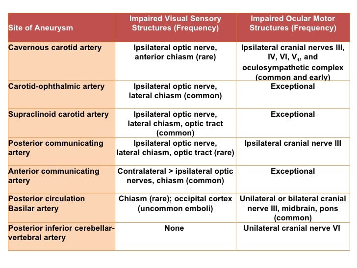
What is the effect of an aneurysm on the optic chiasm?
An aneurysm in the circle of Willis can impinge on the optic chiasm, which may impair vision in one or more visual fields. It can also place pressure on the pituitary stalk (a part of the pituitary gland), disturbing its function. 3
What is the unique feature of the circle of Willis?
One of the unique features of the circle of Willis is that its continuous structure creates a redundant blood supply in the brain. 2 What this means is that the ACOM and PCAs, which do not directly send blood to the brain, connect the ACAs and the ICAs—arteries that directly send blood to the brain.
Why is redundancy not effective?
If the arteries bleed, the built-in redundancy is not particularly effective because blood in the brain causes irritation and damage. Loss of blood from a damaged artery is likely to affect blood flow in other arteries in the circle of Willis as well.
What is the function of the circle of Willis?
Function. Associated Conditions. Rehabilitation. The circle of Willis is a group of blood vessels in the brain that connect with each other , forming a continuous structure that resembles a circle. These nine arteries supply blood to a large portion of the brain. Most of the time, blood can flow through the vessels of the circle ...
Which arteries run along the circle of Willis?
The left and right anterior cerebral arteries (ACAs): These vessels run along the sides of the circle of Willis. The left and right internal carotid arteries (ICAs): The ICAs travel in the front of the neck, through the carotid canal, to enter into the brain.
Which arteries provide blood to the brain?
Several of the arteries of the circle of Willis branch into smaller vessels that directly provide blood to the brain.
What is the posterior arc of the circle of Willis?
Posterior arc of the circle of Willis. The posterior arc of the circle of Willis is formed by the posterior cerebral arteries (PCA), on each side, and the posterior communicating arteries (PComm), which connect the posterior cerebral arteries to their ipsilateral internal carotid arteries.
What is the branch of the internal carotid?
At the point of connection between the anterior cerebral and internal carotid arteries, the internal carotid gives off its lateral terminal branch known as the middle cerebral artery (MCA). Together with the AComm and the middle cerebral arteries, the anterior cerebral arteries form the anterior cerebral circulation.
What is the AComm?
Anteriorly, the short anterior communicating artery (AComm) forms an anastomotic channel between the right and left anterior cerebral arteries (ACA), which are terminal branches of the internal carotid artery within the cranial cavity . The anterior cerebral arteries travel toward the anteromedial aspects of the interhemispheric fissure of the brain, to supply the midline regions of the frontal, parietal and cingulate cortices as well as the corpus callosum.
What is the circle of Willis?
Cerebral arterial circle (Circulus arteriosus cerebri) The circle of Willis ( cerebral arterial circle or circulus arteriosus) is an anastomotic ring of arteries located at the base of the brain. This arterial anastomotic circle connects the two major arterial systems to the brain, the internal carotid arteries and the vertebrobasilar ...
Why is the circle of Willis restricted?
This is due to significant variations in the size of component arteries that limit the normal flow of blood through these regions.
Which arteries supply the occipital lobe of the brain?
The vertebral arteries, basilar artery, posterior cerebral arteries, together with the PComm form the posterior cerebral circulation. The posterior cerebral arteries through their central and cortical branches supply the occipital lobe of the brain, the inferior aspect of the temporal lobes , midbrain, thalamus and choroid plexus of the third and lateral ventricles.
Where are the perforated arteries located?
Several small perforating (central) arteries emerge from the circle of Willis, many of which pass into the brain through the anterior and posterior perforated substances, which are areas of grey matter located at the basal forebrain and the interpeduncular fossa respectively.
What is the most attractive method of angiography?
Among these techniques, transcranial color-coded Duplex sonography is the most attractive, providing both morphological and hemodynamic data ( Fig. 2 ). This method adds B-mode imaging and color coding of the Doppler signal, allowing reliable assessment of the insonated artery and direction of the flow. To improve its sensitivity, Duplex sonography must be combined with another noninvasive method. In this regard, time-of-flight (TOF) magnetic resonance (MR) angiography has been widely used for imaging intracranial vascular structures ( Fig. 3). This technique provides a high signal intensity of the arterial lumen owing to the inflow effect of blood flow during its passage in the acquisition volume. Data are usually transferred to a workstation and subvolumes are generated to isolate the circle of Willis from the surrounding structures. Then, by applying the maximum intensity projection (MIP) algorithm, angiographic images may be displayed in multiple projections to improve the interpretation of images. An additional technique of reconstruction, such as multiplanar reformation, is sometimes helpful to circumvent the problem of vessel overlap and to better define the anatomy of the vessel, but this requires additional postprocessing time. MR angiogram may depict the presence of a vessel segment with a diameter of at least 1 mm and is widely used for the detection of intracranial aneurysms of the circle of Willis. Despite the interest in TOF sequences to assess intracranial arterial anatomy, the imaging coverage often remains limited and does not permit a complete visualization of both the anterior and the posterior circulation from the skull base to the vertex. Moreover, the scan time is long, leading to a frequent degradation of images by motion artifacts, and a signal loss is usually observed in the case of stenosis due to turbulent flow. MR angiographic sequences have been developed during the past 5 years to circumvent these persisting drawbacks. These recent technological advances, including high-performance gradient systems, improve the vascular conspicuity in a large imaging volume in a scan time of less than 30 sec ( Fig. 4 ). However, appropriate timing of the contrast bolus injection is required to optimize the arterial phase of contrast enhancement, and the venous enhancement may create a superimposition of vessels.
What is MR angiogram?
MR angiogram may depict the presence of a vessel segment with a diameter of at least 1 mm and is widely used for the detection of intracranial aneurysms of the circle of Willis.
What is computed tomographic angiography?
Computed tomographic angiography is another noninvasive technique allowing accurate assessment of intracranial circulation. This method is based on a rapid acquisition of the entire volume owing to the continuous rotation of the gantry and simultaneous displacement of the examination table. Data acquisition by using narrow collimation can be reconstructed with overlapping sections, providing high spatial resolution images. MIP and shaded surface display are the main techniques of reconstruction used to provide information about the complex anatomy of the circle of Willis ( Fig. 5 ). The main limitations of this method concern the radiation dose, the need for iodinated contrast material infusion, and the artifacts caused by the skull base. The bone structures may be removed from axial sections by using a manual segmentation or sophisticated software, but this procedure is time-consuming.
What are the arteries in the Circle of Willis?
The Circle of Willis is a wreath of interconnected arteries that surrounds the optic chiasm, the tuber cinereum, and the region between the cerebral peduncles at the ventral surface of the diencephalon. It is formed by anastomotic branches of the two ICAs, the horizontal (A1) segments of the ACAs, ACommA, the two PCommAs, the horizontal segments (P1) of both PCAs, and the BA (Figure 2 ). Penetrating vessels from the arteries of the Circle of Willis supply structures within its wreath, such as the optic chiasm and hypothalamus.
Where are berry aneurysms most likely to occur?
The most likely site of these berry aneurysms is at the junctions of arteries in the circle of Willis.
What is the gold standard for vascular anatomy?
Conventional angiography is considered the gold standard for evaluating vascular anatomy ( Fig. 1 ). This technique requires anesthesia, a femoral arterial approach, a selective catheterism of supra-aortic vessels by using a catheter, and the administration of iodinated contrast material for each selective view.
What imaging techniques are used to determine the anatomical structure of the circle of Willis?
Several imaging techniques may be useful to provide anatomical information of the main branches of the circle of Willis including digital subtraction angiography (DSA), Doppler ultrasound, magnetic resonance (MR) angiography, and computed tomography angiography.
What is the circle of Willis?
The Circle of Willis is a fascinating vascular structure within the brain that keeps blood flowing (maintains vital perfusion) to all areas of the brain if one of the four paired vessels (the right and left carotid and vertebral arteries) are compromised by disease or trauma .
What are the 3 arteries in the circle?
The 3 arteries completing the circle are the paired posterior communicating arteries and the single anterior communicating artery (the short vessel not labeled at the top of the circle, connecting the two anterior cerebral arteries).
What is the ring structure of the brain?
Continue Reading. The movement of blood supply through network of cerebral arteries and veins in the brain is cerebral circulation. The Circle of Willis is a ring structure made of arteries which is located in the inferior brain surface. It is responsible for supplying blood to the brain and its surroundings.
How many arteries are there in the cerebral circulation?
It consists of five arteries and is a part of the cerebral circulation. Further it is a circulatory anastomosis which means it connects two blood vessels, either two arteries or two veins or arteries and veins. The five arteries are: Anterior cerebral artery (left and right) Anterior communicating artery. Intern.
What are the two types of arteries?
These arteries include: 1. anterior & posterior cerebral arteries 2. Anterior and posterior communicating arteries 3. Internal carotid artery. The circle of willis provides collateral circulation in case of damage of one of the arteries, blood can still be supplied to the cerebrum normally.
Why do all the arrows in the brain feed the same circle?
Since all the blood flow to the brain has to pass through this circle before it can go into the brain, this means you could have a block in one of the arteries going to the brain the others can compensate because they all feed the same circle which then relays the blood into the brain (the other arrows). It is sim.
Why is the Circle of Willis important?
It's importance is that it can provide redundant blood flow should certain portions of the blood supply to the brain become occluded. Not sure why this is in Geometry, though. Seniors using loophole to save for retirement.
What is the purpose of the circle of Willis?
Collateral circulation through the circle of Willis helps maintain blood flow through the cerebrum, even when one or both carotid arteries are blocked . The circle of Willis is a continuous loop of arteries in the brain that provides collateral circulation. When you turn your head from side-to-side or up and down, ...
What happens if you turn your head to one side?
Likewise, when you turn your head as far as possible to one side, one of your internal carotid arteries is compressed. Without the circle of Willis, this would stop blood flow to one side of your brain. However, the circle of Willis ensures that blood from the other internal carotid artery is distributed throughout the brain.
What is collateral circulation?
Collateral circulation also provides an alternate path for blood to reach any part of the brain, should one of the major arteries become obstructed. If an internal carotid artery is blocked, for example, the other major arteries partly compensate for the loss of blood flow. Other cerebral arteries may also connect, providing collateral circulation to other parts of the brain. This varies among individuals, however. In fact, a complete circle of Willis may occur in only 40 percent of people. The more collateral circulation you have, the better your chance of surviving a stroke without severe deficits.
What happens when a fatty streak is enlarged?
At least four things begin to happen as the plaque enlarges. First, some of the white blood cells inside the fatty streak begin to die , forming what doctors call a “necrotic core.” Second, the muscle cells of the artery wall begin to grow over the necrotic core. Third, blood cells called platelets begin to stick to the injured endothelium and facilitate the formation of small blood clots on the surface of the plaque. Platelets are an essential component of blood clotting in arteries. Normally, they circulate without doing much. But when they sense an injury that causes bleeding, they clump together to stop it by forming a plug. They also present a sticky surface that attracts actual blood clots formed from circulating proteins. As the plaque enlarges, platelets and blood clots get mixed in with the cells covering the necrotic core to form a fibrous cap. The cap is delicate and prone to rupture, releasing its contents into the bloodstream. This is a highly dangerous situation.
What is the function of platelets in arteries?
Platelets are an essential component of blood clotting in arteries. Normally, they circulate without doing much. But when they sense an injury that causes bleeding, they clump together to stop it by forming a plug. They also present a sticky surface that attracts actual blood clots formed from circulating proteins.
What is the first step in the development of atherosclerosis?
The first is called “initiation.”. The lining of arteries (endothelium) is a smooth, inert surface that blood flows across. High cholesterol levels, high blood pressure, toxins in cigarette smoke, and other factors can damage the endothelium and stimulate the development of atherosclerosis. Injured endothelial cells interact with white blood cells, ...
Which lipoproteins carry cholesterol?
Low-density lipoproteins (LDL) and high-density lipoproteins (HDL) contain high concentrations of cholesterol, and chylomicrons are another dangerous fat. LDL, VLDL, and chylomicrons carry fats from the gut and the liver throughout your body to meet the cells’ metabolic needs. HDL carries fats to the liver, which prepares it for removal from ...
