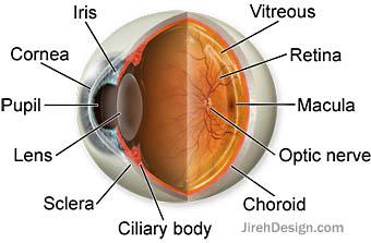
What are the inner and outer parts of the eye?
Eye Parts Description and Functions; Cornea: The cornea is the outer covering of the eye. This dome-shaped layer protects your eye from elements that could cause damage to the inner parts of the eye. There are several layers of the cornea, creating a tough layer that provides additional protection.
What is the inner corner of the eye called?
What are the internal structures of the eye?
- Lens.
- Retina.
- Aqueous humour.
- Optic nerve.
- Vitreous humour.
What is the inside corner of your eye called?
What Is the Inside Corner of Your Eye Called? According to Stanford Children's Health, the inside corner of the eye is called the lacrimal caruncle. This small pink nodule is made of modified oil and sweat glands, and it may become inflamed and itchy in response to allergies.
What are the basic parts of the eye?
BASIC PARTS OF THE EYEThis video describes the 8 most common basic anatomy parts of the eyeball. It includes the cornea, lens, iris, pupil, vitreous, retin...
What is the inner part of the eye called?
retinaThe inner layer of the eye, or retina, is similar to film in a camera. It receives light from an image we are looking at, and converts that light into electrical impulses which are sent through the fibres of the optic nerve to the brain.
What is the surface of the eye called?
The sclera is the tough outer layer of the eyeball (the white of the eye). The slight bulge in the sclera at the front of the eye is a clear, thin, dome-shaped tissue called the cornea. The cornea directs light rays into the eye and helps focus them on the retina.
What is the inner surface of the eyelids called?
Conjunctiva. The conjunctiva is a smooth, translucent mucous membrane. Palpebral conjunctiva lines the posterior surface of the lids as tarsal conjunctiva (from the mucocutaneous junction of the lid margin to the tarsal plate border) and continues as orbital palpebral conjunctiva into the fornix.
What covers the inner surface of the eye and eyelids?
Conjunctiva. The conjunctiva is a transparent mucous membrane that lines the inner surface of the eyelids (palpebral conjunctiva) and the anterior surface of the sclera (bulbar conjunctiva).
What is the surface of the cornea?
The cornea is the clear outer dome of the eye that allows light to enter and become focused through the lens onto the retina. The cornea is an avascular tissue and receives most of its nutrients from the tears, the air, and fluid inside the eye....Regular Hours.Monday8:00 am – 5:00 pmFriday8:00 am – 5:00 pm3 more rows
What covers the outer surface of the eye?
The conjunctiva is the thin, transparent tissue that covers the outer surface of the eye. The cornea is a clear, dome-shaped outer coating that covers the front of the eye. Light passes through the cornea to the lens. The cornea provides much of the eye's focusing power.
What is conjunctiva?
The conjunctiva of the eye provides protection and lubrication of the eye by the production of mucus and tears. It prevents microbial entrance into the eye and plays a role in immune surveillance. It lines the inside of the eyelids and provides a covering to the sclera.
What is conjunctiva and cornea?
The conjunctiva is the clear tissue that covers the white part of the eye and the inside of the eyelids. A healthy conjunctiva is necessary for the eye to function normally, as it helps to create a suitable environment for the cornea, which is responsible for focusing most of the light that enters the eye.
What is sclera and conjunctiva?
The conjunctiva is the membrane that lines the eyelid and loops back to cover the sclera (the tough white fiber layer covering the eye), right up to the edge of the cornea (the clear layer in front of the iris and pupil—see Structure and Function of the Eyes.
What is the retina?
Retina Definition. The retina is the sensory membrane that lines the inner surface of the back of the eyeball. It's composed of several layers, including one that contains specialized cells called photoreceptors. There are two types of photoreceptor cells in the human eye — rods and cones. Rod photoreceptors detect motion, provide black-and-white ...
Where does vision begin?
The Retina: Where Vision Begins. The first step in the process of vision is the conversion of light into signals that can be interpreted in the brain. This takes place in the retina, which is located in the back of the eye.
What is retinal detachment?
A retinal detachment — a pulling away of the retina from the underlying choroid layer of the eye that provides its nourishment — is a medical emergency. If the retina is not surgically reattached as soon as possible, permanent and worsening vision loss can occur. [Read more about retinal detachment .]
What is the cause of distorted vision?
Central serous retinopathy. This is when fluid builds up under the central retina, causing distorted vision. Though the cause of central serous retinopathy (CSR) often is unknown, it tends to affect men in their 30s to 50s more frequently than women, and stress appears to be a major risk factor. Hypertensive retinopathy.
What are the most common retinal problems?
There is a wide variety of retina problems, conditions and diseases. Here is a short list of the more common retina problems: Macular degeneration. Age-related macular degeneration (AMD) is the most common serious, age-related eye disease, affecting 9.1 million Americans. And the prevalence of AMD — which affects one in 14 Americans over age 40 ...
What is the vascular layer of the eye?
The vascular layer of the eye lying between the retina and sclera. This layer furnishes nourishment to outer layers of the retina. A ring of tissue inside the eye composed of ciliary muscle, which is involved in lens focusing and control of the intraocular pressure.
Where is fluid filled space in the eye?
Fluid-filled space inside the eye between the iris and the innermost corneal surface. Junction of the front surface of the iris and the back surface of the cornea, where aqueous fluid filters out of the eye. The vascular layer of the eye lying between the retina and sclera.
What is the material that fills the rear two-thirds of the interior of the eyeball, between the lens
Vitreous. Transparent, colorless, gelatinous material that fills the rear two-thirds of the interior of the eyeball, between the lens and the retina. Zonules. Fibers that suspend the lens from the ciliary body and hold it in position.
What is the retina?
The retina lines the rear two-thirds of the eye and consists of layers that include rods and cones. This part of the eye can be compared to film in a camera. This part of the eye is directly affected when Macular Degeneration, Diabetic Retinopathy, Retinal Tears & Detachments, and Retinal Vascular Disease occur. Sclera.
What is the primary sensory nerve of the eye?
A cataract is a lens that has become cloudy or opaque. Macula. The centralized area of the retina responsible for acute central vision necessary for reading and detail work. Optic Nerve. The primary sensory nerve of the eye.
Which part of the eye produces aqueous?
There are also ciliary processes that produce aqueous. Cornea. The transparent front segment of the eye that covers the iris, pupil and anterior chamber, and provides most of the eye’s optical power. Iris.
Which nerve carries impulses for sight from the retina to the brain?
Optic Nerve. The primary sensory nerve of the eye. It carries impulses for sight from the retina to the brain. Pupil. A black circular opening in the center of the iris that regulates the amount of light that enters the eye. Retina.
What is the innermost layer of the cornea?
The corneal endothelium. This is the innermost layer of the cornea. The back of the endothelium is bathed in the clear aqueous humor that fills the space between the cornea and the iris and pupil. The corneal endothelium is only a single layer of cells thick and measures about 5 microns.
Which layer of the eye protects the cornea from scratches?
The corneal epithelium provides an optimal surface for the tear film to spread across the surface of the eye to keep it moist and healthy and to maintain clear, stable vision. Bowman' s layer. The dense nature of Bowman's layer helps prevent corneal scratches from penetrating into the corneal stroma.
What is corneal ulcer?
A corneal ulcer is a serious abscess-like infection of the cornea that can lead to significant pain, scarring and vision loss. [Read more about corneal ulcers.] Corneal dystrophy. A dystrophy is a weakening or degeneration of a tissue.
What is the ring on the periphery of the cornea called?
Appearance of arcus senilis (corneal arcus). Arcus senilis. As people get older, a white ring often develops in the periphery of the cornea. This is called arcus senilis (also called corneal arcus ), and it's the most common aging change in the cornea.
Why is the cornea wider than the iris?
It lies directly in front of the iris and pupil, and it allows light to enter the eye. Viewed from the front of the eye, the cornea appears slightly wider than it is tall. This is because the sclera (the "white" of the eye) slightly overlaps the top and bottom of the anterior cornea. The horizontal diameter of the cornea typically measures about 12 ...
How thick is the Descemet membrane?
Descemet's (pronounced "DESS-eh-mays") membrane gradually thickens throughout life — it's about 5 microns thick in children and 15 microns thick in older adults. The corneal endothelium.
How thick is the cornea?
The center thickness of the average cornea is about 550 microns, or slightly more than half a millimeter. The cornea has five layers. From front to back, these layers are: The corneal epithelium. This outer layer of the cornea is five to seven cells thick and measures about 50 microns — making it slightly less than 10 percent of the thickness ...
What is the outermost layer of the eye?
Sclera. The outermost layer of the eye consists of the sclera, also known as the white part of the eye. The sclera is made up of tough fibrous tissue and is responsible for giving the eye it’s round shape and protecting the inner structures of the eye. The sclera is thickest in the back of the eye as it provides extra protection to ...
What is the eye?
The eye is a complicated organ (second in its complexity to the brain), and all of its parts need to work in perfect harmony to enable you to see the world around you. The following ocular structures lie within the outer and middle layers of the eye, also known as the anterior area of the eye.
What is the conjunctival connection of the sclera to the eyelids?
The conjunctival connection of the sclera to the eyelids prevents any objects from “getting lost behind the eye” — a common concern of first time contact lens users. The conjunctiva lubricates the eyeball and allows the eyelids to easily slide over the surface of the eye upon blinking.
Why is my conjunctiva red?
Often, the conjunctiva can become red from other conditions as well, such as dry eyes and eye allergies. If you suspect you have an eye condition, contact an eye doctor near you, who can diagnose and treat the condition. SEE RELATED: Eye Anatomy: External Parts of the Eye.
Why does the pressure in my eye rise?
If the ciliary body produces too much fluid, or if the fluid does not drain out of the anterior chamber fast enough, the pressure within the eye can rise. High pressure within the eye can cause glaucoma.
Why is my eye red?
When the conjunctival vessels become swollen, the eye appears red or pink— this is generally caused by an infection called conjunctivitis, also known as “pink eye”.
Which layer of the cornea prevents harmful particles from entering the eye?
This enables light to easily pass through for clear vision. The cornea consists of five layers: Epithelium- As the outermost layer of the cornea, this layer prevents harmful particles from entering the eye and absorbs oxygen and nutrients from the tears. Bowman’s layer- This layer maintains the cornea’s shape.
What are the layers of the eyelid?
The layers are: Subcutaneous connective tissue (the Oculoplastics BCSC book lumps the skin and subcutaneous tissue into one layer, as clinically they are fairly indistinct) Levator palpebrae superioris muscle (not present in the lower eyelid) Müller muscle (inferior tarsal muscle in the lower eyelid)
How many structures are there in the upper eyelid?
There are several ways to mentally organize the multiple layers of the upper eyelid. The Fundamentals BCSC book lists 9 structures, while the Oculoplastics BCSC book lists 7 structures; they are essentially the same lists so there’s no need to fret over which list to memorize.
What is the levator palpebrae superioris?
The levator palpebrae superioris (red) has firm attachments to anterior aspect of the tarsus, approximately 3 mm superior to the eyelid margin. At Whitnall ligament it splits into the levator aponeurosis (blue) and the superior tarsal (Müller) muscle (green), which inserts at the superior border of the tarsus.
What muscle is not present in the lower eyelid?
Levator palpebrae superioris muscle (not present in the lower eyelid) Müller muscle (inferior tarsal muscle in the lower eyelid) Tarsus. Conjunctiva. The eyelid margin is another unique aspect of the eyelids, which is important to understand for surgical landmarks and various pathologies.
What is the function of the orbicularis oculi muscle?
It is innervated by the facial nerve (CN VII). It has multiple functions, including involuntary blinking, voluntarily and forcibly closing the eyelids, and tear drainage.
Which muscle is absent in the superior eyelid?
Eyelid Folds. In non-Asians, the levator palpebrae superioris muscle has some attachments to the upper border of the tarsus, which forms a superior eyelid fold. In non-Asians, the levator palpebrae superioris muscle does not have these attachments, so the superior eyelid fold is minimal or absent.
Why is it important to know the orientation of the eyelid margin?
Knowing the orientation and position of the margin structures is especially important with trauma, where restoration of the anatomy as best as possible is critical.