
What is the interatrial and interventricular SEPTA composed of?
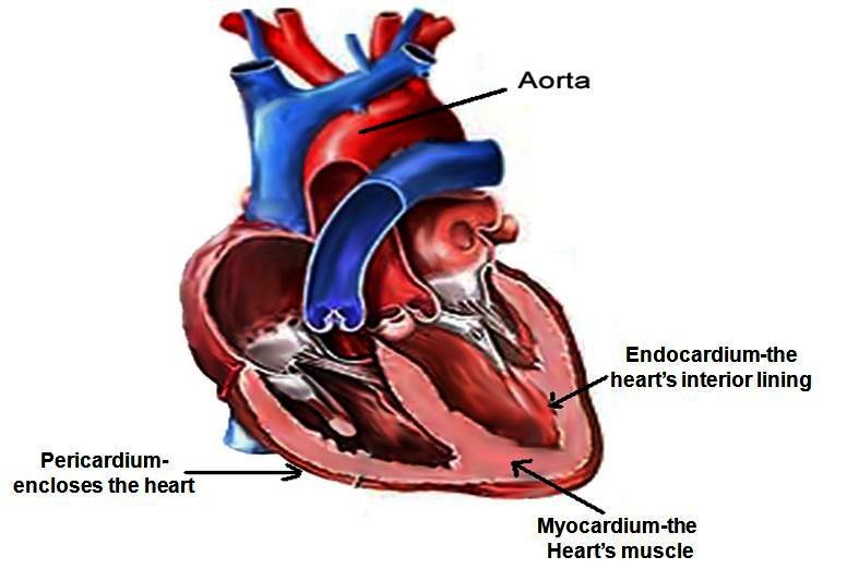
…a partition known as the interatrial septum; the lower chambers, the ventricles, are separated by the interventricular septum. The atria receive blood from various parts of the body and pass it into the ventricles. The ventricles, in turn, pump blood to the lungs and to the remainder of the body.
What are the contents of the intermuscular septum?
The muscle was innervated by the musculocutaneous nerve. It also arises from medial intermuscular septum and is inserted into the coronoid process of ulna. Orbital fat is divided into two compartments by the extraocular muscles and the intermuscular septum. Intraconal fat lies inner to this layer, and extraconal fat is external to it.
What is the job of the ventricular septum?
What are the Similarities Between Atrial and Ventricular Septal Defect?
- Atrial and ventricular septal defects are two types of common heart defects.
- Both conditions are congenital heart defects that present since birth.
- In both cases, the small holes are repaired by the body itself.
- Both conditions sometimes can be occurred in adulthood.
- Genetics and environmental factors are risk factors in both conditions.
What does the interventricular septum intact in an echo mean?
There are 4 chambers of of the heart: 2 atria and 2 ventricles. IAS stands for interatrial septum and IVS stands for interventricular septum, which are the walls separating the atria and ventricles, respectively. In your case, they are intact, without defects, which is normal.

What is the function of interventricular septum?
The interventricular septum separates the ventricles and allows for proper blood flow through the heart: from the right atrium through the tricuspid valve to the right ventricle to the lungs, back to the left atrium, followed by the left ventricle through the mitral valve.
What is the function of the interventricular septum quizlet?
Interventricular septum is a muscular wall that separates right and left ventricles. It prevents the mixing of oxygenated and deoxygenated blood.
What is the function of the interatrial and interventricular septum?
cardiovascular system. …a partition known as the interatrial septum; the lower chambers, the ventricles, are separated by the interventricular septum. The atria receive blood from various parts of the body and pass it into the ventricles. The ventricles, in turn, pump blood to the lungs and to the remainder of the body ...
What makes up the interventricular septum?
The interventricular septum is a complex structure composed of muscular and fibrous tissue. Defects in the septum are extremely common and can occur at a single or multiple locations (Fig. 2-69).
Does the interventricular septum contribute to contraction?
During each cardiac cycle the interventricular septum contracts by shortening longitudinally and becoming thicker.
What separates the two ventricles?
ventricular septumThere is another wall between the two ventricles, and it is called the ventricular septum.
What does interventricular mean?
Definition of interventricular : situated or occurring between ventricles the interventricular septum of the heart interventricular brain hemorrhage.
What is the difference between the interatrial and interventricular septum?
That portion of the septum that separates the two upper chambers (the right and left atria) of the heart is termed the atrial (or interatrial) septum while the portion of the septum that lies between the two lower chambers (the right and left ventricles) of the heart is called the ventricular (or interventricular) ...
What would happen if the interventricular septum has a hole in it?
This hole allows blood from the left ventricle to go back into the right ventricle instead of out of the heart through the aorta. When this happens, too much blood can enter the lungs and may cause problems over time. Sometimes a VSD closes on its own. Other times, treatment is needed to repair the hole.
What does the intraventricular septum contract?
The interventricular septum is quite dynamic during each cardiac cycle. It contracts with the ventricles during systole such that it shortens longitudinally (from the base to the apex) and becomes thicker.
What is the septum quizlet?
the septum is the wall or partition dividing a body space or cavity. interatrial septum. interatrial septum is the portion or wall that separates the right atrium from the left atrium (intra= between , septum = wall)
What is the purpose of the heart septal wall quizlet?
Their purpose is to increase the capacity of the atrium, and so also increase the volume of blood that it is able to contain. The interatrial septum is the wall of tissue that separates the right and left atria of the heart.
What regulates the flow of blood through the heart?
Your heart has four valves that control the flow of blood in and out of the chambers. There are valves between the atrium and the ventricle on each side of your heart. There is also a valve controlling the flow of blood out of each of your ventricles. The valves are designed to keep blood flowing forward only.
Which valves stop backflow in the heart?
Aortic valve: Allows blood to pass from the left ventricle to the aorta; prevents backflow of blood into the left ventricle.
What is the interventricular septum?
The interventricular septum, also known as the ventricular septum, refers to the triangular wall of cardiac tissue that separates the left and righ...
Where is the interventricular septum located?
The interventricular septum is located between the right and left ventricles of the heart. It runs between the interventricular grooves, which are...
What is the blood supply to the interventricular septum?
The posterior interventricular artery, a branch of the right coronary artery, supplies the posterior third of the interventricular septum. The rema...
What is the function of the interventricular septum?
The interventricular septum separates the ventricles and allows for proper blood flow through the heart: from the right atrium through the tricuspi...
What disorders affect the interventricular septum?
Disorders of the interventricular septum may occur as a result of genetic mutation; fetal exposure to a teratogen, a drug that causes mutation in-u...
What are the most important facts to know about interventricular septum?
The interventricular septum is the wall of cardiac muscle and membranous tissue that separates the left and right ventricles. Its purpose is to all...
Where is the interventricular septum located?
Anatomy. The interventricular septum separates the left ventricle and the right ventricle. It is muscular at the apex and tapers to a membranous portion at the heart base near the origin of the aorta. Septal defects may occur in any area of the septum, but are most commonly located in the membranous portion.
How long does it take for a ruptured ventricular septum to appear?
Rupture of the interventricular septum is a relatively rare mechanical complication that occurs 3 to 5 days following infarction but may appear within the first 24 hours or on presentation, particularly in patients treated with thrombolytic therapy.26 The incidence has been reduced from 2.0% to 0.2% in the reperfusion era. 26 Risk factors for rupture in the modern era include advanced age and female gender. 27 Pre-infarct angina and the development of coronary collaterals in the infarct-related artery are associated with reduced risk of rupture. 28 Ventricular septal rupture is seen more frequently in anterior infarctions where the defect is located in the apical septum; with inferior infarcts, the defect is located in the basal inferoposterior septum. Rupture occurs at the margin of necrotic and healthy myocardium and may be a discrete defect that ranges from a few millimeters to several centimeters wide or the rupture may be an irregular and serpiginous connection. Associated right ventricular infarction is common, and when right ventricular dysfunction is present, prognosis is significantly worsened. 29,30
What is right ventricular pacing?
Right ventricular pacing activates the interventricular septum before the left ventricular lateral wall, which is manifested as a left bundle branch block pattern on the surface electrocardiogram (ECG). This results in LV dyssynchrony, with deleterious effects on left ventricular function and adverse clinical outcomes, including heart failure and increase in mortality (Wilkoff et al., 2002 ). Pacing-induced cardiomyopathy is a well-recognized consequence of chronic right ventricular pacing, even among patients with preserved baseline ejection fraction (EF) and occurs in as much as 20% of individuals with chronic right ventricular pacing ( Khurshid et al., 2014 ). It is reported that a pacing burden of 20% is associated with pacing-induced cardiomyopathy ( Khurshid et al., 2014 ). Basic programming features of pacemakers are centered on minimizing ventricular pacing. Different features were developed in dual chamber devices that utilize atrial-only pacing with automatic switching to dual chamber pacing with failed AV conduction or algorithms that automatically prolong AV intervals with shortening of these intervals with no intrinsic AV conduction. These various programming algorithms have decreased significantly the rates of right ventricular pacing in patients without complete AV block ( Stockburger et al., 2015; Sweeney et al., 2007 ).
What is the most common congenital cardiac defect in infants?
Interventricular Septal Defect. Defects in the interventricular septum are the most common congenital cardiac defect in infants, but most of the defects close spontaneously before these children are 10 years old. In adults, these defects are not as common as atrial septal defects.
How to reduce shunt volume?
Reduction of the shunt can be accomplished by pulmonary artery banding, a technique resulting in elevation of the right ventricular systolic pressure. As right ventricular pressure increases, the shunt volume decreases, and the pulmonary circulation is spared the deleterious effects of chronic volume overload.
What is the name of the defect in the heart that results from the absence of the atrioventricular septum
Atrioventricular septal defects result from the absence of the atrioventricular septum, giving rise to large defects in the center of the heart that have an atrial as well as an inlet ventricular component. These are also known as “endocardial cushion” or “atrioventricular canal” defects.
What is the IVS of a VSD?
The IVS is a complex arrangement composed of muscular and fibrous tissues. The defects in the septum are common and may occur at a single or multiple sites. The echocardiographic recognition of a VSD depends on dropout in the IVS and the use of PW or color-flow Doppler to identify the turbulent shunt flow across the defect. Muscular VSDs occur frequently in children, and many of them close instinctively within the first 2 years of life. Nonetheless, muscular defects near the apex may be of considerable size. However, the fibrous part of the IVS (the membranous septum) lies adjacent to the AV annulus, and the TV septal leaflet, and its chordal apparatus lie along the RV aspect of the membranous septum. Consequently, the incorporation of this tissue into a septal aneurysm often causes the spontaneous closure of this type of VSD. The right coronary or noncoronary aortic cusps rarely prolapse into a high-membranous VSD, distorting AV coaptation and causing AR. Supracristal VSDs occur in that part of the IVS placed above the crista supraventricularis and beneath the pulmonary annulus. Echocardiographic views of RVOT are best for noticing this type of defect. Prolapse and distortion of the right coronary aortic leaflet also occur with supracristal VSDs.
What is the name of the part of the membranous portion of the interventricular septum between the left vent
atrial septum ( septum atrio´rum cor´dis) interatrial septum. atrioventricular septum the part of the membranous portion of the interventricular septum between the left ventricle and the right atrium. deviated septum an injury or malformation of the nasal septum so that one part of the nasal cavity is smaller than the other;
What is a deviated septum?
deviated septum an injury or malformation of the nasal septum so that one part of the nasal cavity is smaller than the other; this is fairly common and seldom causes complications. Occasionally the deviation may handicap breathing, block the normal flow of mucus from the sinuses during a cold, or prevent proper drainage of infected sinuses.
What is the name of the partition between the right and left atria of the heart?
interatrial septum ( septum interatria´le cor´dis) the partition separating the right and left atria of the heart; called also atrial septum. interradicular septum interalveolar septum (def. 1). interventricular septum ( septum interventricula´re cor´dis) the partition separating the right and left ventricles of the heart;
What is the name of the triangular membrane that separates the anterior horns of the lateral ventric
pellucid septum ( septum pellu´cidum) the triangular double membrane separating the anterior horns of the lateral ventricles of the brain; called also septum lucidum. septum pri´mum a septum in the embryonic heart, dividing the primitive atrium into right and left chambers. See also congenital heart defect.
What is the meaning of septum?
septum. 1. a wall or partition dividing a body space or cavity. Some are membranous, some are osseous, and some are cartilaginous; each is named according to its location. See also septal defect. adj., adj sep´tal. 2. nasal septum. alveolar septum interalveolar septum.
What is the name of the thin septa that separates adjacent pulmonary alveoli?
Called also interradicular septum. 2. one of the thin septa that separate adjacent pulmonary alveoli, containing connective tissue and the capillary network of the blood supply of the lung. Defs. 1 and 2 called also alveolar septum.
What is the procedure called when you have a sinus resection?
In some cases surgery (called partial or complete submucous resection) may be necessary to relieve the obstruction and reduce irritation and infection in the nose and sinuses. 1. one of the thin plates of bone separating the alveoli of the teeth in the mandible and maxilla. Called also interradicular septum.
What is the ventricular septum?
ventricular septum. Its posterior and upper portion separating the aortic vestibule from the upper portion of the right ventricle and the lower portion of the right atrium. It is a fibrous and thin referred to as the membranous ventricular septum.
Which part of the heart separates the lower chambers?
The interventricular septum is the thick wall that separates the heart’s lower chambers from each other. The ventricular septum is directed backwards and to the right and is curved to the right ventricle. The greater part of it is muscular and thick constituting the muscular. ventricular septum. Its posterior and upper portion separating ...
What is paradoxical septal motion?
Paradoxical septal motion, the abnormal systolic movement of the IVS toward the RV with preserved wall thickening, is a common phenomenon after cardiac surgery. 13 This can be distinguished from dyskinetic septal motion due to ischemia, when wall thickening does not occur. The exact mechanism of paradoxical septal motion is not truly understood. Some explanations include loss of an intact pericardium, which limits excessive heart motion toward the sternum, restriction of the RV from closure of the pericardium or chest wall, and LV underfilling. 13, 14 This abnormal motion is of no pathologic significance and can persist for weeks after surgery.
What is the normal motion of the IVS during systole?
In systole, the IVS both thickens and moves. The normal motion of the contracting IVS is away from the sternum and toward the inferior LV free wall, with shortening from base to apex. From a TEE perspective, there is clockwise rotation of the apex and counterclockwise rotation of the base. Normal motion is not uniform throughout the IVS. When the ventricles contract, the upper IVS acts as a hinge point between the aortic root and the remaining IVS. 1 This motion is the result of the complex arrangement of the ventricular myocardial fibers. 2 During ventricular contraction, myocardial fibers undergo longitudinal shortening, circumferential thinning, and radial thickening. The resulting deformation, also known as strain, cannot be fully appreciated using conventional 2-dimensional echocardiographic imaging: only radial thickening can be seen. Advanced imaging techniques such as Doppler tissue imaging, Doppler strain echocardiography, and 2-dimensional speckle tracking imaging allow for this analysis; information on these techniques can be found in a recent consensus statement by the American Society of Echocardiography. 2
What causes an aneurysm of the IVS?
Aneurysm of the IVS is rare. It is most often associated with congenital perimembranous ventricular septal defects, but can also be caused by increased atrial and ventricular pressure, endocarditis, or can occur after a transmural septal myocardial infarct. 9 Echocardiographically, an aneurysm is characterized by myocardial thinning with dyskinetic motion toward the RV during systole. Thrombus is often present. Aneurysms are most frequently located in the anterior aspect of the IVS below the aortic valve and are best seen in the ME long-axis view or by anteflexing and/or withdrawing the probe in the ME 4-chamber view.
What is the IVS?
The IVS is comprised of the thick muscular septum and the thin fibrous membranous septum and is concave toward the LV ( Fig. 1 ). 1 The muscular septum borders much of the LV and RV cavities.
Why is the IVS important?
The IVS has a central role in the function of both ventricles and it is important for the echocardiographer to understand its structure and function, in normal and pathologic states.
How thick is the LV myocardium?
The normal thickness of the LV myocardium is 6 to 9 mm in women and 6 to 10 mm in men. 4 When performing TEE, the IVS thickness is measured in the TG midpapillary short-axis view at end diastole. This measurement should be compared with the thickness of the inferolateral (posterior) segment ( Fig. 2 ). End diastole can be determined by the onset of the QRS, or the frame where the cardiac cavity is the largest. 4 Measuring the IVS can be difficult: adjacent right-sided (tricuspid valve apparatus or moderator band) and left-sided (false tendon) structures can make it difficult to determine the endocardial borders for caliper placement. In addition, measurement of thickness and function depends on lateral resolution, which is less precise than main beam resolution: difficulty can arise when the epicardial border is not well delineated.
Overview
The interventricular septum (IVS, or ventricular septum, or during development septum inferius) is the stout wall separating the ventricles, the lower chambers of the heart, from one another.
The ventricular septum is directed obliquely backward to the right and curved with the convexity toward the right ventricle; its margins correspond with the anterior and posterior interventricular sulci. The lower part of the septum, which is the major part, is thick and muscular, and its much …
Structure
The interventricular septum is the stout wall separating the ventricles, the lower chambers of the heart, from one another.
The ventricular septum is directed obliquely backward to the right, and curved with the convexity toward the right ventricle; its margins correspond with the anterior and posterior longitudinal sulci. The greater portion of it is thick and muscular and constitutes the muscular interventricular sept…
Development
The muscular part of the interventricular septum derives from the bulboventricular flange which is developed due to differential growth of primitive ventricle and bulbous cordis. Membranous part has a neural crest origin which connects the upper free margin of the bulboventricular flange and anterior and posterior endocardial cushions of atrio ventricular canal. It also gets attached to lower border of spiral septum or the aortico pulmonary septum.
Clinical significance
A ventricular septal defect (VSD), a hole in the interventricular septum is one of the four congenital defects of the condition of tetralogy of Fallot. A VSD can cause a left-to-right shunt of blood flow in the heart, and is one of the most common of the congenital heart defects. This type of shunt is an acyanotic disorder that can result in ventricular hypertrophy.
The alignment of interventricular septum and interatrial septum is disturbed in various congenita…
Additional images
• Heart normal short axis echo
External links
• Histology image: 128_06 at the University of Oklahoma Health Sciences Center - "Heart and semilunar valve"