How to scan the letter “E” on a microscope?
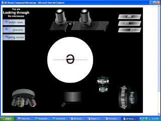
The directions in the lab handout said to place the letter “e” on the slide so that the “e” was right side up and to ake sure the letter was positioned directly over the hole where the light was coming in. The directions said to use the scanning objective, which is the shortest one and use the coarse knob to bring the “e” into focus.
What does “e” look like under a microscope?
When you view “e” under a microscope it just looks like a tube with “nanos” in it. What would happen if you would view a slide containing the letter e on it and you moved the slide from left to right under the scanning lens of the microscope? What is the orientation of the image of the 'e' relative to its actual orientation on the microscope slide?
What is the image of the letter E on the slide?
If the orientation of the letter "e" on the slide is exactly the same as it appears in this response, then the image of the "e" would be an inverted reflection. In lamens terms, the "e" would be upside down inside the microscope. What shape would the bacteria be if they are indeed Escherichia coli when viewed under a microscope?
How do you bring the “E” into focus on a microscope?
The directions said to use the scanning objective, which is the shortest one and use the coarse knob to bring the “e” into focus. It worked slowly and could finally see the letter. I was surprised to see that it was upside-down. I looked on the stage and I had put it there pointed the right way, but it seemed like the microscope reversed the image!