The medial cuneiform, also called the first or internal cuneiform, is located around the middle foot. It is on the inner side of the foot, behind the first metatarsal (which leads to the bones of the big toe) and the in front of the navicular bone. It is shaped similarly to a wedge.
What is the medial aspect of the foot?
What is the medial side of the foot? The navicular is on the medial (internal) side of the foot, in between the talus as well as the cuneiform bones ahead. The navicular kinds joints with 4 bones: the talus as well as the 3 picture writings.
What causes pain on the medial side of the foot?
What causes pain in the middle of foot when pressure applied?
- Cuboid syndrome ( If the pain is on the outer side)
- Navicular fracture (If the pain is on the inner side)
- Midfoot Arthritis
- Extensor tendonitis
- Midtarsal joint sprain
- Direct foot injury
- Cellulitis (Obvious skin redness, pain, and warmth)
What part of the foot is considered the sole of the foot?
The sole is the bottom of the foot. In humans the sole of the foot is anatomically referred to as the plantar aspect. Contents
What is medial foot pain?
Truthfully, there is more than one cause of pain on the medial side of the heel, but the most common would be plantar fasciitis. Plantar fasciitis is a condition associated with inflammatory change within the fascia under the sole of the foot. Usually, the heel pain overrides pain through the arch of the foot, more distally.
What is the medial arch?
What is the function of the medial arch?
How many arches are there in the foot?
What is the medial side of a foot?
The medial arch runs from the heel to the forefoot. The function of this foot arch is to support bodyweight during activities such as standing, walking and running. When the toes touch the ground, this arch stretches to its maximum length and rapidly as the toes are off the ground.
What is the middle part of your foot called?
The midfoot is a pyramid-like collection of bones that form the arches of the feet. These include the three cuneiform bones, the cuboid bone, and the navicular bone. The hindfoot forms the heel and ankle. The talus bone supports the leg bones (tibia and fibula), forming the ankle.
What are areas of the foot called?
The foot is traditionally divided into three regions: the hindfoot, the midfoot, and the forefoot (Figure 2).
What is the lateral side of the foot?
Lateral foot injuries are those on the little toe side of the foot, whereas medial foot injuries are on the big toe side.
What part of the foot is the instep?
The instep is the arched part of the top of the foot between the toes and the ankle.
Why does the middle of my foot hurt?
Pain in the arch of the foot can be caused by a number of underlying conditions. Plantar fasciitis is the most common, but other causes may include posterior tibial tendon dysfunction, cavus foot, and more. Arch pain is a common foot concern.
What is upper part of foot called?
Talus – the bone on top of the foot that forms a joint with the two bones of the lower leg, the tibia and fibula. Calcaneus – the largest bone of the foot, which lies beneath the talus to form the heel bone.
What is the bottom of the foot called in medical terms?
Planta pedis – the bottom of the foot; called also sole.
Why do the bottom of my feet hurt when I walk?
Pain in the bottom of your foot is often caused by exercise, such as running, wearing shoes that are too tight or a condition, such as Morton's neuroma. Some people also have a foot shape that puts extra pressure on the bottom of the foot. Hard or cracked skin or a verruca can also cause this type of pain.
What is it called when the sides of your feet hurt?
Lateral foot pain is pain on the outer side of the foot. This is oftentimes tied to cuboid syndrome, which is when the bone on the outside of the foot shifts out of place. Torn joints and ligaments typically cause the bone to shift. This can happen over time or suddenly due to an ankle sprain.
Why does my foot hurt on the side when I walk?
Pain can be common on the outside of the foot in this location. There are several reasons pain can manifest along the lateral border of your foot. Common foot problems seen in this area tend to be Jones fractures and related stress fractures, peroneal tendonitis, and bursitis.
What is back of foot called?
The two bones that make up the back part of the foot (sometimes referred to as the hindfoot) are the talus and the calcaneus, or heel bone. The talus is connected to the calcaneus at the subtalar joint. The ankle joint allows the foot to bend up and down.
Where is plantar part of foot?
The plantar fascia is the thick tissue on the bottom of the foot. It connects the heel bone to the toes and creates the arch of the foot.
What are foot knuckles called?
The metatarsophalangeal joints (MTP joints), also informally known as toe knuckles, are the joints between the metatarsal bones of the foot and the proximal bones (proximal phalanges) of the toes.
What is the top of the foot called in medical terms?
Talus – the bone on top of the foot that forms a joint with the two bones of the lower leg, the tibia and fibula. Calcaneus – the largest bone of the foot, which lies beneath the talus to form the heel bone.
What is the common name for tarsal?
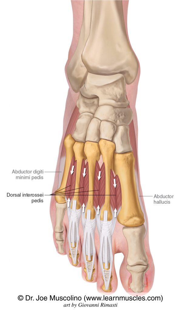
The tarsal bones are called the calcaneus, talus, navicular, cuboid, lateral cuneiform, intermediate cuneiform, and medial cuneiform. Of these, the calcaneus, or 'heel bone,' is the largest.
Which bones anchor the ligaments in the foot?
The largest of the cuneiform bones, it anchors several ligaments in the foot. Intermediate cuneiform: Located between the other two cuneiform bones, it is also wedge-shaped. It articulates with the two other cuneiform bones, the second metatarsal (connected to the bones of the second toe) and the navicular bone.
Which bones are located on the lateral side of the foot?
These bones include: Cuboid: The cuboid bone is one of the seven tarsal bones located on the lateral or outer part of the foot and, as its name suggests, is cube-shaped. It provides stability, connecting the foot and the ankle, and assists in the movement of the toes.
What are the three bones of the foot?
The bones of the foot are divided into three groups: The phalanges. The metatarsal bones. The tarsal bones. The foot is divided into three major structural areas: the forefoot, the midfoot and the hindfoot or rearfoot .
What is the midfoot?
The midfoot is one of three regions of the human foot. Its name is somewhat self-explanatory, referring to the area in the middle of the foot. It encompasses the arch of the foot and is composed of bones, tendons, and ligaments, connecting the forefoot with the hindfoot. PeopleImages / Getty Images.
How many bones are there in the foot?
The human foot is an incredibly complex part of the body, made up of 26 bones—fully 25 percent of the total number of bones in the entire body. It is strong, flexible and durable, able to bear considerable weight, impact force and general wear-and-tear as it propels us when we walk, run, jump, pivot and even simply stand still.
Where is the medial cuneiform located?
It is located on the inside of the foot behind the first metatarsal (a bone of the big toe) and in front of the navicular. The largest of the cuneiform bones, it anchors several ligaments in the foot.
Which bones are lateral cuneiform?
Lateral cuneiform: Located at the center of the front tarsal bones, the lateral cuneiform sits between the third metatarsal, the cuboid, the navicular, and the intermediate cuneiform bones.
What bones are in the foot?
Tibia and Fibula (long bones) The foot is connected to the body where the talus articulates with the tibia and fibula. In a typical foot the tibia is responsible for supporting about 85% of body weight. The fibula accepts the remaining 15%; its main role is to serve as the lateral wall of the ankle mortise (Figure 4).
What are the three regions of the foot?
The foot is traditionally divided into three regions: the hindfoot, the midfoot, and the forefoot (Figure 2). Additionally, the lower leg often refers to the area between the knee and the ankle and this area is critical to the functioning of the foot.
What is the cartilage of the talus?
Because it articulates with so many other bones, 70% of the talus is covered with hyaline cartilage (joint cartilage). The talus connects to the calcaneus on the underside through the subtalar joint, and distally it connects to the navicular through the talonavicular joint.
Where does the hindfoot start?
The Hindfoot begins at the ankle joint and stops at the transverse tarsal joint (a combination of the talonavicular and calcaneal-cuboid joints). The bones of the hindfoot are the talus and the calcaneus.
How many metatarsals are there in the foot?
Each foot contains five metatarsals, numbered 1-5 medial (great toe) to lateral. The first three metatarsals medially are more rigidly held in place than the lateral two. The metatarsals articulate with the mid-foot at their base, a joint called the tarsal-metatarsal (TMT) joint, or Lisfranc joint. The TMT joint is made stable not only by strong ligaments connecting these bones, but also because the second metatarsal is recessed into the middle cuneiform in comparison to the others (Figure 7). The metatarsal heads are the main weight bearing surface and the site where the phalanges attached at the metatarsal-phalangeal (MTP) joint.
How many cuneiform bones are there in the foot?
There are three cuneiform bones in the foot: the medial, medial (intermediate), and lateral cuneiforms (Figure 7). These bones, along with the strong plantar and dorsal ligaments that connect to them, provide a good deal of stability for the foot.
Where is the talar neck located?
The talar head is adjacent to the navicular bone to form the talonavicular joint. The talar neck is located between the body and head of the talus. The talar neck is one of the few areas of the talus not covered with cartilage, and is thus the point of entry for the blood vessels supplying the talus.
Which bones are in the midfoot?
Together with the first and second metatarsals, the navicular, and the intermediate cuneiform, this bone is part of joints in the mid-foot. The medial cuneiform is the largest cuneiform in size, although all of these bones are still relatively small. The cuneiforms are situated between the metatarsal bones and the small navicular bone.
Where is the medial cuneiform located?
Medial cuneiform. The medial cuneiform, also called the first or internal cuneiform, is located around the middle foot. It is on the inner side of the foot, behind the first metatarsal (which leads to the bones of the big toe) and the in front of the navicular bone. It is shaped similarly to a wedge. Together with the first and second metatarsals, ...
Which muscle is responsible for the movement of the foot?
These are the main muscles that facilitate movement in the foot: Tibialis posterior (supports the foot's arch) Tibialis anterior (allows the foot to move upward)
What are the three sections of the foot?
The foot can be divided into three sections: the forefoot, midfoot, and hindfoot. There are bones, joints, muscles, tendons, and ligaments in each section.
Where is the forefoot located?
The forefoot meets the midfoot at the five tarsometatarsal joints.
How many joints are there in the big toe?
Each big toe has two joints, the metatarsophalangeal joint and the interphalangeal joint. The other four toes on each foot have three joints each: the metatarsophalangeal joint at the base of the toe, the proximal interphalangeal joint in the middle of the toe, and the distal phalangeal joint—the joint closest to the tip of the toe.
What are the conditions that affect the feet?
And like any body part that's made up of bone, muscle, and connective tissue, the feet are subject to certain conditions that can affect any other extremity, limb, or the spine, including: 1 Sprains, strains, and pulls affecting muscles or ligaments 2 Tendinitis (when a tendon becomes overstretched or torn) 3 Bone fractures and breaks 4 Osteoarthritis (which is particularly common in feet, especially in the joints that connect the toes to the midfoot) 5 Rheumatoid arthritis
How many bones are there in the forefoot?
Forefoot. This is the very front part of the foot, including the toes, or phalanges. There are 14 toe bones (two per big toe and three per each of the other four), plus five metatarsals. The first metatarsal bone is the shortest and thickest and plays an important role during propulsion (forward movement).
What bones are in the hindfoot?
Hindfoot. There are only two large bones in this section of the foot: the talus and the calcaneus. The largest of these, the calcaneus, forms the heel of the foot. The talus rests on top of the calcaneus and forms the pivoting joint of the ankle.
What are the bones of the foot?
It consists of 28 bones, which can be divided functionally into three groups, referred to as the tarsus, metatarsus and phalanges.
What is the joint between the ankle and the foot called?
Ankle joint. The ankle joint, also known as the talocrural joint, is a hinge joint that involves the tibia and fibula of the leg and the talus of the foot. The body of the talus sits within a deep recess referred to as the mortise. This mortise is formed by the: Medial malleolus of the tibia.
Which nerves innervate the talocalcaneal joint?
The talocalcaneal joint is innervated by branches of the sural, medial plantar and posterior tibial nerves .
How many bands of ligaments are involved in stabilising joints?
There are three bands of ligaments involved in stabilising these joints:
Which ligament is the strongest?
The distal part of this ligament, the inferior transverse ligament, is a yellow band that connects the medial and lateral malleoli. The interosseus tibiofibular ligament is a continuation of the interosseus membrane and is the strongest of the three ligaments.
Which nerve innervates the inferior tibiofibular joint?
The inferior tibiofibular joint is innervated by branches of the deep fibular and sural nerves .
Which tendons are involved in the dorsiflexion of the tibialis anterior?
The tibialis anterior allows dorsiflexion at the ankle joint and is assisted by the tendons of the fibularis tertius, hallucis longus and extensor digitorum longus .
What tendon is strained in the medial arch of the foot?
If you happen to have pain in the medial arch of the foot, then the flexor hallucis tendon is strained, but if you have pain in the Achilles’ area, then you need to determine the real reason of the pain.
Why does my foot hurt under the medial arch?
Pain under the medial arch of the foot: Why? Many people experience some pain in the medial arch of the foot. Causes can include an injury of the flexor hallucis, which is a unity of the tendon and a very strong muscle, or structural abnormalities that grow worse with age and activity and start causing symptoms later in life.
What is the arch of the foot?
The arch of your foot is responsible for keeping us in balance while standing or walking. The tendon spreads itself all the way from the great toe across the medial arch of the foot to the Achilles’ tendon. There are many causes of an injury to the flexor hallucis and they include excess amounts of body weight pressing onto the toes of the foot.
Where do flexor hallucis occur?
These injuries usually occur in the medial arch of the foot. Sometimes, people may mistake injuries of the flexor hallucis for Achilles’ tendonitis or plantar fasciitis. There are some people who may experience injuries of the flexor tendons of other toes and have a pain not only at the medial arch of the foot, ...
Why does my big toe hurt?
For instance, a big toe carries almost one third of the body's weight in the case of a normally aligned foot. If a person's feet are turned out while walking or standing, the big (or great) toe has to carry even more of your body's weight and injuring the flexor hallucis becomes more likely.
What is a hammer toe?
Hammer toe is a deformity that affects the second, third, or fourth toe. The affected toe bends downward and appears misshapen. The toe may appear swollen or discolored, and a person might not be able to straighten it. Sometimes, hammer toe affects multiple toes.
Why does my ball of the foot hurt?
Muscle strains and sprains, minor overuse injuries, and tense muscles can all cause pain on the ball of the foot. This pain usually resolves within a few weeks, and it may get better with massage.
How long does it take for plantar fasciitis to heal?
In most cases, symptoms get better within 3 to 6 months of beginning treatment. Plantar fasciitis is inflammation of the plantar fascia, which is a thick band of tissue that extends from the heel to the toes. The pain is sharp and stabbing, often feeling worse in the morning or when beginning exercise.
Why does my big toe hurt?
Big toe pain. Many of the same conditions that affect the toes generally, such as broken bones, strains, sprains, and arthritis, can cause big toe pain. However, if the pain is only in the big toe, and the top of the toe hurts most, the problem may be an ingrown toenail.
Why does my foot arch hurt?
Foot arch pain. Injuries to the muscles and tendons of the foot, unsupportive shoes, and overuse may cause pain in the arch of the foot. Conditions that affect the heel, such as plantar fasciitis and tendinitis, may also cause foot arch pain. For some people, fallen arches cause foot arch pain.
Why do my feet hurt so bad?
Foot pain is common as it has a wide range of contributing factors, including uncomfortable shoes, too much time standing, athletic injuries, and chronic conditions, such as arthritis. have frequent foot pain in midlife. The specific location of the pain can sometimes be helpful in determining the cause.
What happens if a hammer toe does not resolve?
However, if the hammer toe does not resolve on its own, a person might need surgery.
What is the medial arch?
What Is the Medial Foot Arch? by MASS4D® Insoles. The foot is a unique structure made of 26 bones, 33 joints, and over 100 muscles, ligaments and tendons. It can primarily be divided into three regions — the heel, arch, and forefoot. What’s interesting is that the arch structure of the foot actually consists of three arches: ...
What is the function of the medial arch?
The medial arch runs from the heel to the forefoot. The function of this foot arch is to support bodyweight during activities such as standing, walking and running. When the toes touch the ground, this arch stretches to its maximum length and rapidly as the toes are off the ground. The medial arch also plays a major role in shock absorption ...
How many arches are there in the foot?
What’s interesting is that the arch structure of the foot actually consists of three arches:
