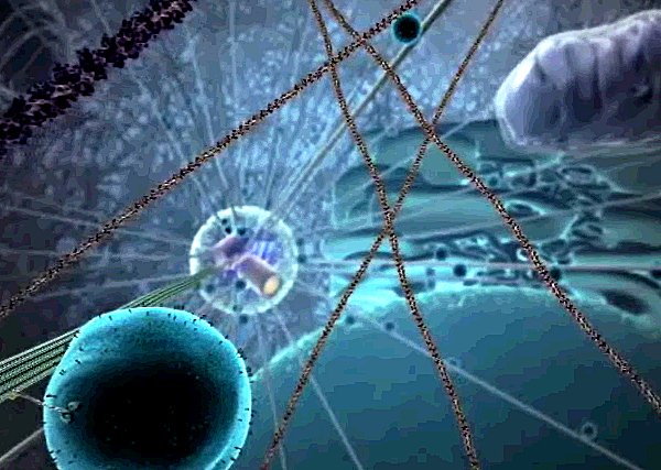
What is the function of microtubule organizing center?
The microtubule-organizing center (MTOC) is a structure found in eukaryotic cells from which microtubules emerge. MTOCs have two main functions: the organization of eukaryotic flagella and cilia and the organization of the mitotic and meiotic spindle apparatus , which separate the chromosomes during cell division .
Where are the microtubules located in a cell?
The microtubules in most cells extend outward from a microtubule-organizing center, in which the minus ends of microtubules are anchored. In animal cells, the major microtubule-organizing centeris the centrosome, which is located adjacent to the nucleusnear the center of interphase(nondividing) cells (Figure 11.39).
What is the role of the centrosome in the microtubule?
This complex acts as a template for α/β-tubulin dimers to begin polymerization; it acts as a cap of the (−) end while microtubule growth continues away from the MTOC in the (+) direction. The centrosome is the primary MTOC of most cell types. However, microtubules can be nucleated from other sites as well.
Where are the endoplasmic reticulum and microtubule-organizing center?
Endoplasmic reticulum, Golgi, the microtubule-organizing center (MTOC), mitochondria, and lysosomes are all concentrated in the rear behind the nuclei of polarized migrating naïve human T-cells and murine T-lymphoblasts ( Campello et al., 2006; Colvin et al., 2010; Sánchez-Madrid and Serrador, 2009 ).

What is another name for the microtubule organizing center?
The centrosome is the main microtubule organizing center in animal cells.
Where is the microtubule organizing center?
In dividing animal cells, a major site of microtubule nucleation and anchoring is the centrosome, which thus forms the microtubule-organizing center (MTOC)—the central point of a radial microtubule array (Bornens 2012, Conduit et al. 2015b).
What are the two microtubule-organizing centers?
Microtubule organization by microtubule-organizing centers such as the centrosome requires γ-tubulin, which exists in the γ-tubulin ring complex (γTuRC) that nucleates microtubules. The γTuRC is a ring-shaped, macromolecular complex whose core components are γ-tubulin and the γ-tubulin complex proteins.
Are centrioles microtubule-organizing centers?
Two centrioles (which are made of microtubules) form a centrosome, which are microtubule organizing centers in animal cells. A centriole is a cylinder of nine triplets of microtubules, held together by supporting proteins.
Why is the centrosome called a microtubule organizing center?
The centrosome is often touted as 'the major microtubule-organizing center of the cell,' generating a radial organization of microtubules well suited for the division of genomic material between daughter cells.
What is the microtubule organizing center quizlet?
The microtubule-organizing center (MTOC) is a structure found in eukaryotic cells from which microtubules emerge. MTOCs have two main functions: the organization of flagella and cilia and the organization of the mitotic and meiotic spindle apparatus, which separate the chromosomes during cell division.
How does the centrosome organize microtubules?
The centrosome acts as a microtubule organizing center (MTOC), orchestrating microtubules into the mitotic spindle through its pericentriolar material (PCM). This activity is biphasic, cycling through assembly and disassembly during the cell cycle.
What is the difference between centrosome and centriole?
A centrosome is an organelle that consists of two centrioles. A centriole is a structure made of microtubule proteins arranged in a particular way. A centriole is always smaller than a centrosome and also forms flagella and cilia. Both centrosomes and centrioles are found in animal cells and some protists.
How are microtubules organized in the cell?
The microtubules in most cells extend outward from a microtubule-organizing center, in which the minus ends of microtubules are anchored. In animal cells, the major microtubule-organizing center is the centrosome, which is located adjacent to the nucleus near the center of interphase (nondividing) cells (Figure 11.39).
Are basal bodies microtubule organizing centers?
The basal body serves as a nucleation site for the growth of the axoneme microtubules. Centrioles, from which basal bodies are derived, act as anchoring sites for proteins that in turn anchor microtubules, and are known as the microtubule organizing center (MTOC).
What is a centrosome?
A centrosome is a cellular structure involved in the process of cell division. Before cell division, the centrosome duplicates and then, as division begins, the two centrosomes move to opposite ends of the cell.
What organelle helps in the organization of microtubules during cell division?
Centrioles are paired barrel-shaped organelles located in the cytoplasm of animal cells near the nuclear envelope. Centrioles play a role in organizing microtubules that serve as the cell's skeletal system. They help determine the locations of the nucleus and other organelles within the cell.
How are microtubules organized in the cell?
The microtubules in most cells extend outward from a microtubule-organizing center, in which the minus ends of microtubules are anchored. In animal cells, the major microtubule-organizing center is the centrosome, which is located adjacent to the nucleus near the center of interphase (nondividing) cells (Figure 11.39).
Are basal bodies microtubule organizing centers?
The basal body serves as a nucleation site for the growth of the axoneme microtubules. Centrioles, from which basal bodies are derived, act as anchoring sites for proteins that in turn anchor microtubules, and are known as the microtubule organizing center (MTOC).
Where is the Pericentriolar material?
the centrosomeThe pericentriolar material (PCM) refers to the proteinaceous material that surrounds the centrioles — two small microtubule-based cylinders — and with them constitutes the centrosome, the main microtubule- organizing center (MTOC) found in animal cells.
Do all microtubules originate from the centrosome?
The centrosome is critical to mitosis as most microtubules involved in the process originate from the centrosome. The minus ends of each microtubule begin at the centrosome, while the plus ends radiate out in all directions.
What is a microtubule?
Jump to navigation Jump to search. Polymer of tubulin that forms part of the cytoskeleton. Microtubule and tubulin metrics. Microtubules are polymers of tubulin that form part of the cytoskeleton and provide structure and shape to eukaryotic cells.
What is the inner space of a microtubule?
The inner space of the hollow microtubule cylinders is referred to as the lumen . The α and β-tubulin subunits are identical at the amino acid level, and each have a molecular weight of approximately 50 kDa.
How are microtubules formed?
They are formed by the polymerization of a dimer of two globular proteins, alpha and beta tubulin into protofilaments that can then associate laterally to form a hollow tube, the microtubule. The most common form of a microtubule consists of 13 protofilaments in the tubular arrangement.
Why is the centrosome important?
Thus the centrosome is also important in maintaining the polarity of microtubules during mitosis.
What is the role of microtubules in eukaryotic cells?
Microtubules are one of the cytoskeletal filament systems in eukaryotic cells. The microtubule cytoskeleton is involved in the transport of material within cells, carried out by motor proteins that move on the surface of the microtubule. Microtubules are very important in a number of cellular processes.
How many protofilaments are there in a microtubule?
Typically, microtubules are formed by the parallel association of thirteen protofilaments, although microtubules composed of fewer or more protofilaments have been observed in various species as well as in vitro.
Where do microtubules form in mitosis?
Most of the microtubules that form the mitotic spindle originate from the centrosome. Originally it was thought that all of these microtubules originated from the centrosome via a method called search and capture, described in more detail in a section above, however new research has shown that there are addition means of microtubule nucleation during mitosis. One of the most important of these additional means of microtubule nucleation is the RAN-GTP pathway. RAN-GTP associates with chromatin during mitosis to create a gradient that allows for local nucleation of microtubules near the chromosomes. Furthermore, a second pathway known as the augmin/HAUS complex (some organisms use the more studied augmin complex, while others such as humans use an analogous complex called HAUS) acts an additional means of microtubule nucleation in the mitotic spindle.
Why is the organization of microtubules important?
The organization of microtubule networks is crucial for controlling chromosome segregation during cell division, for positioning and transport of different organelles, and for cell polarity and morphogenesis.
Why are microtubules important?
The organization of microtubule networks is crucial for controlling chromosome segregation during cell division, for positioning and transport of different organelles, and for cell polarity and morphogenesis.
Which cell compartments can nucleate, stabilize, and tether microtubules?
In addition, other microtubules, as well as membrane compartments such as the cell nucleus, the Golgi apparatus, and the cell cortex, can nucleate, stabilize, and tether microtubule minus ends.
Which complex is responsible for the organization of microtubules?
Microtubule organization by microtubule-organizing centers such as the centrosome requires γ-tubulin, which exists in the γ-tubulin ring complex (γTuRC) that nucleates microtubules. The γTuRC is a ring-shaped, macromolecular complex whose core components are γ-tubulin and the γ-tubulin complex proteins.
What stage of actin meshwork disassembles microtubules?
An isotropic actin meshwork maintains microtubule organization until stage 10B when this meshwork disassembles and microtubules align in parallel bundles close to the cortex of the oocyte and initiate minus-end-directed, kinesin-dependent ooplasmic streaming ( Dahlgaard, Raposo, Niccoli, & St Johnston, 2007; Serbus, Cha, Theurkauf, & Saxton, 2005 ). Ooplasmic streaming along the microtubule bundles mixes nurse cell and oocyte contents that are not yet anchored in the oocyte ( Fig. 1 F). Active nurse cell-to-oocyte transport ceases and nurse cells “dump” their contents into the oocyte as the actin cytoskeleton within the nurse cells constricts followed by nurse cell death ( Fig. 1 G). While osk RNA localization at the posterior begins well before ooplasmic streaming and nurse cell dumping, green fluorescent protein (GFP) tagging of osk RNA with the MS2/MCP system allowed observation of the localization process live (multiple MS2 RNA stem loops inserted into osk RNA were bound by MS2 coat protein fused to a fluorescent protein like green fluorescent protein (GFP)). This revealed a 45% increase in osk RNA accumulation in the oocyte after stage 10 ( Sinsimer, Jain, Chatterjee, & Gavis, 2011; Snee, Harrison, Yan, & Macdonald, 2007 ). This suggests that osk RNA is continuously produced and transported into the oocyte even after the microtubule network dependent, directed transport into and within the oocyte ceases. Late accumulation and sustained maintenance of osk RNA are mediated by the RNA-binding protein Rump and its associated factor Lost, the actin cytoskeleton, and require Oskar protein translation (see below) ( Fig. 1 G; Babu, Cai, Bahri, Yang, & Chia, 2004; Sinsimer et al., 2011; Suyama, Jenny, Curado, Pellis-van Berkel, & Ephrussi, 2009 ).
What is the centrosome?
12.6.3 Asymmetric segregation of the centrosome and the primary cilium membrane. The centrosome is the main microtubule organizing center in animal cells. It consists of a pair of centrioles (an older mother centriole and a newer daughter centriole) surrounded by amorphous pericentriolar material.
What is the role of MCAK in mitotic chromosome segregation?
In vertebrates, MCAK regulates the microtubule–kinetochore attachments that mediate mitotic chromosome segregation during anaphase ( Ems-McClung & Walczak, 2010; Kline-Smith et al., 2004 ). In the absence of MCAK in human cell culture lines, improper syntelic (kinetochores of both sister chromatids attached to the same pole) and merotelic (kinetochore of one sister chromatid attached to both poles) microtubule–kinetochore attachments are observed. In wild-type cells, MCAK associates with kinetochores prior to anaphase, and its depolymerase activity may destabilize inappropriate microtubule–kinetochore attachments. In klp-7 ( −) /MCAK mutant C. elegans oocytes, the persistence of such improper attachments during oocyte meiosis might lead to abnormal tension within the assembling spindle, and this imbalance in forces has been proposed to interfere with the coalescence of ASPM-1 foci into a bipolar structure ( Connolly et al., 2015 ). Consistent with such a model, partial knockdown of components of the Ndc80 complex that mediates microtubule–kinetochore attachment rescues spindle bipolarity in klp-7 ( −) mutants.
What is the function of MCAK?
MCAK, a kinesin-related protein whose abundance is highest during the early stages of mitosis, has been shown to regulate microtubule detachment. Abnormal increases or decreases in the frequency of detachment interfere with spindle function and inhibit cell division.
Where are the endoplasmic reticulum, Golgi, and mitochondria located?
Endoplasmic reticulum, Golgi, the microtubule-organizing center (MTOC), mitochondria, and lysosomes are all concentrated in the rear behind the nuclei of polarized migrating naïve human T-cells and murine T-lymphoblasts ( Campello et al., 2006; Colvin et al., 2010; Sánchez-Madrid and Serrador, 2009 ). Several studies have addressed the roles of these organelles and of endo/exocytosis in T-cell motility.
Where does endocytosis occur?
Both endo- and exocytosis have been shown to occur in the rear of migrating T-cells and have been implicated in migration. Samaniego et al. (2007) show enrichment of the heavy chain of clathrin in the uropods of HSB-2 cells and human T-lymphoblasts migrating on ICAM-1. The authors propose that rearward cortical flow may help to localize clathrin-mediated endocytosis at the rear . Inhibition of myosin II with blebbistatin in HSB-2 cells results in disruption of the polarized location of clathrin and in an approximately 60% inhibition of endocytosis accompanied by long, unretracted tails. A 50% downregulation of clathrin heavy chain in HSB-2 cells results in marked reduction of endocytosis and of chemotaxis to CXCL-12. Whether downregulation of clathrin modified cell polarization, adhesion, and rear release was not studied. The authors postulate that endocytosis localized in the rear may promote rear retraction by removing superfluous plasma membrane and/or may reinforce polarized signaling, possibly by internalizing chemokine and adhesion receptors in specific cellular areas. However, these concepts will have to be verified in primary T-cells.
What is a microtubule?
Microtubule Definition. Microtubules are microscopic hollow tubes made of the proteins alpha and beta tubulin that are part of a cell ’s cytoskeleton, a network of protein filaments that extends throughout the cell, gives the cell shape, and keeps its organelles in place. Microtubules are the largest structures in the cytoskeleton ...
Where are microtubules found?
Microtubules give structures like cilia and flagella their structure. Cilia are small protuberances of a cell. In humans, they are found on cells lining the trachea, where they prevent materials like mucus and dirt from entering the lungs.
What is the role of microtubules in mitosis?
The mitotic spindle organizes and separates chromosomes during cell division so that the chromosomes can be partitioned into two separate daughter cells. Its components include microtubules, the MTOC, and microtubule-associated proteins (MAPs).
What are the tail-like appendages that allow cells to move?
Flagella are tail-like appendages that allow cells to move. They are found in some bacteria, and human sperm also move via flagella . Microtubules also allow whole cells to “crawl” or migrate from one place to another by contracting at one end of the cell and expanding at another.
Which protein complex helps attach chromosomes to microtubules in the mitotic spindle?
Kinetochore – A protein complex that helps attach chromosomes to microtubules in the mitotic spindle.
What are the functions of microtubules in the cytoskeleton?
As part of the cytoskeleton, microtubules help move organelles inside a cell’s cytoplasm, which is all of the cell’s contents except for its nucleus. They also help various areas of the cell communicate with each other. However, even though microtubules help components of the cell to move, they also provide the cell with shape and structure.
Why are microtubules in dynamic equilibrium?
They are said to be in a state of dynamic equilibrium because their structure is maintained even though the individual molecules themselves are constantly changing. Microtubules are polar molecules, with a positively charged end that grows relatively fast and a negatively charged end that grows relatively slow.
