What are the three branches of the aortic arch?
The aortic arch gives rise to three arterial branches:
- Brachiocephalic artery, which supplies blood flow to the right arm and right carotid artery to the right side of the brain
- Left carotid artery, which provides circulation to the left side of the brain
- Left subclavian artery, which provides circulation to the left arm
What is the first branch off the aortic arch?
- First aortic arch - regresses early, but a remnant forms a portion of the maxillary artery.
- Second aortic arch - regresses early, but a remnant forms portions of the hyoid and stapedial arteries.
- Third aortic arch - contributes to the formation of the common carotid arteries bilaterally and the proximal internal carotid arteries bilaterally.
What is a three vessel aortic arch?
What are the 3 main branches of the aortic arch and which main body regions are supplied by each branch? Three vessels come out of the aortic arch: the brachiocephalic artery, the left common carotid artery, and the left subclavian artery. These vessels supply blood to the head, neck, thorax and upper limbs.
Is a left sided aortic arch normal?
The normal left aortic arch results from regression of the distal right fourth arch between the right subclavian artery and the descending aorta, including the right ductus arteriosus, and the right dorsal aorta distal to the origin of the seventh intersegmental artery (the distal subclavian artery precursor) ( Fig 4 ).

What is the most common aortic arch anomaly?
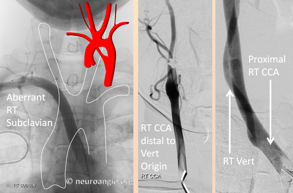
ARSALeft Aortic Arch with an Aberrant Right Subclavian Artery. An ARSA originating from normal left sided aortic arch is the most common aortic arch anomaly, with an incidence of 0.5-2% (5).
What are aortic arch anomalies?
Aortic arch anomalies are a type of congenital heart condition, which means it is a disease or abnormality that is present from birth. The aorta arises from the left ventricle and carries oxygenated blood from the heart to the body. There are many different variations of aortic arch anomalies.
What are the three aortic arch?
Background. The aortic arch is a continuation of the ascending aorta, being located in the superior mediastinum. Three branches, the brachiocephalic trunk, left common carotid artery and left subclavian artery usually branch from the aortic arch.
How many types of aortic arches are found?
The Aortic Arches All six pairs are not present simultaneously; they develop and regress at different stages. First aortic arch - regresses early, but a remnant forms a portion of the maxillary artery. Second aortic arch - regresses early, but a remnant forms portions of the hyoid and stapedial arteries.
How is aortic arch syndrome diagnosed?
Diagnosis. It is difficult to diagnose aortic arch conditions early because symptoms typically emerge only once an artery has narrowed. To rule out any other diseases with similar symptoms, a physician will review a patient's complete medical history and then perform a thorough physical exam.
What is a Type 1 aortic arch?
If the origins of all the great vessels arise within the arc segment of the aortic arch subtended by the first parallel reference line, it is termed a type I arch. If the origins of all the great vessels are included in the arc segment of the aortic arch subtended by the second index line, it is termed a type II arch.
What are the three branches of the aortic arch quizlet?
What are the three main branches off of the aortic arch?...Brachiocephalic trunk (right common carotid and right subclavian)Left common carotid.Left subclavian artery.
What do the aortic arches become?
The aortic arches or pharyngeal arch arteries (previously referred to as branchial arches in human embryos) are a series of six paired embryological vascular structures which give rise to the great arteries of the neck and head. They are ventral to the dorsal aorta and arise from the aortic sac.
What three vessels originate from the aortic arch and a normal configuration?
NORMAL ANATOMY The three main branches of the aortic arch are the brachiocephalic (innominate) artery (dividing into the right subclavian and common carotid arteries), the left common carotid artery, and the left subclavian artery.
What causes aortic arch?
What causes an aortic arch aneurysm? The most common causes of aortic arch aneurysms are smoking and high blood pressure. You're also at increased risk as you age due to a buildup of plaque in your arteries, known as atherosclerosis.
What is the function of aortic arches?
The aortic arch is the segment of the aorta that helps distribute blood to the head and upper extremities via the brachiocephalic trunk, the left common carotid, and the left subclavian artery. The aortic arch also plays a role in blood pressure homeostasis via baroreceptors found within the walls of the aortic arch.
What does left sided aortic arch mean?
Left aortic arch The ductus remains patent on the left side and connects the aorta distal to the left subclavian artery origin to the proximal left pulmonary artery. The descending aorta is usually on the same side as is the aortic arch, and it is due to the persistence of ipsilateral dorsal aorta (23).
Where is your aortic arch?
The aortic arch, arch of the aorta, or transverse aortic arch (English: /eɪˈɔːrtɪk/) is the part of the aorta between the ascending and descending aorta. The arch travels backward, so that it ultimately runs to the left of the trachea.
What is the aortic arch?
Location. The aortic arch is the part of the aorta between the ascending aorta and thoracic descending aorta. The sharpness of the angle can be different among individuals. The aortic arch gives rise to three arterial branches: Brachiocephalic artery, which supplies blood flow to the right arm and right carotid artery to the right side of the brain.
What is the aorta at the top of the thorax called?
This part is called the thoracic descending aorta. The average overall length of the aorta in the thorax—ascending, aortic arch, and descending—is around 33.2 cm or about 13 inches in adult men.
Why is the aortic arch important?
In some people, the angle of the aortic arch coupled with certain medical conditions can lead to aortic dissection where the ascending aorta meets the aortic arch.
What causes a bulge in the aorta?
Aortic dissection occurs when a tear in the tunica intima allows blood to be pushed between the tunica intima and the tunica media. The build-up of blood causes a separation of the two layers and a bulge is created on the side of the aorta.
What is the genetic condition of the aorta?
Arteriosclerosis (hardening of the arteries) Weakening of the aortic wall (aneurysm) Narrowing of the aorta that restricts blood flow (aortic stenosis or coarctation) Marfan syndrome and Turner's syndrome are two uncommon genetic conditions that can lead to an increased risk of aortic dissection.
What muscle is responsible for the heart's ability to expand?
The muscle tone of the aorta plays a big part in the ability of the heart to fully expand and in the overall control of blood pressure in the body. It also helps create back pressure on blood ejected from the ventricles during systole, which pushes blood into the coronary arteries to provide circulation to the heart muscle.
What is the difference between the aorta and the other arteries?
The only difference between the aorta and other arteries is its size . The overall structure of the aorta is identical to other arteries and subject to the same conditions such as hardening and weakening of the arterial walls. Common to all arterial walls are three main layers:
What is the second most common pattern of human aortic arch branching?
This pattern has erroneously been referred to as a “bovine arch.”
What is the aortic arch?
The term “bovine aortic arch” is a common misnomer in the medical literature, and the human aortic arch branching patterns to which it is ascribed are not commonly found in cattle. We suggest a descriptive aortic arch naming scheme that facilitates communication between practitioners and excludes the use of misnomers such as “bovine arch.”
What is the most common anatomical appearance in humans?
Standard Aortic Arch. This is the most common anatomic appearance in humans ( Fig 1 ). There are 3 great vessels that originate from the arch: the innominate artery, left common carotid artery, and left subclavian artery.
What is the name of the vessel that comes off the aortic arch?
The presence of a single long vessel originating from the aortic arch is a common occurrence in animals with deep chests. The general thinking among veterinary anatomists is that the long distance from the aortic arch to the thoracic inlet is the reason that all the great vessels come off the arch as a single vessel, the brachiocephalic trunk.
What is the true bovine arch?
True Bovine Arch. A true bovine aortic arch bears no resemblance to any of the common human aortic arch variations. In cattle, a single great vessel originates from the aortic arch. 7 This large brachiocephalic trunk gives rise to both subclavian arteries and a bicarotid trunk.
What is the final configuration of the aortic arch?
The final configuration of the aortic arch and its branches is probably related to different growth rates in the various arteries and the associated “migration” and “merging” of the branches. 3 The second most common variant of aortic arch branching occurs when the left common carotid artery has a common origin with the innominate artery.
What animal has a bovine aortic arch?
By its name, the bovine aortic arch in humans would presumably resemble the aortic arch branching pattern found in the family of ruminant animals, including cattle and buffalo. However, the bovine aortic arch configuration ascribed to the most common human aortic arch variants bears no resemblance to the aortic arch branching pattern found in ...
What is the most common variant of the aortic arch?
Bovine arch. Bovine arch is the most common variant of the aortic arch and occurs when the brachiocephalic (innominate) artery shares a common origin with the left common carotid artery . A bovine arch is apparent in ~15% (range 8-25%) of the population and is more common in individuals of African descent.
Which artery is not consistent with truncus bicaroticus?
The term truncus bicaroticus is also not consistent and can be misleading as the branching pattern not only includes the right and left common carotid artery, but also the right subclavian artery.
What is a bovine arch?
A true bovine arch (as found in cattle) has a common single brachiocephalic trunk which trifurcates into bilateral subclavian arteries and a single bicarotid trunk. This variation is very rare in humans.
What percentage of the population has a bovine arch?
A bovine arch is apparent in ~15% (range 8-25%) of the population and is more common in individuals of African descent. A related variant, also known as truncus bicaroticus, is the origin of the left common carotid artery from the brachiocephalic artery but not sharing a true common origin, which occurs in ~9% of the population.
Is the truncus bicaroticus branching pattern consistent?
The term truncus bicaroticus is also not consistent and can be misleading as the branching pattern not only includes the right and left common carotid artery, but also the right subclavian artery. Bovine arch is a common misnomer.
What are congenital variants and anomalies of the aortic arch?
Congenital variants and anomalies of the aortic arch are important to recognize as they may be associated with vascular rings, congenital heart disease ( CHD ), and chromosomal abnormalities, and can have important implications for prognosis and management, including surgical and percutaneous interventions.
Which arch is the definitive distal aortic arch?
The distal left dorsal aorta forms the definitive distal aortic arch, which passes posterior to the esophagus to a descending aorta beginning to the right of the vertebral column ( 8 ).
What is the most common congenital anomaly of the aortic arch?
Left aortic arch with aberrant right subclavian artery is the most common congenital anomaly of the aortic arch with a prevalence of 0.5%–2% ( 14 ). This anomaly results from regression of the right arch (between the right common carotid and right subclavian arteries) including the right ductus arteriosus ( Fig 6 ).
How does the aortic system develop?
Each primitive aorta consists of a ventral and a dorsal segment ( Fig 1 ). The two ventral aortae fuse to form the aortic sac, and the two dorsal aortae fuse to form the midline descending aorta. Six paired primitive, or pharyngeal, aortic arches develop between the ventral and dorsal aortae. The dorsal aortae also give rise to several intersegmental arteries. The primitive arches appear and regress one after another in a cranial-to-caudal order, and are not all present at the same time. The mature aortic arch system is formed as some of the primitive arches regress, whereas others persist and develop. The mechanism for determining persistence or regression of aortic arch segments is not completely known; however, migration of neural crest cells into the pharyngeal arches may play a role ( 18 ).
How are vascular rings formed?
A vascular ring is formed when vessels (or their atretic portions) completely encircle the trachea and esophagus, with the potential for airway and/or esophageal compression ( 1 ). Vascular rings can be challenging to diagnose if they contain atretic portions that are not opacifiable by contrast medium, and therefore not readily detectable at cross-sectional imaging. Features and signs that should raise suspicion for the presence of a vascular ring completed by a non–contrast medium opacified atretic component, include a distorted subclavian artery, a retroesophageal diverticulum of Kommerell (or, Kommerell diverticulum), a ductal diverticulum (or, ductal dimple) contralateral to the aortic arch, and a descending aorta contralateral to the arch or circumflex aorta ( 1 ).
What imaging is used to diagnose aortic arch variants?
Echocardiography, cardiac magnetic resonance (MR) imaging, and computed tomographic (CT) angiography are important imaging modalities used to identify and diagnose aortic arch variants and anomalies. These noninvasive imaging modalities have largely replaced catheter angiography and barium esophagrams in the diagnosis of suspected vascular rings.
What is double aortic arch?
Arch variants that form a vascular ring, such as double aortic arch, can result in respiratory distress due to tracheal compression. Certain arch anomalies are strongly associated with congenital heart disease, including right aortic arch with mirror image branching.
How do aortic arches develop?
Variations of aortic arch occur when the development of normal arterial pattern is disturbed. Aortic arches develop initially by vasculogenesis, soon after the neural crest cells have invaded the early pharyngeal arches. Angiogenic mesenchyme forms the endothelial lining of the vessels, and the neural crest contributes to the outer layer of the walls. The first aortic arch artery is a part of the original vascular circuit that links the truncus arteriosus to the paired dorsal aorta. As the heart descends, the aortic sac gives rise to paired aortic arches at successive caudal levels, which pass lateral to the pharynx to join the dorsal aorta. The persistence of arch arteries that normally disappear, either in part or in full, or disappearance of some arch arteries that should in the normal course persist, leads to the anomalous pattern [3] .
What is the AA in medical terms?
The anatomic and morphologic variation of the aortic arch (AA) and its branches assumes importance in diagnostic and surgical procedures of the neck and thorax [1]. Radiological evaluation of anatomy and pathology involving the aorta has undergone considerable refinement in recent years [2]. Non-invasive cross-sectional imaging, including ultrasonography, computerised tomography (CT), and magnetic resonance imaging (MRI) have replaced traditional aortography [2]. This has limited the role of catheter angiography, which is now restricted to patients who require catheter-based interventions.
Can aortic arch be found out before intervention?
However, there are many countries including India where such facilities may still be not widely available. The purpose of this study was to assess the prevalence of these anatomical variants in patients undergoing Computerised Tomography (CT) chest with contrast.