What will the neural tube become?
the neural tube, which will become the brain and spinal cord. In humans, it begins in the 3rd week after fertilization and requires that the top layers of the embryonicgermdiscelevateasfoldsand fuseinthemidline.Thephenomenonis complex, involves numerous cell pro-cesses, and is often disrupted, resulting in neural tube defects (NTDs), such as
What does the neural tube become?
What does neural tube become? The neural tube is the embryonic structure that ultimately forms the brain and spinal cord. It is formed in a process called neurulation, in primary and secondary neurulation processes.
What is the development of the neural tube?
The neural tube itself is formed from the ectoderm at a very early stage. Anteriorly (i.e., toward the head) it extends above the open end of the cylinder and is enlarged to form the brain. It is not in immediate contact with the epidermis, for the… …the midline to form the neural tube, which will develop into the central nervous system.
When does the neural tube form?
The neural tube forms very early in embryonic development — just one month after conception, sometimes before the mother knows she is pregnant. It starts as a flat, ribbon-like structure that rolls together, lengthwise, to form the tube that will normally grow into the brain and spinal cord.

How is the neural tube formed and when?
It starts during the 3rd and 4th week of gestation. This process is called primary neurulation, and it begins with an open neural plate, then ends with the neural plate bending in specific, distinct steps. [1] These steps ultimately lead to the neural plate closing to form the neural tube.
Is neural tube formed from mesoderm?
From this level caudalward, secondary neurulation forms the remainder of the cord. In this phenomenon, the neural tube forms from mesoderm cells that coalesce and then epithelialize [Schoenwolf, 1979].
Does neural tube come from ectoderm?
Neural Plate and Neural Tube As the notochord develops, it induces the overlying embryonic ectoderm, located at or adjacent to the midline, to thicken and form an elongatedneural plate of thickened epithelial cells (seeFig. 4.8C andD). The neuroectoderm of the plate gives rise to theCNS, the brain and spinal cord.
How neural plate is formed?
The neural plate is formed during gastrulation when epiblast cells located rostral to and beside Hensen's node and the cranial portion of the primitive streak respond to signals from the node by a process known as neural induction.
Which primary germ layer forms the neural tube?
ectodermThe ectoderm is also sub-specialized to form the (2) neural ectoderm, which gives rise to the neural tube and neural crest, which subsequently give rise to the brain, spinal cord, and peripheral nerves. The endoderm gives rise to the lining of the gastrointestinal and respiratory systems.
Does the notochord become the neural tube?
During formation, the notochord induces the overlying ectoderm to form the neural plate. Primary neurulation involves the formation and infolding of the neural plate to form the neural tube that eventually becomes the spinal cord down to the level of the lumbosacral junction and occurs days 18 to 27 after ovulation.
Is neural plate mesoderm or ectoderm?
ectodermal cellsStretched over the notochord, the ectodermal cells on the dorsal portion of the embryo are ultimately the ones that form the neural plate.
What develops from the ectoderm?
The tissues derived from the ectoderm are: some epithelial tissue (epidermis or outer layer of the skin, the lining for all hollow organs which have cavities open to a surface covered by epidermis), modified epidermal tissue (fingernails and toenails, hair, glands of the skin), all nerve tissue, salivary glands, and ...
What is formed from ectoderm?
The ectoderm gives rise to the skin, the brain, the spinal cord, subcortex, cortex and peripheral nerves, pineal gland, pituitary gland, kidney marrow, hair, nails, sweat glands, cornea, teeth, the mucous membrane of the nose, and the lenses of the eye (see Fig. 5.3).
What is a neural tube?
The neural tube forms the early brain and spine. These types of birth defects develop very early during pregnancy, often before a woman knows she is pregnant. The two most common NTDs are spina bifida (a spinal cord defect) and anencephaly (a brain defect).
What are the layers of the neural tube?
The neural tube consists of three cellular layers from inner to outer: the ventricular zone (ependymal layer), the intermediate zone (mantle layer), and the marginal zone (marginal layer).
Where is the neural tube embedded in?
In vertebrates the neural tube lies immediately above the notochord and extends beyond its anterior tip. The neural tube is the rudiment of the brain and spinal cord; its lumen gives rise to the cavities, or ventricles, of the brain and to the… …and fuse, thereby creating a neural tube.
What develops from the mesoderm?
As organs form, a process called organogenesis, mesoderm interacts with endoderm and ectoderm to give rise to the digestive tract, the heart and skeletal muscles, red blood cells, and the tubules of the kidneys, as well as a type of connective tissue called mesenchyme.
Which of the following is formed from mesoderm?
Cells derived from the mesoderm, which lies between the endoderm and the ectoderm, give rise to all other tissues of the body, including the dermis of the skin, the heart, the muscle system, the urogenital system, the bones, and the bone marrow (and therefore the blood).
What arises from the mesoderm?
The mesoderm is responsible for the formation of a number of critical structures and organs within the developing embryo including the skeletal system, the muscular system, the excretory system, the circulatory system, the lymphatic system, and the reproductive system.
Which structure is not formed from mesoderm?
So, the correct option is 'Nervous System'
What is the neural tube?
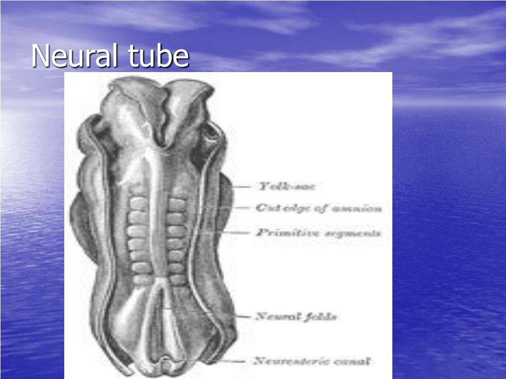
The neural folds pinch in towards the midline of the embryo and fuse together to form the neural tube. In secondary neurulation, the cells of the neural plate form a cord-like structure that migrates inside the embryo and hollows to form the tube. Each organism uses primary and secondary neurulation to varying degrees.
Which part of the neural tube is associated with sensation?
The dorsal part of the neural tube contains the alar plate, which is associated primarily with sensation. The ventral part of the neural tube contains the basal plate, which is primarily associated with motor (i.e., muscle) control.
How does Shh affect the ventral neural tube?
These transcription factors are grouped into two protein classes based on how Shh affects them. Class I is inhibited by Shh , whereas Class II is activated by Shh. These two classes of proteins then cross-regulate each other to create more defined boundaries of expression. The different combinations of expression of these transcription factors along the dorsal-ventral axis of the neural tube are responsible for creating the identity of the neuronal progenitor cells. Five molecularly distinct groups of ventral neurons form from these neuronal progenitor cells in vitro. Also, the position at which these neuronal groups are generated in vivo can be predicted by the concentration of Shh required for their induction in vitro. Studies have shown that neural progenitors can evoke different responses based on the length of exposure to Shh, with a longer exposure time resulting in more ventral cell types.
What are the neural tube patterns?
The neural tube patterns along the dorsal-ventral axis to establish defined compartments of neural progenitor cells that lead to distinct classes of neurons. According to the French flag model of morphogenesis, this patterning occurs early in development and results from the activity of several secreted signaling molecules. Sonic hedgehog (Shh) is a key player in patterning the ventral axis, while bone morphogenic proteins (BMPs) and Wnt family members play an important role in patterning the dorsal axis. Other factors shown to provide positional information to the neural progenitor cells include fibroblast growth factors (FGFs) and retinoic acid. Retinoic acid is required ventrally along with Shh to induce Pax6 and Olig2 during differentiation of motor neurons.
What is the primary neurulation of the ectoderm?
Primary neurulation divides the ectoderm into three cell types: The internally located neural tube. The externally located epidermis. The neural crest cells, which develop in the region between the neural tube and epidermis but then migrate to new locations. Primary neurulation begins after the neural plate forms.
What is the neural groove in the embryo?
The center of the neural plate remains grounded, allowing a U-shaped neural groove to form. This neural gro ove sets the boundary between the right and left sides of the embryo. The neural folds pinch in towards the midline of the embryo and fuse together to form the neural tube.
What is the name of the neural tube that is open both cranially and caudally?
The mesencephalon stays as the midbrain. The rhombencephalon develops into the metencephalon (the pons and cerebellum) and the myelencephalon (the medulla oblongata ). For a short time, the neural tube is open both cranially and caudally. These openings, called neuropores, close during the fourth week in humans.
When do NTDs occur?
NTDs occur when the neural tube does not close properly. The neural tube forms the early brain and spine. These types of birth defects develop very early during pregnancy, often before a woman knows she is pregnant.
What are the two most common NTDs?
The two most common NTDs are spina bifida (a spinal cord defect) and anencephaly (a brain defect).
What is neural tube defect?
Neural tube defects, also known as spinal dysraphisms, are a category of neurological disorders related to malformations of the spinal cord, such as spina bifida, anencephaly, meningocele, myelomeningocele and tethered spinal cord syndrome .
How are neural tube defects diagnosed?
If a child is born with a neural tube defect, such as spina bifida, a thorough evaluation by a pediatric neurosurgeon can help determine the best treatment option.
What happens if the neural tube does not close correctly?
If the seam of the neural tube does not close correctly, portions of the spine, the covering of the spinal cord (meninges) or the cord itself can push outside of the back as the fetus grows.

Overview
In the developing chordate (including vertebrates), the neural tube is the embryonic precursor to the central nervous system, which is made up of the brain and spinal cord. The neural groove gradually deepens as the neural fold become elevated, and ultimately the folds meet and coalesce in the middle line and convert the groove into the closed neural tube. In humans, neural tube closure …
Development
The neural tube develops in two ways: primary neurulation and secondary neurulation.
Primary neurulation divides the ectoderm into three cell types:
• The internally located neural tube
• The externally located epidermis
• The neural crest cells, which develop in the region between the neural tube and epidermis but then migrate to new locations
Structure
Four neural tube subdivisions each eventually develop into distinct regions of the central nervous system by the division of neuroepithelial cells: the forebrain (prosencephalon), the midbrain (mesencephalon), the hindbrain (rhombencephalon) and the spinal cord.
• The prosencephalon further goes on to develop into the telencephalon (cerebrum) and the diencephalon (the optic vesicles and hypothalamus).
Dorsal-ventral patterning
The neural tube patterns along the dorsal-ventral axis to establish defined compartments of neural progenitor cells that lead to distinct classes of neurons. According to the French flag model of morphogenesis, this patterning occurs early in development and results from the activity of several secreted signaling molecules. Sonic hedgehog (Shh) is a key player in patterning the ventral axis, …
See also
• Neural fold
• Neural plate
• Neurulation
• Neural tube defects
• Cdx protein family
External links
• Swiss embryology (from UL, UB, and UF) iperiodembry/carnegie03
• Embryology at UNSW Notes/week3_5
• Diagram at embryology.med.unsw.edu.au
• Diagram at brainviews.com