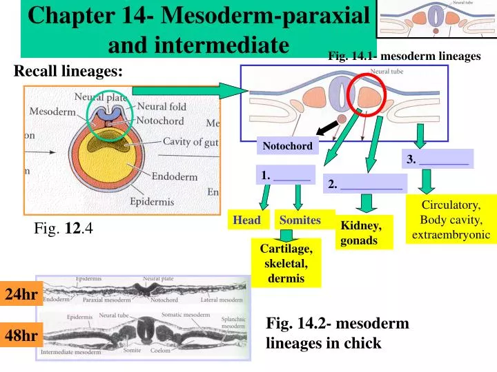
Paraxial mesoderm, also known as presomitic or somitic mesoderm is the area of mesoderm in the neurulating embryo that flanks and forms simultaneously with the neural tube.
What does paraxial mesoderm look like?
* Chordamesoderm: yellow, at notochord. * Paraxial mesoderm: red, at somite. * Intermediate mesoderm: purple, near Wolffian duct. * Lateral plate mesoderm: purple, near "Somatic mesoderm" and "Splanchic mesoderm". Chick embryo of thirty-three hours’ incubation, viewed from the dorsal aspect. (Paraxial mesoderm labeled at left.)
What are the progenitors of the paraxial mesoderm?
Head paraxial mesoderm contains progenitors for many tissues in addition to skeletal muscle. These include cartilages and bones associated with the braincase, loose connective tissues such as meninges and adipocytes, and endothelial cells.
What types of tissue are derived from the segmented paraxial mesoderm?
Many kinds of tissue derive from the segmented paraxial mesoderm by means of the somite. Among these are: the sclerotome, which forms cartilage, the syndetome, which forms tendons, the myotome, which forms skeletal muscle, the dermatome, which forms the dermis as well as skeletal muscle, and endothelial cells.
How do somites form from the paraxial mesoderm?
As the primitive streak continues to regress, somites form from the paraxial mesoderm by "budding off" rostrally. In certain model systems, it has been shown that the daughter cells of stem cell-like progenitor cells which come from the primitive streak or site of gastrulation migrate out and localize in the posterior paraxial mesoderm.
What does the paraxial mesoderm give rise to?
The paraxial mesoderm gives rise to the axial skeleton. The lateral plate mesoderm gives rise to the appendicular skeleton.
Where is the paraxial mesoderm?
The presomitic paraxial mesoderm is located adjacent to the notochord and neural tube and constitutes a longitudinal column of cells on either side of the notochord.
What does the lateral plate mesoderm form?
The lateral plate mesoderm (LPM) forms the progenitor cells that constitute the heart and cardiovascular system, blood, kidneys, smooth muscle lineage and limb skeleton in the developing vertebrate embryo.
What are the different types of mesoderm?
On day 20, mesoderm cells around the notochord differentiate into three specialized types of mesoderm called the paraxial mesoderm, intermediate mesoderm, and lateral plate mesoderm, each of which goes on to make different tissues and organs.
Where does the mesoderm come from?
Gastrulation is an early stage of development during which an embryo, then a single-layered ball of cells called a blastula, reorganizes itself into a three-layered ball of cells, called a gastrula. During this process, the primary germ layers, endoderm and ectoderm, interact to form the third, called mesoderm.
What is a somite paraxial mesoderm?
Paraxial mesoderm, also known as presomitic or somitic mesoderm is the area of mesoderm in the neurulating embryo that flanks and forms simultaneously with the neural tube.
What are the two layers of the lateral plate mesoderm?
Development. The lateral plate mesoderm will split into two layers, the somatopleuric mesenchyme, and the splanchnopleuric mesenchyme. The somatopleuric layer forms the future body wall. The splanchnopleuric layer forms the circulatory system.
What part of mesoderm forms the heart?
Formation of the chick heart from the splanchnic lateral plate mesoderm. The endocardium forms the inner lining of the heart, the myocardium forms the heart muscles, and the epicardium will eventually cover the heart.
What does parietal mesoderm form?
The parietal layer together with overlying ectoderm forms the lateral body wall folds. The visceral layer forms the walls of the gut tube. Mesoderm cells of the parietal layer form the mesothelial membranes or serous membranes which line the peritoneal, pleural and pericardial cavities.
What is mesoderm and its function?
The mesoderm is a germ layer that arises during gastrulation, and is present between the ectoderm, which will turn into skin and central nervous system cells, and the endoderm, which will produce the gut and the lungs (4).
What are the 3 germ layers?
Three primary germ layers Gastrulation is a key phase in embryonic development when pluripotent stem cells differentiate into the three primordial germ layers: ectoderm, mesoderm and endoderm.
Which structure is not formed from mesoderm?
So, the correct option is 'Nervous System'
What develops from the neural crest?
The cardiac neural crest cells can develop into melanocytes, neurons, cartilage, and connective tissue (of the third, fourth, and sixth pharyngeal arches).
What derives from the neural crest?
The cranial neural crest gives rise to the majority of the head connective and skeletal structures, nerves and pigment cells.
What does the intermediate mesoderm develop into?
The intermediate mesoderm generates the urogenital system—the kidneys, the gonads, and their respective duct systems. Saving the gonads for our discussion of sex determination in Chapter 17, we will concentrate here on the development of the mammalian kidney.
What does somatic mesoderm give rise to?
The somatic mesoderm, which is adjacent to the ectoderm and amnion, gives rise to the bones, ligaments, blood vessels, and connective tissue of the limbs.
What are the progenitors of the head mesoderm?
These include cartilages and bones associated with the braincase, loose connective tissues such as meninges and adipocytes, and endothelial cells. In contrast to somites, wherein these progenitor populations are largely segregated, it appears based on mapping studies that these diverse precursors are either intermingled or contiguous in head mesoderm.
What is the Pax gene?
Pax Genes in the Paraxial Mesoderm. The paraxial mesoderm arises from the primitive streak. The first noncompartmentalized epithelial somite undergoes several morphological changes and differentiates into a ventral mesenchymal part consisting of the sclerotome and a dorsal epithelial compartment, the dermomyotome.
Is prenatal muscle development a biphasic process?
In amniotes, prenatal skeletal muscle development is a biphasic process . A primary embryonic myogenesis takes place to generate primary muscle fibers, between embryonic day (E)9.5 and E14.5 in mice. This is followed by a secondary fetal myogenesis which gives rise to the bulk of skeletal-muscle fibers present at birth. However, thanks to lineage tracing studies, as reported by Relaix et al. (2005), Schienda et al. (2006), and Lescroart et al. (2015), we now know that the muscles of the body derive from paraxial mesoderm where muscle specification takes place at earlier stages of the development.
Does a barrier between the brain and paraxial mesoderm prevent myogenesis?
Placing a barrier between the brain and paraxial mesoderm at this region does not prevent myogenesis, but the developing muscle cells lack molecular features that define their specific identity. Together, these experiments suggest that a rich tableau of local signals is necessary for early eye muscle differentiation, with both general myogenic and individual eye muscle-specific components.
Where is the paraxial mesoderm located?
Paraxial mesoderm is formed bilaterally adjacent to the neural tube in the mouse embryo and becomes progressively segmented along the rostral-to-caudal axis to generate the somites.
What are the progenitors of the head paraxial mesoderm?
These include cartilages and bones associated with the braincase, loose connective tissues such as meninges and adipocytes, and endothelial cells.
What is the Pax gene?
Pax Genes in the Paraxial Mesoderm. The paraxial mesoderm arises from the primitive streak. The first noncompartmentalized epithelial somite undergoes several morphological changes and differentiates into a ventral mesenchymal part consisting of the sclerotome and a dorsal epithelial compartment, the dermomyotome.
How long does it take for a paraxial mesoderm to differentiate?
The first pair of somites form at about 20 days; thereafter at a rate of about three per day until 42–44 pairs are formed. Not all therefore exist at the same time, and all will differentiate further. The age of an embryo is often related to the number of somite pairs present. From the beginning of the fourth week the somites undergo further differentiation to form dermomyotomes (form connective tissue and skeletal muscle) and sclerotomes (form bone and cartilage) ( Fig. 1.12A). Cells from the sclerotomes surround the notochord and spinal cord, and give rise to the vertebral column. Details of the formation of vertebrae, and the contribution made by the sclerotomes, may be found in Chapter 4.
Which mesoderm extends caudally without interruption to the first somite?
Prechordal mesoderm is contiguous laterally with paraxial mesoderm, which extends caudally without interruption to the first somite. In a transverse plane, mesenchymal paraxial mesoderm is contiguous laterally with lateral splanchnic mesoderm, which bends to the ventral side, beneath the floor of the pharynx.
Which compartment of the skeleton contains the progenitors of the skeletal muscles, dermis, and?
The somites soon become subdivided into a ventral compartment (the sclerotome) that contributes to the axial skeleton and a dorsal epithelial compartment called dermomyotome which maintains expression of Pax3 and contains the progenitors of the skeletal muscles, dermis, and brown fat (Chal & Pourquie, 2009 ).
Where are somite cells located?
Somite cells are held in fixed positions relative to the dorsal and ventral parts of the adjacent neural tube (hindbrain and spinal cord) and overlying surface epithelium , all of which provide combinations of positive and negative regulators of early myogenesis and skeletogenesis.
What is the position of the segmental plate mesoderm?
The segmental plate mesoderm is determined as to its position along the anterior-posterior axis before somitogenesis. When segmental plate mesoderm that would ordinarily form thoracic somites is transplanted into a region in a younger embryo (caudal to (more...) Differentiation within the somite.
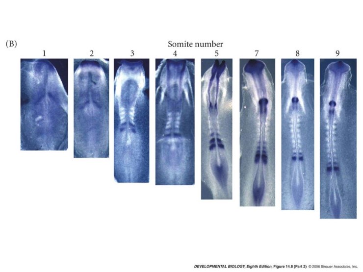
What is the expression pattern of Somite X?
Somite X is marked. The expression of the hairygene (purple) is seen in the caudal half of this somite, (more...) Thus, the expression pattern of the hairygene correlates with the positioning of the place where a somite will separate from the unsegmented mesoderm.
What is the purpose of gastrulation?
One of the major tasks of gastrulation is to create a mesodermal layer between the endoderm and the ectoderm. As shown in Figure 14.2, the formation of mesodermal and endodermal organs is not subsequent to neural tube formation, but occurs synchronously. The notochord extends beneath the neural tube from the base of the head into the tail. On either side of the neural tube lie thick bands of mesodermal cells. These bands of paraxial mesoderm are referred to as the segmental plate(in birds) and the unsegmented mesoderm(in mammals). As the primitive streak regresses and the neural folds begin to gather at the center of the embryo, the paraxial mesoderm separates into blocks of cells called somites. Although somites are transient structures, they are extremely important in organizing the segmental pattern of vertebrate embryos. As we saw in the preceding chapter, the somites determine the migration paths of neural crest cells and spinal nerve axons. Somites give rise to the cells that form the vertebrae and ribs, the dermis of the dorsal skin, the skeletal muscles of the back, and the skeletal muscles of the body wall and limbs.
What is the term for the segmental plate in birds?
These bands of paraxial mesoderm are referred to as the segmental plate (in birds) and the unsegmented mesoderm (in mammals). As the primitive streak regresses and the neural folds begin to gather at the center of the embryo, the paraxial mesoderm separates into blocks of cells called somites.
How do somites form?
The important components of somitogenesis (somite formation) are periodicity, epithelialization, specification, and differentiation. The first somites appear in the anterior portion of the trunk, and new somites “bud off” from the rostral end of the paraxial mesoderm at regular intervals (Figures 14.2C,Dand 14.3). Somite formation begins as paraxial mesoderm cells become organized into whorls of cells called somitomeres. The somitomeres become compacted and bound together by an epithelium, and eventually separate from the presomitic paraxial mesoderm to form individual somites. Because individual embryos can develop at slightly different rates (as when chick embryos are incubated at slightly different temperatures), the number of somites present is usually the best indicator of how far development has proceeded. The total number of somites formed is characteristic of a species (50 in chicks, 65 in mice, and about 500 in some snakes).
Where do epaxial muscles form?
Myotome derivatives of the mouse embryo. The epaxial muscles form from the region of the dermamyotome closest to the neural tube. The hypaxial muscles form from the region of dermamyotome furthest from the neural tube. The epaxial myotome will form the (more...)
Which layer of the dermamyotome is the central region of the dermamyot?
The central region of the dorsal layer of the dermamyotome is called the dermatome, and it generates the mesenchymal connective tissue of the back skin: the dermis. (The dermis of other areas of the body forms from other mesenchymal cells, not from the somites.)
What is the paraxial mesoderm derived from?
The early paraxial mesoderm is referred to as the presomitic mesoderm, and consists of bilateral streaks of mesenchymal cells adjacent to the notochord 17. The presomitic mesoderm is derived from the primitive streak or neuromesodermal progenitors in the tail bud, as shown in studies with mouse and bird embryos 18,19.
Where does the paraxial mesoderm lie?
The notochord extends beneath the neural tube from the base of the head into the tail. On either side of the neural tube lie thick bands of mesodermal cells. These bands of paraxial mesoderm are referred to as the segmental plate (in birds) and the unsegmented mesoderm (in mammals).
What is the intermediate mesoderm derived from?
The intermediate mesoderm is a germ layer aptly named for its intermediate position between the paraxial and lateral plate mesoderm. The paired cylindrical masses of the intermediate mesoderm are arranged along the posterior aspect of the embryo, laterally to the paraxial mesoderm.
Where do endothelial cells come from?
Endothelial cells (ECs), which line blood and lymphatic vessels, are generally described to come from the lateral plate mesoderm despite experimental evidence for a broader source of origin, including the paraxial mesoderm (PXM).
Where do ECs come from?
Endothelial cells (ECs), which line blood and lymphatic vessels, are generally described to come from the lateral plate mesoderm despite experimental evidence for a broader source of origin, including the paraxial mesoderm (PXM). Current dogma suggests that following specification from mesoderm, local environmental cues establish the distinct molecular and functional characteristics of ECs in different vascular beds. Here we present evidence to challenge this view, showing that lymphatic EC fate is imprinted during transition through the PXM lineage. We show that PXM-derived cells form the lymphatic endothelium of multiple organs and tissues, with a more restricted contribution to blood vessel endothelium. By deleting Prox1 specifically in PXM-derived cells, we show that this lineage is indispensable for lymphatic vessel development. Collectively, our data establish lineage history as a critical determinant of EC specialization, a finding with broad implications for our understanding of vascular development and heterogeneity.
Where is the paraxial mesoderm located?
a A dorsal view of the mesoderm fate in the posterior of an amniote embryo. The paraxial mesoderm forms in the primitive streak and from NMPs. Through the elongation of the embryo axis, the PSM is located from posterior to anterior according to the body axis produced by competing gradients of Wnt, FGF, and RA. The anterior PSM forms somite at the determination front depending on the FGF gradient and Notch signaling. b A flowchart of the in vitro differentiation process from PSCs to sclerotomes, with critical inducers and specific markers in each stage. In the future, a next step will be to differentiate the induced sclerotome into sclerotome derivatives, such as bone, tendon, and vessels. RA retinoic acid, NMP neuromesodermal progenitor, PS primitive streak, aPS anterior primitive streak, pPSM posterior presomitic mesoderm, aPSM anterior presomitic mesoderm, nt neural tube, n notochord, LPM lateral plate mesoderm. Asterisks in each of the differentiation stages in a correspond to the stages highlighted by asterisks in b.
What is the process of the paraxial mesoderm?
In this process, key developmental processes, including initiation of the segmentation clock, formation of the determination front, and the mesenchymal–epithelial transition, are sequentially coordinated.
How is somite formed?
The somite is derived from the anterior presomitic mesoderm through a series of dynamic morphogenetic events that involve cyclical signaling. The periodicity of somitomere formation is produced by the segmentation clock that operates in the presomitic mesoderm. A study with mouse embryos demonstrated that this segmental prepattern is defined at the “determination front”, which creates future somitic boundaries 31. This process proceeds according to a “clock and wavefront model”: a clock determines the time, and a wavefront determines the place for the segmentation 32. Mesenchymal–epithelial transition (MET) is another essential process for somitogenesis, as it is involved in epithelial somite formation 33. Studies with mouse embryos demonstrate that during these processes, Msgn1 is downregulated, but several other markers, including Mesp2, Paraxis, Pax3, Foxc1/2, and Meox1/2, are upregulated 9, 34, 35, 36.
What is the BMP gradient?
The BMP gradient is a critical factor in the determination of the mediolateral axis during mesoderm development and in somite specification 10, 63. In line with this, some reports showed that a BMP inhibitor improved somite–sclerotome induction, possibly by both promoting medial fate and protecting the mesodermal population from lateralization 10, 82, 83, 84, 85, 86.
What are the two major populations of the paraxial mesoderm?
Mature somites contain two major populations: the sclerotome and dermomyotome. The sclerotome gives rise to the vertebrae and associated ribs, tendons, and other tissues, such as vascular cells of the dorsal aorta, intervertebral blood vessels, and meninges 12, 13. The dermomyotome produces two components: the myotome and the dermatome. The myotome gives rise to the musculature of the back, rib cage, ventral body wall, and limbs. The dermatome gives rise to the dermis of the back, although the term dermo myotome is sometimes used to describe this region because a recent study showed that this central region of the dermomyotome also gave rise to muscles in chick embryos 14.
Why is MET necessary for somitogenesis?
MET is necessary to form the epithelial layer of the somite during somitogenesis, since the presomitic mesoderm is composed of only mesenchymal cells. A study with mouse embryos demonstrated that without MET, neither the epithelial somite nor the dermomyotome can properly form; the absence of MET leads to abnormalities of the axial skeleton, such as numerous patterning defects of the musculature in the axial skeleton, limbs, and body wall 33. During MET in the future somatic boundaries, the outer epithelial layer assumes apical–basal polarity and expresses N-cadherin, β-catenin, and F-actin in apical adherens junctions 50. This process is intimately regulated in a spatial and temporal manner along the anterior–posterior axis, as evidenced by a study in birds 50.
Where are Pax1 and Pax9 expressed?
Pax1 and Pax9, transcription factors of the Pax family, are specifically expressed in the large part of sclerotomes. Homozygous Pax1 -null newborn mice showed severe abnormalities in the axial skeleton 67. On the other hand, homozygous Pax9 mutant mice showed skeletal defects in the limbs and the skull, but exhibited no obvious defects in the axial skeleton 68. In addition, Pax1/Pax9 double mutant mice showed much more severe phenotypes than Pax1 single homozygous mutants; Pax1/Pax9 double mutants completely lack tissues derived from the medial part of the sclerotome, such as the vertebral bodies, the intervertebral discs, and the proximal parts of the ribs 69. The condensation of the ventromedial sclerotome around the notochord was also prevented in the double mutants, resulting in an impairment of chondrogenesis and vertebral formation 69. Moreover, a rescue experiment in mice showed that Pax1 compensated for Pax9 function, whereas Pax9 did not compensate for Pax1 function 69.