The pharyngeal plexus lies on the posterolateral wall of the pharynx, mainly over the middle pharyngeal constrictor and is the main motor and sensory nerve supply to the muscles of the pharynx and soft palate and acts to coordinate swallowing and speech 1-7.
Where is the pharyngeal plexus located?
The pharyngeal plexus is a network of nerve fibers innervating most of the palate and pharynx. (Larynx, which is innervated by superior and recurrent laryngeal nerve from vagus nerve (CN X), is not included). It is located on the surface of the middle pharyngeal constrictor muscle.
What is the glossopharyngeal plexus?
pharyngeal plexus. 1. A nerve plexus along the posterior surface of the middle pharyngeal constrictor muscle. The plexus contains sympathetic, parasympathetic, and somatic (branchial) axons from the pharyngeal branch of the vagus nerve, the glossopharyngeal and external laryngeal nerves, and the sympathetic trunk.
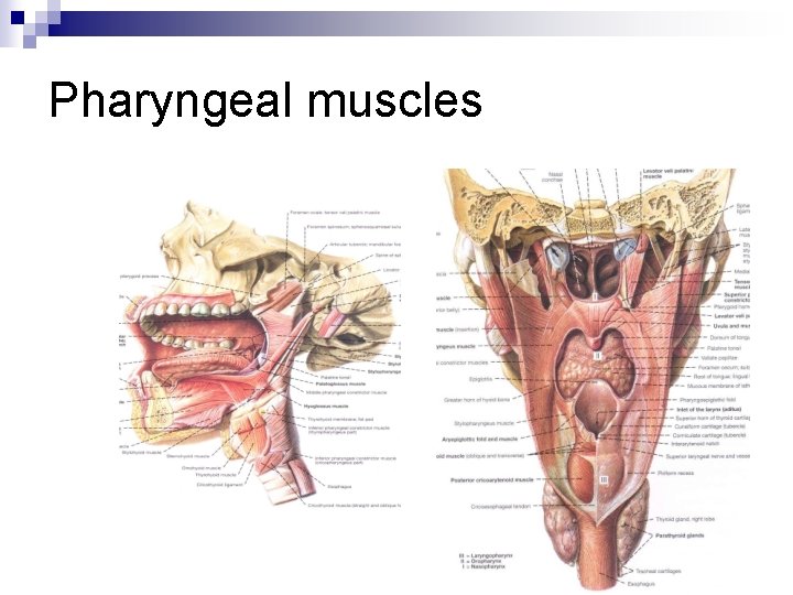
What are the muscles of the pharynx?
Muscles of the pharynx, viewed from behind, together with the associated vessels and nerves. (Pharyngeal plexus visible but not labeled.) The pharyngeal plexus is a network of nerve fibers innervating most of the palate and pharynx. ( Larynx, which is innervated by superior and recurrent laryngeal nerve from vagus nerve (CN X), is not included)
What is the function of the middle pharynx plexus?
The plexus lies along the middle pharyngeal constrictor muscle and is responsible for sensory and motor innervation. SENSORY INNERVATION The three regions of the pharynx each receive a unique cranial nerve supply.

What is the meaning of pharyngeal plexus?
Medical Definition of pharyngeal plexus : a plexus formed by branches of the glossopharyngeal, vagus, and sympathetic nerves supplying the muscles and mucous membrane of the pharynx and adjoining parts.
Where is pharyngeal plexus?
pharynxThe plexus is located in the retropharyngeal space close to the longus capitis and colli muscles, the prevertebral fascia, the vertebral bodies of the second and third cervical vertebrae posteriorly, and the posterior wall of the pharynx, and is closer to the superior and middle constrictor anteriorly [18, 37].
What does the pharyngeal nerve do?
The pharyngeal branch of the vagus innervates the superior, middle, and inferior pharyngeal constrictors. These muscles constrict the walls of the pharynx during swallowing.
What does the pharyngeal plexus innervate?
The pharyngeal plexus provides sensory innervation of the oropharynx and laryngopharynx from CN IX and CN X. (The nasopharynx above the pharyngotympanic tube and the torus tubarius is innervated by CN V2).
What is the pharyngeal plexus made up of?
The neural contributions of the cranial nerves IX, X, and superior sympathetic ganglion intertwine to form the pharyngeal plexus that can be injured during ACDF procedures.
What nerve causes difficulty in swallowing?
Glossopharyngeal and vagus nerve dysfunction are typically the cause of chronic dysphagia when no structural lesion is found. The glossopharyngeal nerve innervates the stylopharyngeus muscle, which elevates the larynx and pulls it forward during the pharyngeal stage of swallowing.
Which nerve is responsible for swallowing?
The vagal nerve (VN), the tenth cranial nerve, provides both motor and sensory innervation, and plays an important role in the pharyngeal phase of swallowing [4, 6].
Where is pharyngeal?
ThroatThe pharynx (plural: pharynges) is the part of the throat behind the mouth and nasal cavity, and above the oesophagus and trachea (the tubes going down to the stomach and the lungs)....PharynxPart ofThroatSystemRespiratory system, digestive system14 more rows
Where is pharyngeal?
ThroatThe pharynx (plural: pharynges) is the part of the throat behind the mouth and nasal cavity, and above the oesophagus and trachea (the tubes going down to the stomach and the lungs)....PharynxPart ofThroatSystemRespiratory system, digestive system14 more rows
Where does the vagus nerve begin and end?
The vagus nerve runs from the brain through the face and thorax to the abdomen. Exits the brain from the medulla oblongata of the brainstem and travels laterally exiting the skull through the jugular foramen.
Where are located nuclei of VIII pair of cranial nerves?
These nuclei are the most lateral of all the cranial nuclei. They are located medially to the inferior peduncle of the cerebellum in the lateral angle of the rhomboid fossa.
Where is the pharyngeal plexus located?
The pharyngeal plexus lies within the external fascia of the pharynx 3 on the posterolateral wall of the pharynx in the retropharyngeal space 7. It lies mainly over the middle pharyngeal constrictor muscle 4,5 although it spreads its branches over the dorsolateral surface of the both the superior and middle constrictors, and lower branches descend onto the inferior constrictor 6. The pharyngeal plexus sends some branches between the middle and inferior constrictors and other branches between the superior and middle constrictors 4,6. It is formed by the pharyngeal branches from the vagus and glossopharyngeal nerves and the cervical sympathetic 3-7.
What nerves form the pharyngeal plexus?
It is formed by the pharyngeal branches from the vagus and glossopharyngeal nerves and the cervical sympathetic 3-7. The pharyngeal branch of the glossopharyngeal nerve arises prior to ...
What causes pharyngeal plexus injury?
Unilateral right sided pharyngeal plexus injury has been reported following use of an orophryngeal pack during third molar surgery in which the patient exhibited sensory loss over the right side of the pharynx, soft palate, and posterior third of the tongue and weakness of the right side of the pharynx and soft palate without vocal cord injury 7. A case of isolated transient pharyngeal plexus injury has been reported following posterior plating of a C2 fracture in which the patient presented with dense pharyngeal paralysis and inability to initiate swallowing but with true vocal cord movement preserved 14 .
Which muscle receives its nerve supply solely through the pharyngeal plexus?
The levator veli palatini muscle has been reported by varying sources to receive its nerve supply solely through the pharyngeal plexus or doubly through the pharyngeal plexus and branches of the facial nerve 11-13. The muscle of the uvula has been reported to receive both dual innervation from the pharyngeal plexus and lesser palatine nerve, ...
Where does the pharyngeal branch of the vagus join the pharyngeal?
The pharyngeal branch of the vagus emerges from the upper part of the inferior vagal ganglion and passes forward between the internal and external carotid arteries parallel with and below the glossopharyngeal nerve to join the pharyngeal plexus at the upper border of the middle pharyngeal constrictor muscle 1,2,4.
Which muscles are innervated by the pharyngeal plexus?
The superior, middle and lower pharyngeal constrictor muscles, palatopharyngeus, salpingopharyngeus, levator veli palatini, palatoglossus and the muscle of the uvula are all innervated by branches of the pharyngeal plexus 1-5. The innervations of levator veli palatini and uvular muscle remain controversial 11-13.
Which part of the pharynx is responsible for swallowing?
Pharyngeal plexus. The pharyngeal plexus lies on the posterolateral wall of the pharynx, mainly over the middle pharyngeal constrictor and is the main motor and sensory nerve supply to the muscles of the pharynx and soft palate and acts to coordinate swallowing and speech 1-7.
Which CN forms the pharyngeal plexus?
Pharyngeal nerves from CN IX and CN X and a small contribution from CN V-2 form the pharyngeal plexus (Figure 27-3B).
Which plexus is the sensory innervation of the oropharynx?
Oropharynx. Sensory neurons from CN IX enter the pharyn-geal plexus and provide the sensory innervation for the oropharynx.
What is the pharyngeal plexus?
The pharyngeal plexus is an essential anatomical structure , but the contributions from the glossopharyngeal and vagus nerves and the superior cervical ganglion that give rise to the pharyngeal plexus are not fully understood. The pharyngeal plexus is likely to be encountered during various anterior cervical surgical procedures of the neck such as anterior cervical discectomy and fusion. Therefore, a detailed understanding of its anatomy is essential for the surgeon who operates in and around this region. Although the pharyngeal plexus is an anatomical structure that is widely mentioned in literature and anatomy books, detailed descriptions of its structural nuances are scarce; therefore, we provide a comprehensive review that encompasses all the available data from this critical structure. We conducted a narrative review of the current literature using databases like PubMed, Embase, Ovid, and Cochrane. Information was gathered regarding the pharyngeal plexus to improve our understanding of its anatomy to elucidate its involvement in postoperative spine surgery complications such as dysphagia. The neural contributions of the cranial nerves IX, X, and superior sympathetic ganglion intertwine to form the pharyngeal plexus that can be injured during ACDF procedures. Factors like surgical retraction time, postoperative hematoma, surgical hardware materials, and profiles and smoking are related to postoperative dysphagia onset. Thorough anatomical knowledge and lateral approaches to ACDF are the best preventing measures.
Where is the pharyngeal plexus located?
... The pharyngeal plexus (PP) is positioned in the posterior portion of the middle pharyngeal constrictor muscle; the plexus is formed by branches of the nerve IX, X and the sympathetic upper cervical ganglion [37]. The PP is found in the retropharyngeal space near the neck and longus capitis muscles, the connective tissue of the prevertebral fascia of C2-C3 (posteriorly), and the posterior edge of the pharynx. ...
What muscles are in the right pharyngeal plexus?
Posterior view of the right pharyngeal plexus and associated nerves, arteries, and muscles. Medially, the superior (SPC), middle (MPC), and inferior (IPC) muscles are seen . Traveling from lateral to medial, the stylopharyngeus muscle (SP) is seen. For reference, note the hyoid bone (H) at the level of the C3 vertebra and the thyroid cartilage (TC) at the level of C4 and C5 vertebrae. Over, primarily, the posterior surface of the middle pharyngeal constrictor muscle, note the contributions to the pharyngeal plexus from the vagus (A), glossopharyngeal (B), and sympathetic trunk (C). ICA, internal carotid artery; CCA, common carotid artery; SCG, superior cervical ganglion; * = superior laryngeal nerve; C1 = C1 spinal nerve; C2 = C2 spinal nerve, CN IX = glossopharyngeal nerve; CN X = vagus nerve; CN XII = hypoglossal nerve (after Henle)
What is ganglioneuroma in the neck?
Ganglioneuroma is a rare, benign, non-invasive tumor emerging from the sympathetic system. Of these tumors, only 8% occur in the neck. In this report, we present a case of a 13-year-old girl with a 2-year history of enlarging neck mass. Her only complaint, aside from neck swelling, was dysphagia. Physical and radiological examinations revealed a large mass centered in the right carotid space. A transcervical approach was used to excise the tumor emerging from the sympathetic ganglia. The patient developed temporary Horner's syndrome postoperatively. In a few weeks, she was completely asymptomatic. Histological examination was compatible with ganglioneuroma. Surgical excision is the only definite treatment of cervical ganglioneuroma and is also the only way to confirm the diagnosis. Injury during surgery may result in significant morbidity.
What are the brainstem nuclei involved in swallowing?
Anatomical connections are reported between the cerebellum and brainstem nuclei involved in swallow such as the nucleus tractus solitarius, nucleus ambiguus, and Kölliker-fuse nuclei. Despite these connections, a functional role of the cerebellum during swallow has not been elucidated. Therefore, we examined the effects of cerebellectomy on swallow muscle recruitment and swallow–breathing coordination in anesthetized freely breathing cats. Electromyograms were recorded from upper airway, pharyngeal, laryngeal, diaphragm, and chest wall muscles before and after complete cerebellectomy. Removal of the cerebellum reduced the excitability of swallow (i.e., swallow number), and muscle recruitment of the geniohyoid, thyroarytenoid, parasternal (chestwall), and diaphragm muscles, but did not disrupt swallow–breathing coordination. Additionally, diaphragm and parasternal muscle activity during swallow is reduced after cerebellectomy, while no changes were observed during breathing. These findings suggest the cerebellum modulates muscle excitability during recruitment, but not pattern or coordination of swallow with breathing.
What are the five diaphragms?
One of these is the five diaphragm model (tentorium cerebelli, tongue, thoracic outlet, diaphragm, and pelvic floor ), whose foundations are part of another historical model: respiratory-circulatory. The myofascial continuity, anterior and posterior, supports the notion the human body cannot be divided into segments but is a continuum of matter, fluids, and emotions. In this first part, the neurological relationships of the tentorium cerebelli and the lingual muscle complex will be highlighted, underlining the complex interactions and anastomoses, through the most current scientific data and an accurate review of the topic. In the second part, I will describe the neurological relationships of the thoracic outlet, the respiratory diaphragm and the pelvic floor, with clinical reflections. In literature, to my knowledge, it is the first time that the different neurological relationships of these anatomical segments have been discussed, highlighting the constant neurological continuity of the five diaphragms.
Which nerve is the left lateral view?
Left lateral view of the superior laryngeal nerve, a branch of the vagus nerve. Shown here are its two terminal branches, internal and external. The external branch innervates the cricothyroid and inferior pharyngeal constrictor muscles