What causes osteoarthritis of the pisotriquetral?
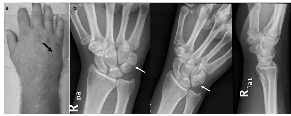
Osteoarthritis of the pisotriquetral joint is most often caused by acute and chronic trauma and instability. The symptoms of osteoarthritis of the pisotriquetral joint are pain over the pisiform, with pressure and grinding of the joint. There may be ulnar nerve symptoms, and attrition or rupture of the flexor profundus tendon to the little finger.
What are the symptoms of pisotriquetral joint disease?
Side-to-side passive motion of the pisiform occasionally led to pain and crepitus. Degenerative arthritis and calcifications in the pisotriquetral joint were confirmed by a wrist radiograph. In 5 patients, local injection with anesthetic temporarily resolved the symptoms.
What is the synovial joint between the pisiform and triquetrum?
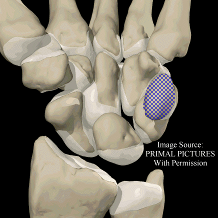
[TA] the synovial joint between the pisiform and triquetrum; it is separate from the other intercarpal joints. Synonym(s): articulatio ossis pisiformis [TA], articulation of pisiform bone, pisotriquetral joint.
What do we know about pisotriquetral instability?
Pisotriquetral instability is an often-overlooked condition that can lead to ulnar-sided wrist pain and dysfunction. Various case series and biomechanical studies have been published regarding the diagnosis and treatment of this condition. We review current methods for examining, diagnosing, and treating pisotriquetral instability.

Where is the Pisotriquetral joint?
The pisotriquetral joint is the smallest of the four joints of the wrist. Although separate, it is often connected to the radiocarpal joint through a fenestration. The gross anatomy and kinematics of the pisotriquetral joint have been well described.
What is Pisotriquetral?
Abstract. Pisotriquetral (PT) osteoarthritis (OA) and enthesopathy of the flexor carpi ulnaris (FCU) are pathologies of the hypothenar eminence which both often remain undiagnosed, but can cause ulnar wrist pain. This study determined the prevalence of these pathologies in an older donor population.
What causes pain in the pisiform bone?
Pain in the area of the pisiform can be because of a wide variety of pathologies including tendinitis at the insertion FCU, arthritis of the pisotriquetral joint, subluxation of the pisiform with associated synovitis, fracture of the triquetrum or pisiform, rheumatism, or osteonecrosis.
Where is the pisiform Triquetral joint?
wristThe pisiform joint is a joint between the pisiform and triquetrum. Vertical section through the articulations at the wrist, showing the synovial cavities. Shown is the right hand, palm down (left) and palm up (right). It includes the pisohamate ligament and pisometacarpal ligament.
How long does it take for a pisiform fracture to heal?
Most patients with a pisiform fracture can be treated with cast immobilization for 4 to 6 weeks. Conservative management for non-displaced triquetrum body fractures or dorsal chip fractures involves a short arm cast for 4 to 6 weeks.
How is pisiform pain treated?
Pisiform area pain treatment by pisiform excision Wrist strength and mobility was maintained by doing a subperiosteal dissection and removal of the pisiform bone. This technique preserves the insertion of the FCU tendon and its distal extension, the piso hamate and the piso metacarpal ligaments.
Can you get arthritis in the pisiform bone?
Arthritis beneath the pisiform bone (pisotrequetral arthritis) causes sharp pain on the outer (ulnar) side of the wrist on movement, and is one of the diagnoses that needs considering in ulnar wrist pain.
What does a broken pisiform feel like?
This injury presents as chronic wrist pain, grip weakness, and/or restriction of wrist movements. Pisiform fractures may also be associated with tenderness in the affected area.
How hard is it to break your pisiform?
Pisiform fractures are an uncommon injury accounting for only 0.2% of all carpal fractures. They are managed by immobilisation in either a plaster cast or a wrist splint. This fracture can be easily missed on first presentation due the superimposition of adjacent carpal bones.
What is the wrist bone that sticks out called?
The pisiform bone (/ˈpaɪsɪfɔːrm/ or /ˈpɪzɪfɔːrm/), also spelled pisiforme (from the Latin pisifomis, pea-shaped), is a small knobbly, sesamoid bone that is found in the wrist. It forms the ulnar border of the carpal tunnel.
What type of bone is the pisiform?
The pisiform is the smallest of the carpals. Because it develops within a tendon, it is actually a sesamoid bone. There are other, much smaller sesamoid bones found embedded in flexor tendons, for example, at some metacarpophalangeal and interphalangeal joints.
What tendon attaches to the pisiform?
flexor carpi ulnaris tendonThe pisiform bone is a sesamoid bone which lies embedded within the flexor carpi ulnaris tendon, providing a smooth surface for it to glide over. It acts as an important attachment site for both the flexor carpi ulnaris and abductor digiti minimi muscles.
What is Pisiformectomy?
Conclusions Pisiformectomy is a surgery used sparingly in cases with refractory pain associated with arthrosis of the pisotriquetral joint or enthesopathy of the flexor carpi ulnaris/pisiform interface.
What is the STT joint in the hand?
What is STT arthritis? This is arthritis occurring between three bones in the wrist; the Scaphoid, the Trapezium and the Trapezoid. It can lead to pain and stiffness in the thumb and wrist.
What is a pisiform excision?
Pisiform excision is a relatively safe procedure for patients with chronic ulnar-sided wrist pain due to pisotriquetral osteoarthritis, FCU tendinitis, or ulnar neuropathy when a conservative treatment is insufficient. Mixed diagnoses are often encountered in clinical practice.
What are the carpal bones?
The proximal row of carpal bones (moving from radial to ulnar) are the scaphoid, lunate, triquetrum, and pisiform, while the distal row of carpal bones (also from radial to ulnar) comprises the trapezium, trapezoid, capitate, and hamate.
Which ligaments are primary stabilizers?
Conclusions: The pisiform ligament complex has primary and secondary stabilizers to the PT joint. The primary stabilizers are the PH, PM, and ulnar PT ligaments. The transverse carpal and radial PT ligaments are secondary stabilizers. Injuries of the primary stabilizers of the PT joint may lead to instability that predisposes to degenerative joint disease.
Which ligaments attach to the pisiform?
Results: The ligaments that attached to the pisiform were the pisometacarpal (PM), pisohamate (PH), radial PT, ulnar PT, and transverse carpal ligament. The PH ligament was shorter, wider, and thicker than the PM ligament. The transverse carpal ligament attachment in the pisiform was insubstantial. In 10 limbs degenerative changes were present, most of them peripheral. Biomechanical testing showed that the primary stabilizers of the PT joint were the PM, PH, and ulnar PT ligaments and that these were responsible for resisting proximal, ulnar, and radial forces, respectively. The PH distance increased along with the pisiform sagittal motion during wrist flexion on oblique x-rays after transection of the PM and ulnar PT ligaments. Concomitantly this distance decreased on the anteroposterior x-rays during radial deviation. The PH distance increased along with the pisiform frontal motion after transection of the PH and radial PT ligaments.
What causes pain in the pisotriquetral joint?
Osteoarthritis of the pisotriquetral joint is most often caused by acute and chronic trauma and instability. The symptoms of osteoarthritis of the pisotriquetral joint are pain over the pisiform, with pressure and grinding of the joint. There may be ulnar nerve symptoms, and attrition or rupture of the flexor profundus tendon to the little finger.
What is the treatment for pisotriquetral arthritis?
Conservative treatment of pisotriquetral arthritis consists of local injections of steroid into the pisotriquetral joint along with nonsteroid al anti-inflamma tory drugs (NSAIDs) and protective splinting. When conservative therapy fails, consideration should be given to pisiform excision.
Why does my pisiform joint hurt?
Pisotriquetral arthritis. Chronic pain in the pisiform area may be caused by tendinitis of the insertion of the flexor carpi ulnaris, bony fractures , or osteoarthrosis of the pisotriquetral joint, which some report as a frequent site of osteoarthritis slightly less common than the scaphotrapezial osteoarthrosis [52].
Is pisiform pain common?
Although pain and tenderness on the palmar and ulnar aspects of the wrist in the area of the pisiform bone is fairly common, refractory pisotri -quetral osteoarthritis was unusual enough for Green to be able to make a case report of simple excision of the pisiform back in 1979 [53].
What is the pain in the pisiform bone?
Pain and tenderness on the palmar and ulnar aspects of the wrist in the area of the pisiform bone is fairly common. Chronic pain in the pisiform area (or wrist pain ) may be caused by tendonitis of the flexor carpi ulnaris, bony fractures or osteoarthritis of the pisotriquetral joint.
What are the symptoms of osteoarthritis of the pisotriquetral joint?
The symptoms of osteoarthritis of the pisotriquetral joint are pain over the pisi form, with pressure and grinding of the joint . There may be ulnar nerve symptoms such as numbness and tingling in the little finger and along the outside of the ring finger.
What causes pain in the pisotriquetral joint?
Osteoarthritis of the pisotriquetral joint is most often caused by acute and chronic trauma and instability. The symptoms of osteoarthritis of the pisotriquetral joint are pain over the pisiform, with pressure and grinding of the joint. There may be ulnar nerve symptoms such as numbness and tingling in the little finger and along the outside of the ring finger.
What is the bone that makes your wrist hurt?
Causes of wrist pain. The pisiform bone is one of the eight carpal (wrist) bones. It is a small pea-shaped sesamoid bone located where the ulna (one of the bones in the forearm) joins the wrist (on the little finger side).
What is the treatment for pisotriquetral arthritis?
Treatment for pisotriquetral arthritis. Conservative treatment of pisotriquetral arthritis consists of local injections of steroid into the pisotriquetral joint along with nonsteroidal anti-inflammatory drugs (NSAIDs) and a protective splint.
Can you speak to a physiotherapist with AXA?
And remember if you have health cover with AXA Health, you can speak to a qualified physiotherapist for help with any musculoskeletal problems as soon as symptoms occur, and without the need for a GP referral, through our Working Body service.
What are the supportive structures of the PT joint?
Dissection of the supportive structures of the PT joint was performed; these included the pisohamate (PH) ligament, the pisometacarpal (PM) ligament, and the transverse carpal ligament (TCL). The PH and PM ligaments were measured for length, width, and thickness by using precision calipers (SPI, Garden Grove, CA). The lengths of the PH and PM ligaments were measured in full passive wrist flexion and extension. The attachments of the PH and PM ligaments to the pisiform were observed and recorded and the angle formed between the 2 ligaments was measured. The TCL insertion and its relation to the PH ligament and the pisiform were observed. The radial and ulnar PT ligaments were identified, examined, and compared. The PT joint capsule was identified and its integrity was assessed. The articular surface of the pisiform was examined for degenerative changes and their patterns, if any.
What causes ulnar sided wrist pain?
Pisotriquetral (PT) joint instability is a common cause of ulnar-sided wrist pain. Several reports have documented that PT joint injury and dysfunction can result in instability and degenerative changes. 1, 2, 3, 4, 5, 6, 7 In a cadaver study from our institute we investigated the anatomy of the soft-tissue attachments of the pisiform by using biomechanical testing of the PT joint. We identified structures of variable ligamentous stiffness around the pisiform. 8 Recent studies have attempted to explain the anatomy of the PT joint better and how ligament injury and instability may relate to degenerative changes. 9, 10, 11, 12, 13 In a more recent study of healthy volunteers we evaluated the normal radiographic kinematics of the pisiform, identified limits for its physiologic excursion, and generated baseline parameters for the PT joint. 14
What ligaments attach to the pisiform?
The ligaments that attached to the pisiform were the pisometacarpal (PM), pisohamate (PH), radial PT, ulnar PT, and transverse carpal ligament. The PH ligament was shorter, wider, and thicker than the PM ligament. The transverse carpal ligament attachment in the pisiform was insubstantial. In 10 limbs degenerative changes were present, most of them peripheral. Biomechanical testing showed that the primary stabilizers of the PT joint were the PM, PH, and ulnar PT ligaments and that these were responsible for resisting proximal, ulnar, and radial forces, respectively. The PH distance increased along with the pisiform sagittal motion during wrist flexion on oblique x-rays after transection of the PM and ulnar PT ligaments. Concomitantly this distance decreased on the anteroposterior x-rays during radial deviation. The PH distance increased along with the pisiform frontal motion after transection of the PH and radial PT ligaments.
How were pisiforms exposed?
The pisiform and PT joint were exposed through a palmar approach preserving all supporting anatomic structures. The soft tissues that were attached directly to the pisiform were exposed and dissected. The pisiform was identified and its proximal and distal poles were marked with a 27-gauge needle. The hamate also was marked in a similar fashion at its midsubstance, which is located 3 mm proximal and radial to the hook of the hamate (hamulus). A 1.4-mm (0.045-inch) K-wire was placed midway between both poles of the pisiform. The initial biomechanical and radiographic studies then were performed.
How many cadaver arms were used in the study?
Twelve cadaver arms were used. The study had 3 components: (1) anatomic dissection of the PT joint ligaments and patterns of degenerative changes, (2) biomechanical sequential sectioning of the supporting ligaments, and (3) radiographic assessment of PT joint motion in several planes both before and after ligament sectioning.
How far is the gap between the wrist and the triquetrum?
Pretransection radiographs showed an average gap distance of 1.5 mm in the neutral wrist position and 1 mm in full extension between the pisiform and triquetrum. This PT joint space averaged 2.5 mm in full flexion. There was an average increase of 0.5 mm in the distance post-ligament transection radiographs during full flexion.
Which is thicker, ulnar or radial PT?
There were 2 components for this ligament: radial and ulnar. The ulnar PT ligament substance always was thicker than the radial PT ligament. The radial PT ligament was a distinct structure from the TCL in all specimens.
What ligaments attach to the pisiform?
The ligaments that attached to the pisiform were the pisometacarpal (PM), pisohamate (PH), radial PT, ulnar PT, and transverse carpal ligament. The PH ligament was shorter, wider, and thicker than the PM ligament. The transverse carpal ligament attachment in the pisiform was insubstantial. In 10 limbs degenerative changes were present, most of them peripheral. Biomechanical testing showed that the primary stabilizers of the PT joint were the PM, PH, and ulnar PT ligaments and that these were responsible for resisting proximal, ulnar, and radial forces, respectively. The PH distance increased along with the pisiform sagittal motion during wrist flexion on oblique x-rays after transection of the PM and ulnar PT ligaments. Concomitantly this distance decreased on the anteroposterior x-rays during radial deviation. The PH distance increased along with the pisiform frontal motion after transection of the PH and radial PT ligaments.
What is the PLC of the wrist?
The pisiform ligament complex (PLC) is a group of ligaments attached to the pisiform that contribute to its stability in different planes. Pisiform ligament complex syndrome is defined as palmar ulnar wrist pain in the vicinity of the pisiform caused by injury to any of the PLC components. Injuries of this ligament complex can be acute or chronic. Acute injuries usually are caused by a fall on the outstretched hand with the force directed to the pisiform. Chronic PLC injuries may be caused by
What is the PLC?
The pisiform ligament complex (PLC) is a group of ligaments attached to the pisiform that contribute to its stability in different planes. Pisiform ligament complex syndrome is defined as palmar ulnar wrist pain in the vicinity of the pisiform caused by injury to any of the PLC components. Injuries of this ligament complex can be acute or chronic. Acute injuries usually are caused by a fall on the outstretched hand with the force directed to the pisiform. Chronic PLC injuries may be caused by
Which ligament inserts on the palmar aspect of the pisiform?
In cadaver models, transection of the pisohamate ligament results in 2 to 3 times the pisiform ulnar translation compared with intact specimens.7 The pisometacarpal ligament inserts on the palmar aspect of the pisiform and is longer and thinner than the pisohamate ligament.2,7 It is the primary stabilizer against proximal displacement.
Which bone is associated with the proximal carpal row?
Akin to a sesamoid bone, the pisiform provides a lever arm to the tendon.The pisiform is included in the proximal carpal row20–22 and articulates with the triquetrum, forming the pisotriquetral joint, a synovial joint that communicates with the radiocarpal joint in the majority of hands.21,22In 1995, Pevny et al20 published a detailed description of the ligamentous and tendinous attachments to the pisiform.
Which ligaments are primary stabilizers?
The pisiform ligament complex has primary and secondary stabilizers to the PT joint. The primary stabilizers are the PH, PM, and ulnar PT ligaments. The transverse carpal and radial PT ligaments are secondary stabilizers. Injuries of the primary stabilizers of the PT joint may lead to instability that predisposes to degenerative joint disease.
How many segments of the median nerve were measured in CTS?
Antidromic nerve conduction velocities were measured in 3 segments of the median nerve: forearm, wrist, and palm. Differences and ratios in nerve conduction velocities were computed between the forearm and wrist and between the palm and wrist segments.

Causes
- Trauma- Where the pisotriquetral joint suffers acute or chronic trauma.
- Tendinitis from flexor carpi ulnaris insertion.
- Age. The condition is also degenerative and therefore, it can be prevalent among the older population. Old age may damage the articular cartilage.
- Trauma- Where the pisotriquetral joint suffers acute or chronic trauma.
- Tendinitis from flexor carpi ulnaris insertion.
- Age. The condition is also degenerative and therefore, it can be prevalent among the older population. Old age may damage the articular cartilage.
- Bone fractures.
Symptoms
- Pain over the pisiform that can worsen with grinding of the joint and pressure.
- Irritation when you perform tasks that involve the wrist.
- Tenderness or swelling over the pisiform.
- Some ulnar nerve symptomscan be experienced, such as weakness, tingling, and numbness within the little finger. You may also experience these symptoms along the outside of your rin…
- Pain over the pisiform that can worsen with grinding of the joint and pressure.
- Irritation when you perform tasks that involve the wrist.
- Tenderness or swelling over the pisiform.
- Some ulnar nerve symptomscan be experienced, such as weakness, tingling, and numbness within the little finger. You may also experience these symptoms along the outside of your ring finger.
Diagnosis
- Diagnosis is mainly based on a clinical examination. Radiographs may be used to reveal its evidence but, they are not sufficient. They can also be misinterpreted; hence, other diagnostic tools are involved. MRI can be used to reveal any degeneration of the wrist tendon and sometimes, the inflammation. CT scans and Microscopy assessment can also be done to show …
Treatment and Management
- There are several treatment options for the condition that include conservative measures. Medication– Nonsteroidal anti-inflammatory drugs can be administered for treatment. Pain relievers such as acetaminophen are recommended for limited use or as needed. Corticosteroids can also be used to reduce inflammation. Steroid Injections– The pisotriquetral joint can be inje…
The Gout Eraser™: The All-Natural Guide For Permanent Gout Removal
- The Gout Eraser™ is a short, to the point guide on how to reverse gout symptoms without ever leaving your home. The guide goes into extensive detail on exactly what you need to do to safely, effectively and permanently get rid of gout, and you are GUARANTEED to see dramatic improvements in days if not hours.