
What is another name for the posterior nasal aperture?
It is also known as the posterior nasal aperture, and the internal or posterior naris (plural: nares) [2]. The thin trapezoidal bone, vomer, located behind and beneath the nasal septum separates the two choanae in the middle [3].
What is numerical aperture in microscope?
numerical aperture An expression designating the light-gathering power of microscope objectives. It is equal to the product of the index of refraction nof the object space and the sine of the angle usubtended by a radius of the entrance pupil at the axial point on the object, i.e. nsin u.
What is the opening of the nasal cavity?
The nasal cavity opens anteriorly onto the face through the anterior nasal aperture and posteriorly into the nasopharynx by way of the posterior nasal apertures.
What is the Apertura of the nose?
A funnel-shaped opening, especially one of the internal openings of the nose into the PHARYNX. The right or left opening from the nasal cavity into the nasopharynx. Synonym (s): apertura nasalis posterior.
What is the name of the bone that divides the nasal cavity?
How large is the nasal cavity?
What are the three segments of the pharynx?
About this website
What does the nasal aperture do?
The pyriform or nasal aperture, is the pear-shaped bony inlet of the nose formed by the nasal and maxillary bones. It forms the boundary between the anterior nasal vestibule (of the nasal cavity) and the posterior nasal cavity proper.
What is anterior nasal aperture?
The anterior nasal aperture (piriform or pyriform aperture) is a heart- or pear-shaped opening it the skull. Its long axis is vertical, and narrow end upward; in the recent state it is much contracted by the lateral nasal cartilage and alar cartilages of the nose.
What is the posterior portion of the nose called?
The nasal cavity is a large, air-filled space above and behind the nose in the middle of the face. The nasal septum divides the cavity into two cavities, also known as fossae.
What is posterior nasal packing?
Posterior nasal packing is a procedure to control the bleeding from the behind (posterior aspect) of the nose. Posterior bleeding is much less common than anterior bleeding and usually is treated by the ear, nose, and throat surgeon (an otolaryngologist).
What bones contribute to the nasal aperture?
Nasal septum The anterior nasal aperture is simply the area where the anterior bony aspects of both the maxilla and the nasal bone terminate and form an opening into the cartilaginous nasal vestibule. The structure is also referred to as the piriform aperture.
What is nasal notch?
Medical Definition of nasal notch : the rough surface on the anterior lower border of the frontal bone between the orbits which articulates with the nasal bones and the maxillae.
What structure is located in the posterior aspect of the nasal cavity?
What structure is located in the posterior aspect of the nasal cavity? The turbinates, which extend into the nasal passageway, function by: improving filtration, warming, and humidification of inhaled air.
What is the bottom of the nose called?
apexFrom the outside, the nose has a pyramid shape. The nasal root is the part of the nose that connects with your forehead. The apex, at the "bottom" of the nose, is where the openings of the nostrils (nares) are located. The outside of the nose is made up of the nasal bone, firm, flexible cartilage, and fatty tissue.
What are sections of the nose called?
Nostrils (nares): These are the openings to the nasal cavities that are on the face. Septum: The septum is made of bone and firm cartilage. It runs down the center of your nose and separates the two nasal cavities. Sinuses: You have four pairs of sinuses.
How do you place posterior nasal packing?
D. The gauze pack should rest in the posterior nasal cavity. It is secured in position by maintaining tension on the catheter with a padded clamp or gauze roll placed anterior to the nostril. Ties protruding from the mouth (used to remove the pack) should be secured to the patient's cheek.
How do you insert a posterior nasal pack?
2:185:13Posterior packing and summary of the management of Epistaxis - YouTubeYouTubeStart of suggested clipEnd of suggested clipOnce all prior packing has been removed the procedure can begin insert the catheter into the nostrilMoreOnce all prior packing has been removed the procedure can begin insert the catheter into the nostril parallel to the floor of the nasal cavity pack. The side with the greatest bleeding.
What is anterior and posterior epistaxis?
Anterior nosebleeds originate toward the front of the nose and cause blood to flow out through the nostrils. This is the most common type of nosebleed and it is usually not serious. ● Posterior nosebleeds originate toward the back of the nasal passage, near the throat.
What is the function of the internal nostrils?
Their primary purpose is to transfer the air inhaled by the nostrils and purified by the nasal cavity down into the nasopharynx, so it can then pass into the next parts of the airways, the larynx, trachea, and bronchi to enter the lungs.
Which bone forms the upper and back borders of the internal nares?
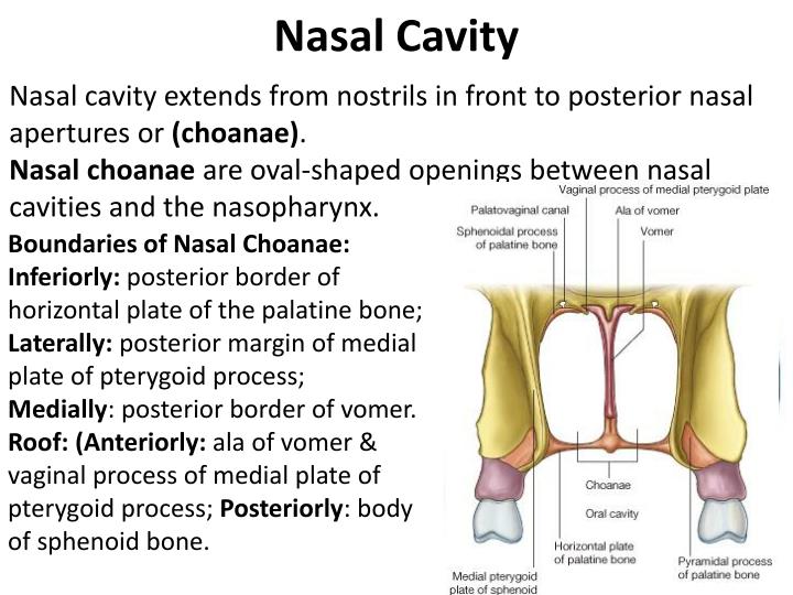
They are surrounded in the front and the lower side by the palatine bones (horizontal plate), while the sphenoid bone forms the upper and back borders of the internal nares.
What is the name of the bone that divides the nasal cavity?
The vomer is a small, thin, plow-shaped, midline bone that occupies and divides the nasal cavity. It articulates inferiorly on the midline with the maxillae and the palatines, superiorly with the sphenoid via its wings, and anterosuperiorly with the ethmoid. Thus, the bone forms the posteroinferior part of the nasal septum, which divides the nasal cavity.
How large is the nasal cavity?
The human nasal cavity has a total volume of about 16–19 ml, and a total surface area of about 150–180 cm 2 (measured using a computed tomography scan). The nasal septum divides the nasal cavity along the center into two halves, one opening to the facial side and one to the rhinopharynx, through the anterior and via the posterior nasal apertures, respectively. The nasal septum is not very accessible for the penetration of drugs into the human system because it consists mostly of cartilage and skin. The volume of each cavity is approximately 7.5 ml, having a surface area around 75 cm 2[46]. The most efficient area for drug administration is the lateral walls of the nasal cavity, which consist of highly vascularized mucosa.
What are the three segments of the pharynx?
14 It can be divided into three segments: the nasopharynx (exten ds from the base of skull to the soft palate), the oropharynx (extends from the soft palate to the pharyngoepiglottic fold), and the hypopharynx (extends from the pharyngoepiglottic fold to the UES) ( Figure 33.2 ). It is predominately a muscular structure, although the epiglottis, arytenoid, cuneiform, corniculate, and cricoid cartilage form part of the anterior wall. Furthermore, the hyoid and thyroid bones provide attachment to some of its muscles. Muscles of the pharynx can be broadly viewed as intrinsic and extrinsic. Intrinsic muscles constitute the superior, middle, and inferior pharyngeal constrictors along with the thyropharyngeus (oblique) and cricopharyngeus (horizontal fibers). A triangular area of relatively scanty muscle fibers exists between the oblique and horizontal muscle fibers (Killian’s triangle), which is the site of Zenker’s diverticulum and is seen in patients with difficulty swallowing who have impaired relaxation or opening of the UES. 15,16 Extrinsic muscles of the pharynx can be categorized into three subgroups: (1) the elevators and tensors of the palate including the levator veli palatine, tensor veli palatine, and palatoglossus; (2) the geniohyoid, mylohyoid, stylohyoid, thyrohyoid, digastric, stylopharyngeus, and palatopharyngeus that cause superior and anterior displacement of the larynx during swallow; and (3) the aryepiglottic, thyroarytenoid, and oblique arytenoid muscles that close the laryngeal inlet. These muscles are supplied by branches of cranial nerves V (trigeminal), VII (facial), IX (glossopharyngeal), X (vagus) ansae cervicalis, and XII (hypoglossal). Pharyngeal muscles are richly innervated with a nerve–muscle fiber innervation ratio of 1:2 to 1:6, as compared to 1:2000 for human gastrocnemius muscle 14 and 1:9 for extra-ocular eye muscles, 17 which is important for the “fine” control required for its function.