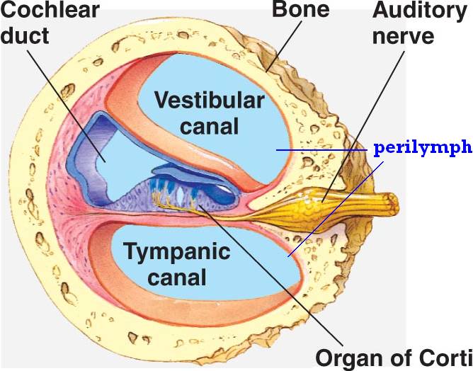
What overlies the organ of the Corti?
the structure that overlies the organ of Corti is the tectorial membrane a blow to the back of the head would result in a loss of vision the lens focuses light on the photoreceptor cells by changing shape the image of an object reaching the retina is a miniture image of the original, but it is upside down and backwards
What does the organ of Corti perform?
Learn about this topic in these articles:
- anatomy of the inner ear. The side of the triangle is formed by two tissues that line the bony wall of the cochlea: the stria vascularis, which lines the outer ...
- function in inner ear. ...
- place in peripheral nervous system. ...
- role in vertebrate hearing
What are the major functions of the organ?
Understanding the 11 Body Organ Systems
- Circulatory System. The circulatory system transports oxygen nutrients to all corners of the body and carries away byproducts of metabolism. ...
- Lymphatic System. ...
- Respiratory System. ...
- Integumentary System. ...
- Endocrine System. ...
- Gastrointestinal (Digestive) System. ...
- Urinary (Excretory) System. ...
- Musculoskeletal System. ...
- Nervous System. ...
- Reproductive System. ...
Does the organ of Corti contains mechanoreceptors?
What does the organ of Corti contain? hair cells. Which part contains mechanoreceptors for sensation of hearing? Hearing or audition involves the transduction of sound waves into neural signals via mechanoreceptors in the inner ear.

What is the primary organ of hearing?
The ear is the organ of hearing and balance.
What is the primary function of the cochlea?
The cochlea is a hollow, spiral-shaped bone found in the inner ear that plays a key role in the sense of hearing and participates in the process of auditory transduction. Sound waves are transduced into electrical impulses that the brain can interpret as individual frequencies of sound.
What is organ Corti quizlet?
Organ of Corti: The true organ of hearing, a spiral structure within the cochlea containing hair cells that are stimulated by sound vibrations.
How does the organ of Corti function to produce nerve impulses?
The organ of Corti contains sensory cells with hairlike projections, called hair cells, that are deformed by the progress of the wave. The hair cells trigger nerve impulses that travel along the cochlear nerve, a branch of the auditory nerve, to the brain, where they are interpreted as sound.
Where is the organ of Corti located?
the inner earThe organ of Corti is located in the scala media of the cochlea of the inner ear between the vestibular duct and the tympanic duct and is composed of mechanosensory cells, known as hair cells.
What is the function of the cochlea quizlet?
Its function is to transmit sound from the air to the ossicles inside the middle ear, and then to the oval window in the fluid-filled cochlea.
What is the function of the organ of Corti quizlet?
Contains tiny hairs which acts as hearing receptors, converts sound vibrations into nerve impulses. You just studied 2 terms!
Which of the following is part of the organ of Corti quizlet?
Organ of Corti (Inner Ear)
What does Corti mean?
noun phrase. : a complex epithelial structure in the cochlea that contains thousands of hair cells, rests on the internal surface of the basilar membrane, and in mammals is the chief part of the ear by which sound waves are perceived and converted into nerve impulses to be transmitted to the brain.
Does the organ of Corti help with balance?
The maculae and the cristae are the sensory epithelium of the vestibular system (balance) and the organ of Corti is the sensory epithelium of the cochlea.
How is sound processed in the ear?
Sound waves enter the outer ear and travel through a narrow passageway called the ear canal, which leads to the eardrum. The eardrum vibrates from the incoming sound waves and sends these vibrations to three tiny bones in the middle ear. These bones are called the malleus, incus, and stapes.
Does organ of Corti helps in maintaining equilibrium?
It helps to maintain equilibrium of body.
What is the function of the organ of Corti?
The function of the organ of Corti, for a soft sound (such as speech), can schematically be summed up in 5 sequences: (1) Sound waves, transmitted by the perilymph, make the basilar membrane vibrate up and down.
What is the organ of the corti?
Organ of Corti: overview. The organ of Corti, named after Alfonso Corti who first described it, is the sensorineural organ of the cochlea. It is composed of sensory cells called hair cells, nerve fibers that connect to them, and supporting structures.
What is the organ of corti?
The organ of Corti (Fig. 24.2C), the sensory epithelium resting upon the basilar membrane (for a review see Slepecky, 1996 ), senses mechanical vibration of incoming sound and converts it to action potentials. It is made up of two types of sensory cells ( Fig. 24.2C–G ): outer hair cells (OHCs) and inner hair cells (IHCs) and several types of supporting cells ( Fig. 24.2D ). The OHCs are arranged in three parallel rows ( Fig. 24.2D, F ), whereas the IHCs are in a single row ( Fig. 24.2D, F, G). At their apical end, the hair cells are provided with stereocilia in a typical ‘W’ pattern. The organ of Corti is overlain by the gel-like tectorial membrane, which is indirectly connected to the osseous spiral lamina through the spiral limbus. Only the stereocilia of the OHCs appear to be in contact with the tectorial membrane. Shearing movements between the basilar membrane with the sensory epithelium and the tectorial membrane cause receptor potentials to be produced in the hair cells, by means of deflections of their stereocilia (reviewed in Nobili et al., 1998 ). Sensory transduction in the cochlea has been studied for many years, and over the last 30 years it has been recognized that the IHCs ( Fig. 24.2G) act as the primary receptor cell (Markin and Hudspeth, 1995; Russell, 1983 ), whereas the OHCs act as motor cells that can convert membrane potential into a mechanical force (reviewed in Nobili et al., 1998 ).
Where does the organ of the corti develop?
The mammalian organ of Corti develops within the dorsal wall of the elongating cochlear duct, a structure composed of polarized, pseudostratified epithelial cells that first emerges from the ventromedial pole of the otocyst at ~ E12 in mice and attains its characteristic coiled configuration, albeit not full length, by E15 ( Morsli, Choo, Ryan, Johnson, & Wu, 1998 ). Two regions can be distinguished within the dorsal wall of the cochlear duct from which the organ of Corti develops, the greater and the lesser epithelial ridges (GER and LER, respectively, see Fig. 6 E14.5), regions which differ initially in their thickness. The IHCs with their associated supporting cells and those lying more medial (i.e., the cells of the spiral sulcus and spiral limbus) originate from the GER, while the outer hairs, Dieters’ cells, Hensen's cells, and the cells of Claudius and Boettcher originate from the LER.
What organs have lost their compact structure?
The mammalian organ of Corti has lost the compact structure that is so obviously a feature of all the other auditory and all vestibular receptor organs (Figs. 17 and 18). There are large extracellular spaces around the outer hair cells as well as between the tunnel rods in the organ of Corti, and without some structural provision for support, the organ would almost certainly collapse. The supporting cells have become specialized to provide the necessary stiffness by the presence of the “tonofilaments” which are arranged in bundles within the cytoplasm. These filaments were observed by early histologists; recent ultrastructural studies have confirmed their presence. One of the supporting elements, the tunnel rod, is a new phylogenetic development; there is no comparable structure in the pigeon or in most reptiles. Larsell, McCrady, and Larsell (1944) studied the embryonic development of the tunnel rods in the opossum pouch young. The 48-day-old pouch animal shows a transition from nondifferentiated epithelium in the apex to well-developed tunnel rods in the lower turns. The first evidence for differentiation is a slit between two tall columnar cells. This is the beginning of the tunnel opening which apparently becomes gradually widened at later stages. Tonofilaments are found in well-differentiated tunnel rods in the region of the upper first and lower second coils but not above and not below this region at this stage of development.
Which system innervates afferent fibers?
There are also two systems of efferent fibers: the lateral efferent system (lateral olivocochlear system; LOC) that innervates afferent fibers under the IHCs and the medial efferent system (medial olivocochlear system; MOC) that innervates the OHCs ( White and Warr, 1983 ).
Which organ is overlain by the gel-like tectorial membrane?
The organ of Corti is overlain by the gel-like tectorial membrane, which is indirectly connected to the osseous spiral lamina through the spiral limbus. Only the stereocilia of the OHCs appear to be in contact with the tectorial membrane.
What organ contains sensory hair cells?
The organ of Corti of the mammalian inner ear contains sensory hair cells and supporting cells in the auditory sensory epithelia. These cells are arranged to form a checkerboard-like cellular pattern. However, cellular and molecular mechanisms that produce this characteristic arrangement of cells had remained unknown for a long time. In the mouse organ of Corti, hair cells express nectin-1 and supporting cells express nectin-3; in addition, both cells express nectin-2 (Fig. 5 ). The trans -interaction between nectin-1 and -3 mediates the heterotypic adhesion between these two types of cells and establishes the checkerboard-like cellular pattern; this pattern is lost due to aberrant attachment between sensory hair cells in both nectin-1-deficient mice and nectin-3-deficient mice ( Togashi et al., 2011 ). Moreover, in the nectin-3-deficient mice, abnormal positioning of the kinocilium and misorientation and dysmorphology of the hair bundles are observed in aberrantly attached sensory hair cells ( Fukuda et al., 2014 ). Thus, the trans -interaction between nectin-1 and -3 is critical not only for the checkerboard-like cellular pattern formation but also for positioning of the kinocilium and the morphology and orientation of stereociliary bundle in hair cells.
Where is the cochlea located?
The cochlea includes three chambers, and the organ of Corti is located in the scala media. A single row of inner hair cells is located on the medial side of the epithelium, whereas three rows of outer hair cells are located more laterally.
Which organ is larger, the basilar membrane or the corti?
The organ of Corti is larger and the basilar membrane on which it sits is longer as it gets further away from the base of the cochlea. This difference in size is consistent with the fact that different frequencies of sound result in greater vibrations of the organ of Corti depending on where along the length of the cochlea you are measuring. ...
Which organ is spiraling towards the top of the cochlea?
The organ of Corti is spiraling towards the top of the cochlea to the right. Stereocilia are on the top and radial fibers of the basilar membrane are seen on the bottom. The outer hair cells sit in a cup formed by a supporting cell. The supporting cells send out a narrow filament that angles towards the base of the cochlea.
What organ is photogenic?
The photogenic appearance of the organ of Corti has long been appreciated by the popular press. Pictures obtained with electron microscopes are routinely published showing the organ of Corti ’s colonnade appearance.
What is the organ of the inner ear called?
The Organ of Corti - The Temple of Hearing. The sensory epithelium of the inner ear is called the organ of Corti after the Italian scientist who first described it. Its orderly rows of outer hair cells is unique among the organs of the body. Figure 5 shows a short section of the organ of Corti as it spirals in the cochlea.
What is the eighth nerve?
The nerve is made up of the neuronal projections that connect the hair cells with the brain and is called the eighth nerve because it is one of 12 nerves that come off the brain in the skull. The spiral shaped cochlea originates from one of the balance organs and contains the sensory epithelium for hearing. Heading.
What is the hearing organ called?
The Cochlea. The hearing organ in mammals is a spiraling structure called the "cochlea" from the Greek word for snail. It spirals out from the saccule (one of the balance organs). There are two and a half turns in the human cochlea and if you were to unwind the cochlea it would stretch to nearly an inch in length.
Where is the central axis of the spiraling cochlea?
The central axis of the spiraling cochlea is to the left of the drawing. Eighth nerve fibers pass through a bony shelf on their way to the hair cells (orange). The organ of Corti is made up of hair cells and supporting cells (purple and blue, respectively) that sit on a flexible basilar membrane which is anchored to the bony shelf on ...
What is the Organ of Corti?
Sound is processed by the central nervous system thanks to afferent signals originating in the inner ear. These signals are created in response to fluid vibrations and structures in the inner and middle ear.
Organ of Corti
As stated above, the organ of Corti is contained within a primary component in the inner ear, the cochlea, and it is the last stop of sound vibration before it is transformed into a nervous impulse. The pathway sound travels will be highlighted below, beginning from its origin as vibration in the air to its final location in the organ of Corti.
Auditory Nerve
The auditory nerve, also known as the vestibulocochlear nerve, acoustic nerve, and cranial nerve VIII, receives signals from the inner ear and transmits them to the brain.
