What does the choroid do in the eye?
Other important functions of the choroid include:
- Providing nutrients for the retina, macula and optic nerve.
- Regulating the temperature of the retina.
- Helping control pressure within the eye.
- Absorbing light and limiting reflections within the eye that could harm vision. ...
What is the function of the choroid layer?
There are four different layers of the choroid:
- Bruch’s membrane – Thin layer of tissue located on the innermost part of the choroid.
- Choriocapillaris – Layer made up of capillaries (tiny blood vessels that connect arteries to veins).
- Sattler’s layer – Layer of medium blood vessels.
- Haller’s layer – Outermost layer of the choroid that contains large blood vessels.
Is choroid the pigmented layer of the eye?
The choroid is the vascular layer of the eye that lies between the retina and the sclera. The choroid is thickest in the back of the eye, where it is about 0.2 mm, and narrows to 0.1 mm in the peripheral part of the eye. 1 It contains the retinal pigmented epithelial cells and provides oxygen and nourishment to the outer retina.
What is choroid plexus of third ventricle and its function?
Function. Associated Conditions. Tests. The choroid plexus is a thin structure that lines most of the the ventricles of the brain. It is a protective barrier that produces cerebrospinal fluid (CSF), a fluid that provides nourishment and cushioning for the brain and spinal cord. 1 . Cysts or tumors can form in the choroid plexus ...
What are two functions of the choroid?
The choroid has several functions: Its vasculature is the major supply for the outer retina; impairment of the flow of oxygen from choroid to retina may cause Age-Related Macular Degeneration. The choroidal blood flow, which is as great as in any other organ, may also cool and warm the retina.
What does the choroid produce?
The choroid forms the uveal tract, which includes the iris and the ciliary body. The dark-colored melanin pigment in the choroid absorbs light and limits reflections within the eye that could degrade vision.
What is the main function of the choroid quizlet?
Nourishes the retina and assists with absorption of light to prevent its scattering within the eye. What is the primary function of the choroid layer? space between the lens and cornea filled with aqueous humor.
What does the choroid coat contain and what is the function of this layer?
Choroid is the vascular layer of the eye. Also referred to as choroid coat or choroidea, it is a thin layer of tissue which is part of the middle layer of the eye wall, found between the sclera and the retina. The choroid is filled with blood vessels, which brings nutrients and oxygen to the outer layers of the retina.
What happens if the choroid is damaged?
Degeneration of the blood vessels of the choroid is followed by damage to the retina, which usually leads to loss of peripheral vision that can progress to eventual blindness. Central vision is usually preserved until late in life.
Is choroidal detachment painful?
Serous choroidal detachments are typically associated with a low pressure in the eye and are usually only mildly uncomfortable. In contrast, hemorrhagic choroidal detachments are commonly painful, and often associated with a high intraocular pressure.
What are the two major functions of the choroid quizlet?
MatchNourishment/waste removal of the outer 1/3rd of the retina.Thermoregulation (heat dissipation during visual processing)Conduit for blood vessels & nerves.Absorption of excess light.
What is the function of choroid plexus and where is it located?
The choroid plexus (ChP) is a secretory tissue found in each of the brain ventricles, the main function of which is to produce cerebrospinal fluid (CSF).
What are the functions of the choroid and ciliary body?
The choroid provides oxygen and nourishment to the outer layers of the retina. Along with the ciliary body and iris, the choroid forms the uveal tract.
What cells are in the choroid?
Choroidal epithelial cells are one of the three types of ependymal cells, themselves a type of glial cell. They cover the surface of the choroid plexus and produce cerebrospinal fluid (CSF). 1.
What controls blood pressure in the choroid?
Choroidal blood flow is controlled by parasympathetic, sympathetic and sensory input. Neurogenic control helps match choroidal blood flow to retinal need. Neurogenic control stabilizes choroidal blood flow during blood pressure variation.
What is secreted by the choroid plexus?
Abstract. The epithelial cells of the choroid plexus secrete cerebrospinal fluid (CSF), by a process that involves the movement of Na(+), Cl(-) and HCO(3)(-) from the blood to the ventricles of the brain.
What are two functions of the choroid quizlet?
MatchNourishment/waste removal of the outer 1/3rd of the retina.Thermoregulation (heat dissipation during visual processing)Conduit for blood vessels & nerves.Absorption of excess light.
Where does the choroid plexus secrete CSF?
brain ventricleC. Each of the choroid plexuses secretes CSF into the respective brain ventricle (Figures 1 and 3). Once in the ventricle, the CSF is in contact with the ependymal epithelium, which lines the ventricles and constitutes a leaky barrier between the ventricle and the brain interior.
What does the choroid plexus do quizlet?
The choroid plexus is the major site for the formation of cerebrospinal fluid. Vascularized areas within the subarachnoid space are other possible secretion sites.
What happens if you rupture a choroidal nevi?
The injury leads to a loss of the photoreceptors in the macula and loss of central vision. If the rupture is not in the macula, central vision is retained. Choroidal nevi are a collection of pigmented or non-pigmented cells in the choroid, the vascular layer under the retina. Most choroidal nevi only need to be monitored.
What is choroidal detachment?
Diseases and Disorders of the Choroid. Hemorrhagic choroidal detachment is a hemorrhage in the choroidal space caused by the rupture of choroidal vessels. 4 Although it can occur spontaneously, it is extremely rare. It usually occurs as a consequence of eye trauma. It can also occur rarely during eye surgery.
What is choroidal rupture?
Choroidal rupture is a complete break in the choroid, Bruch's membrane, and the retinal pigment epithelium that occurs as a result of blunt eye trauma, such as getting hit with a fist . 5 Unfortunately, many choroidal ruptures involve the center of the retina, called the macula. The macula allows us to have high quality, central vision.
What are some examples of choroidal dystrophies?
Choroidal dystrophies are a group of inherited diseases that affect the choroid. 3 Choroideremia , gyrate atrophy, central areolar choroidal dystrophy, diffuse choroidal atrophy, and pigmented paravenous retinochoroidal atrophy are examples of choroidal dystrophies. Severe vision loss can occur in some of these dystrophies.
What is the vascular layer of the eye?
The choroid is the vascular layer of the eye that lies between the retina and the sclera. The choroid is thickest in the back of the eye, where it is about 0.2 mm, and narrows to 0.1 mm in the peripheral part of the eye. 1 It contains the retinal pigmented epithelial cells and provides oxygen and nourishment to the outer retina. The choroid forms the uveal tract, which includes the iris and the ciliary body.
What is the treatment for hemorrhagic choroidal detachment?
A hemorrhagic choroidal detachment can produce profound symptoms. Treatment consists of topical steroid eye drops, cycloplegic eye drops, and eye-pressure-lowering eye drops. Ultimately, depending on the severity of the detachment, surgery may be recommended.
What happens when the optic nerve is compromised?
3 When the macula and optic nerve are compromised or negatively impacted, the result is often a severe decrease in vision and sometimes even total blindness.
What is the innermost choroid composed of?
The choriocapillaris of the innermost choroid are composed of richly anastomotic, fenestrated capillaries. The capillaries of the choriocapillaris are separate and distinct from the capillary bed of the anterior optic nerve. Venous drainage from the choriocapillaris is primarily through the four vortex veins. ...
How many layers are there in the choroid?
Morphologically the choroid can be divided into four layers: suprachoroidea, large-vessel layer, medium-sized vessel and tapetum layer, and choriocapillaris. The suprachoroidea is the potential space between the choroidal stroma and is attached loosely to the sclera by the lamina fusca.
What is the role of VEGF in eye growth?
Recent studies in chick also provide an interesting perspective on the potential role of members of the VEGFs family in eye growth regulation. They are best known for their roles in angiogenesis, and VEGF antagonists such as bevacizumab, an antibody against human VEGF, are now widely used clinically in the treatment of wet age-related maculopathy. However, recent years have seen an expansion of their clinical use to include other macular pathologies, including myopic maculopathy. 115 Thus, the findings that members of the VEGF family and their receptors are expressed in chick choroid, and intravitreal injection of bevacizumab inhibits both the development of form-deprivation myopia and the choroidal thickening during the recovery from form-deprivation myopia in chicks implies a fundamental role for this family in regulating choroidal function. 116,117
What is the vascular network of the retina?
Choroid. The choroid is a thin, pigmented vascular network consisting of three layers (from inner to outer): choriocapillaris, stroma, and lamina fusca. The choriocapillaris provides nutrients to the RPE and the outer third of the retina.
What is the outer choroid?
The outer choroid, known as Haller's layer, is composed of large caliber, non-fenestrated, vessels. The inner choroid is referred to as Sattler's layer, and is composed of significantly smaller vessels. The choriocapillaris of the innermost choroid are composed of richly anastomotic, fenestrated capillaries.
What is the tapetum of an ungulate?
In ungulates, the tapetum is fibrous and composed of regularly arranged collagen fibers and occasional fibrocytes. Herbivores are born with mature eyes and well-developed tapeta.
Which artery sends branches back into the choroid after penetrating the anterior sclera?
Anterior ciliary arteries, which send branches back into the choroid after penetrating the anterior sclera
What is the membrane that covers the sclera?
The conjunctiva is the membrane covering the sclera (white portion of your eye). The conjunctiva also covers the interior of your eyelids.
What is the white part of the eye?
The sclera is sometimes known as the "whites" of the eye. It covers more than 80% of the eyeball's surface. 2
What is the episclera?
The episclera is a thin layer of tissue that lies on top of the sclera. The episclera has tiny blood vessels that supply the sclera with nutrients.
How many fibers are in the optic nerve?
The optic nerve is a bundle of about 1.2 million nerve fibers that transmit visual information to the central nervous system (brain). 7
How do eyes work?
The eyes work in the same way as cameras. When you focus on an object, light is reflected and enters the eye through the cornea. As the light passes through, the dome-shaped nature of the cornea bends light , enabling the eye to focus on fine details.
Where are light rays focused?
Light rays are focused on the macula lutea when an eye is looking directly at an object.
How many muscles are there in the eye?
The eye has six muscles. These muscles arise from the eye socket (orbit) and work to move the eye up and down, side to side, or in a circular motion.
What causes chorioretinitis?
The cause of chorioretinitis varies by patient. Doctors aren’t always sure what causes this condition. Cases of chorioretinitis are usually divided into two groups: infectious (caused by an infection) and non-infectious (not caused by an infection.)
What is a chorioretinal scar?
A chorioretinal scar is a small area of pigment or fibrous tissue in the back of the eye. It may or may not affect vision, depending on where it is located in the eye.
What is Choroidal Melanoma?
Choroidal melanoma is a cancer that affects part of the eye. It develops in the choroid, the sponge-like membrane at the back of the eye between the sclera (the white of the eye) and the retina. (The retina is the light-sensitive structure at the back of the eye. It sends visual information to the brain.) The choroid is rich in blood vessels and supplies nutrients to the retina.
How is choroidal melanomas treated?
Medium and large choroidal melanomas usually are treated with radiation or surgery . Radiation therapy may be given in different ways. Local radiation is delivered via a small, metal, dish-shaped device containing radioactive iodine. The device is stitched to the sclera so that the radiation can target the tumor precisely. In some institutions, external beam radiation therapy may be used. Beams of radiation target the tumor from outside the body.
What is the role of choroidal melanomas in the brain?
It sends visual information to the brain.) The choroid is rich in blood vessels and supplies nutrients to the retina. Over time, many choroidal melanomas enlarge and cause the retina to detach. This can lead to vision loss. The tumors also can spread (metastasize) to other parts of the body.
Why is there no known way to prevent choroidal melanoma?
Because the causes of choroidal melanoma are not well understood , there is no known way to prevent it.
What does it mean when your eyes are blurry?
having blurry vision. seeing spots. seeing flashing lights. having severe eye pain. Having these symptoms doesn't mean you have choroidal melanoma. These symptoms can be caused by other conditions that are more common—and noncancerous. In fact, seeing spots and flashing lights are very common symptoms.
How long do you have to be monitored for choroidal melanoma?
If you are treated for choroidal melanoma, you will need to be monitored for the rest of your life. This helps doctors determine whether the cancer has spread to other parts of the body.
What is the test to see if you have a tumor in your eye?
Most of the time, no other tests are needed. But specialized tests can confirm the diagnosis. These tests include. ultrasound. A small probe placed on the eye directs sound waves toward the tumor.
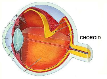
What is the choroid in the eye?
(Choroid labeled at right, second from the bottom.) The choroid, also known as the choroidea or choroid coat, is the vascular layer of the eye, containing connective tissues, and lying between the retina and the sclera.
Where is the choroid located?
The choroid provides oxygen and nourishment to the outer layers of the retina. Along with the ciliary body and iris, the choroid forms the uveal tract .
How many layers are there in the choroid?
The structure of the choroid is generally divided into four layers (classified in order of furthest away from the retina to closest): Haller's layer - outermost layer of the choroid consisting of larger diameter blood vessels; Sattler's layer - layer of medium diameter blood vessels; Choriocapillaris - layer of capillaries; and.
What are the two circulations of the eye?
There are two circulations of the eye: the retinal (in the retina) and uveal, supplied in humans by posterior ciliary arteries, originating from the ophthalmic artery that derived from the internal carotid artery. The arteries of the uveal circulation, supplying the uvea and outer and middle layers of the retina, ...
When was the Choroid first described?
The choroid was first described by Democritus (c. 460 – c. 370 BCE) around 400 BCE, calling it the "chitoon malista somphos" (more spongy tunic [than the sclera ]). Democritus likely saw the choroid from dissections of animal eyes.
Which artery enters the eyeball without passing the optic nerve?
The arteries of the uveal circulation, supplying the uvea and outer and middle layers of the retina, are branches of the ophthalmic artery and enter the eyeball without passing with the optic nerve. The retinal circulation, on the other hand, derives its circulation from the central retin al artery, also a branch of the ophthalmic arter y, ...
Who first described the choroid?
About 100 years later, Herophilos (c. 335 – 280 BCE) also described the choroid from his dissections on eyes of cadavers.
