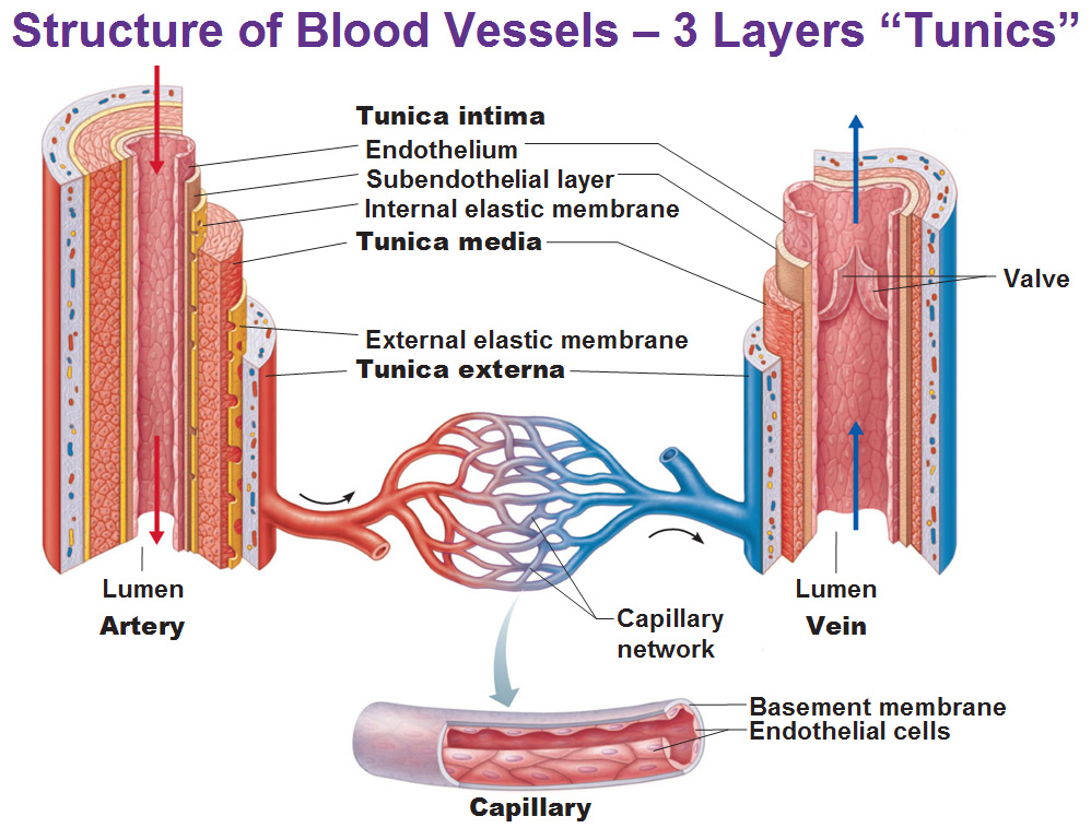
What is main purpose of the smooth muscle tunica media of artery? -regulating blood flow and blood pressure, -smooth muscle contracts when a small artery or arteriole is damaged (vascular spasm) to help limit loss of blood through the injured vessel.
What is the purpose of the tunica media?
The walls of this “pipeline” are formed, from inside-out by:
- Tunica intima or simply “intima” which is made of one layer of epithelial cells and is in contact with the blood. ...
- Tunica media is, as its name refers, the middle layer of the aorta. ...
- Tunica externa or adventitia is the outer layer of the aorta. The adventitia is formed mainly of collagen. ...
What is the function of tunica media in the artery?
Tunica Media
- Mechanobiology of the Arterial Wall. ...
- Vascular Pharmacology. ...
- The biology of vascular calcification. ...
- Management of CNS Vasculitis and Takayasu's Arteritis. ...
- Extracellular Matrix and Egg Coats. ...
- Thorax. ...
- Atherosclerosis and Coronary Artery Disease
What is the primary function of the tunica externa?
Tunica Externa. The outermost layer is the tunica externa or tunica adventitia, composed entirely of connective fibers and surrounded by an external elastic lamina which functions to anchor vessels with surrounding tissues.
What is the structure of the tunica media?
The tunica media, or middle coat, is made up principally of smooth (involuntary) muscle cells and elastic fibres arranged in roughly spiral layers. The outermost coat, or tunica adventitia, is a tough layer consisting mainly of collagen fibres that act as a supportive element.

What is the function of the tunica externa?
Function. The tunica externa provides basic structural support to blood vessels. It prevents vessels from expanding too much from internal blood pressure, particularly arteries. It is also relevant in controlling vascular flow in the lungs.
What is the function of the tunica media quizlet?
The tunica media, the middle layer, is usually the thickest. Strengthens the vessels and prevents blood pressure from rupturing them. It consists of smooth muscle, collagen, and in some cases elastic tissue. Produces vasomotion, changes in the diameter of a blood vessel.
What makes the tunica media unique?
The middle coat (tunica media) is distinguished from the inner (tunica intima) by its color and by the transverse arrangement of its fibers. In the smaller arteries it consists principally of smooth muscle fibers in fine bundles, arranged in lamellæ and disposed circularly around the vessel.
What is the role of the tunica media in maintaining the continuous flow of blood?
In the tunica media of the aorta, there is a large amount of elastic tissue. This is essential for the aorta to stretch to accommodate the output of blood from the left ventricle and then to recoil to maintain an adequate diastolic pressure and generate a more continuous flow of blood.
What is tunica media of artery quizlet?
The TUNICA MEDIA (medius=middle) is the middle layer of the vessel wall. It's composed predominantly of circulatory arranged layers of smooth muscle cells that are supported by elastic fibers.
What is the tunica media composed of quizlet?
The tunica media consists of 40-70 layers of elastic sheets alternating with smooth muscle. The external elastic lamina is between the media and externa.
What are the main components of the tunica media?
The tunica media, or middle coat, is made up principally of smooth (involuntary) muscle cells and elastic fibres arranged in roughly spiral layers. The outermost coat, or tunica adventitia, is a tough layer consisting mainly of collagen fibres that act as a supportive element.
Why is the tunica media thicker in arteries?
Arteries experience a pressure wave as blood is pumped from the heart. This can be felt as a "pulse." Because of this pressure the walls of arteries are much thicker than those of veins. In addition, the tunica media is much thicker in arteries than in veins.
What does the tunica media consist of?
The tunica media is composed chiefly of circumferentially arranged smooth muscle cells. Again, the external elastic lamina often separates the tunica media from the tunica adventitia. Finally, the tunica adventitia is primarily composed of loose connective tissue made up of fibroblasts and associated collagen fibers.
Does the tunica media contains the endothelium?
The tunica intima, the innermost layer, consists of an inner surface of smooth endothelium covered by a surface of elastic tissues. The tunica media, or middle coat, is thicker in arteries, particularly in the large arteries, and consists of smooth muscle cells intermingled with elastic fibres.…
What are tunics in blood vessels?
The tunica media or a middle layer composed of elastic and muscular tissue which regulates the internal diameter of the vessel. The tunic intima or an inner layer consisting of an endothelial lining which provides a frictionless pathway for the movement of blood.
arteries
The tunica media, or middle coat, is made up principally of smooth (involuntary) muscle cells and elastic fibres arranged in roughly spiral layers. The outermost coat, or tunica adventitia, is a tough layer consisting mainly of collagen fibres that act as a supportive element. The large…
description
The tunica media, or middle coat, is thicker in arteries, particularly in the large arteries, and consists of smooth muscle cells intermingled with elastic fibres. The muscle cells and elastic fibres circle the vessel. In larger vessels the tunica media is composed primarily of elastic fibres.…
What is the tunica media?
In large and medium-sized vessels, radially outward from the EC layer is the tunica media, consisting of VSMCs and ECM components including elastin and collagen. The dynamic contraction and relaxation of VSMCs allows for the tone of the blood vessel to be adjusted to the physiological demands of the relevant tissue and to maintain blood pressure and perfusion. Collagen provides strength to the vessel wall, and elastin is largely responsible for its elasticity, such that upon receiving cardiac output in systole, the arterial wall stretches to increase the lumen volume, and subsequently, in diastole, it recoils to help maintain blood pressure. The capillary wall is substantially thinner than that of larger vessels, facilitating the transfer of substances to and from the vascular compartment. Capillary mural cells consist of pericytes rather than VSMCs. Pericytes, VSMCs, and the ECM play critical roles in many vascular diseases, but there are strikingly few studies of the development of these components in comparison to the vast number of investigations of the morphogenesis of EC networks and tubes.
What is the function of SMCs in the tunica media?
SMCs are the major cell type of the tunica media and through dynamic cell contraction and relaxation regulate vascular tone and hence, blood flow. The contraction–relaxation state of SMCs is dictated by a spectrum of contractile and cytoskeletal proteins.
What is TA characterized by?
TA is characterized by adventitial thickening and cellular infiltration of the tunica media along with destruction of elastin and vascular smooth muscle. Myofibroblast proliferation results in intimal hyperplasia which is then followed by fibrosis of the tunica media and intima with eventual stenosis. Aneurysm formation ensues when local destruction of the media predominates.
What is the vascular media?
The vascular media in both pulmonary and systemic arteries is a complex structure, comprised of multiple phenotypically and functionally distinct SMC populations that may subserve distinct cellular functions in health and disease (22–24).
What is the function of SMCs?
SMCs are the major cell type of the tunica media and through dynamic cell contraction and relaxation regulate vascular tone and hence, blood flow . The contraction–relaxation state of SMCs is dictated by a spectrum of contractile and cytoskeletal proteins. During embryogenesis, α-smooth muscle actin (SMA) is considered the first SMC marker to be expressed and ultimately is the most abundant protein in SMCs. For instance, the developing pulmonary artery forms in a field of cells expressing the undifferentiated mesenchyme marker platelet-derived growth factor receptor (PDGFR)-β (Greif et al., 2012 ). Shortly thereafter PDGFR-β + cells adjacent to the nascent EC tube downregulate PDGFR-β and upregulate SMA ( Greif et al., 2012 ). Early developing SMCs also express SM22α (also known as transgelin), which influences the actin cytoskeleton by stabilizing actin filaments. In addition to SMCs, SMA and SM22α are expressed in other cell types as well, whereas smooth muscle myosin heavy chain (SMMHC) and smoothelin are expressed later in SMC differentiation and are generally considered to be specific to the SMC lineage. Yet, our recent studies suggest that SMMHC is also expressed in alveolar myofibroblasts of adult mice exposed to hypoxia ( Sheikh et al., 2014 ).
What is TA characterized by?
TA is characterized by adventitial thickening and cellular infiltration of the tunica media along with destruction of elastin and vascular smooth muscle. Myofibroblast proliferation results in intimal hyperplasia which is then followed by fibrosis of the tunica media and intima with eventual stenosis. Aneurysm formation ensues when local destruction of the media predominates.
Is the vascular media a complex structure?
The vascular media in both pulmonary and systemic arteries is a complex structure, comprised of multiple phenotypically and functionally distinct SMC populations that may subserve distinct cellular functions in health and disease (22–24). For the pulmonary circulation, SMC heterogeneity has been described in both site-specific and regional (along the longitudinal axis) patterns.
What is the tunica media?
Anatomical terminology. The tunica media ( New Latin "middle coat"), or media for short, is the middle tunica (layer) of an artery or vein. It lies between the tunica intima on the inside and the tunica externa on the outside.
What is the middle coat of the tunica?
It lies between the tunica intima on the inside and the tunica externa on the outside. The middle coat (tunica media) is distinguished from the inner (tunica intima) by its color and by the transverse arrangement of its fibers. In the smaller arteries it consists principally of smooth muscle fibers in fine bundles, ...
