Urea Hydrolysis
Hydrolysis
Hydrolysis usually means the cleavage of chemical bonds by the addition of water. When a carbohydrate is broken into its component sugar molecules by hydrolysis (e.g. sucrose being broken down into glucose and fructose), this is termed saccharification. Generally, hydrolysis or …
Urease
Ureases, functionally, belong to the superfamily of amidohydrolases and phosphotriesterases. Ureases are found in numerous bacteria, fungi, algae, plants, and some invertebrates, as well as in soils, as a soil enzyme. They are nickel-containing metalloenzymes of high molecular weight.
Is Staphylococcus aureus urea hydrolysis test positive?
This is an indication of a positive test. Therefore, this ensures that Unknown “A” is indeed Staphylococcus aureus. After doing a gram stain on the bacterium, it was determined that this bacterium was a gram negative. The next step was to continue with another test. A Mannitol agar plate was obtained and inoculated with the bacterium.
Why is RNA is susceptible to hydrolysis?
RNA is susceptible to this base-catalyzed hydrolysis because the ribose sugar in RNA has a hydroxyl group at the 2′ position. This feature makes RNA chemically unstable compared to DNA, which does not have this 2′ -OH group and thus is not susceptible to base-catalyzed hydrolysis.
How to do urea test?
b) Urea Slant method:
- Sterilize the loop in the blue flame of the Bunsen burner till red hot and then allowed to cool.
- Take out a loopful organism from the tryptic broth culture tube with the cooled loop aseptically.
- Again flame the neck of the tube and replace the tube in the test tube rack.
- Take a sterile urea slant tube remove the cap and flame the neck of the tube.
Why do we test for urea electrolytes?
These conditions include:
- Hyponatraemia (low sodium)
- Hypernatraemia (high sodium)
- Hypokalaemia (low potassium)
- Hyperkalaemia (high potassium)
- Metabolic acidosis (low bicarbonate)
- Metabolic alkalosis (high bicarbonate)
- Dehydration (high urea and/or creatinine)
- Renal (kidney) impairment or failure (high urea and/or creatinine)
- Gastrointestinal bleeding (high urea)

What is the purpose of the urease test?
The urease test is used to determine the ability of an organism to split urea, through the production of the enzyme urease.
What is a urea breath test?
Urea breath test is a common non-invasive test to detect Helicobacter pylori also based on urease activity. This is highly sensitive and specific test.
What bacteria produce urease?
It is also useful to identify Cryptococcus spp., Brucella , Helicobacter pylori, and many other bacteria that produce the urease enzyme. Directly, this test is performed on gastric biopsy samples to detect the presence of H. pylori.
What is the color of the mucosa in a biopsy?
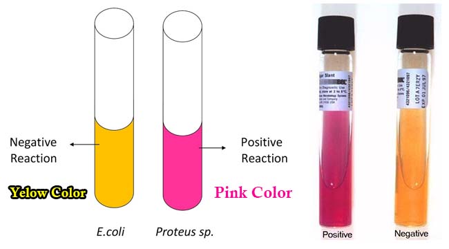
The test is performed at the time of gastroscopy. A biopsy of mucosa is taken from the antrum of the stomach, and is placed into a medium containing urea and an indicator such as phenol red. The urease produced by H. pylori hydrolyzes urea to ammonia, which raises the pH of the medium, and changes the color of the specimen from yellow (NEGATIVE) to red (POSITIVE).
What are the limitations of urea?
Limitations of Urease Test 1 Some organisms rapidly split urea ( Brucella and H. pylori ), while others react slowly. 2 It is recommended that biochemical and/or serological tests be performed on colonies from pure culture for complete identification. 3 To facilitate growth and the urea hydrolysis reaction, do not use inoculum from a broth suspension. 4 After prolonged incubation times a false-positive alkaline reaction may be seen. To rule out this occurrence, check the test with a control (an uninoculated tube of Urea Agar) along with the inoculated tube during prolonged incubation. 5 Do not heat the Urea Agar Slants, as urea decomposes very readily when heated. 6 To detect Proteus species, the Urea Agar, Slants must be examined within 6 hours of inoculation for a reaction. 7 Urea Agar should not be used to determine the quantitative rate of urease activity, as organisms vary in their capability and rate of hydrolysis. 8 Failure to incubate this medium with loose caps may cause erroneous results to occur. 9 Urea is light sensitive and can undergo autohydrolysis. Store at 2 to 8u0001C in the dark.
How many drops of urea agar to inoculate slant?
Streak the surface of a urea agar slant with a portion of a well-isolated colony or inoculate slant with 1 to 2 drops from an overnight brain-heart infusion broth culture.
What is the CLO test?
It is a rapid, cheap and simple test that detects the presence of urease in or on the gastric mucosa. It is also known as the CLO test (Campylobacter-like organism test). This test uses a procedure called gastric endoscopy and biopsy to collect stomach lining cells. The test is performed at the time of gastroscopy.
What is the urease test?
Urease broth is a differential medium that tests the ability of an organism to produce an exoenzyme, called urease, that hydrolyzes urea to ammonia and carbon dioxide. The broth contains two pH buffers, urea, a very small amount of nutrients for the bacteria, and the pH indicator phenol red. Phenol red turns yellow in an acidic ...
Can enterics hydrolyze urea?
Many enterics can hydrolyze urea; however, only a few can degrade urea rapidly. These are known as “rapid urease-positive” organisms. Members of the genus Proteus are included among these organisms. Urea broth is formulated to test for rapid urease-positive organisms.
What is the purpose of urea test?
Urease test is used to identify organisms that are capable of hydrolyzing urea to produce ammonia and carbon dioxide.
What media is used to detect urease?
Both urea agar slants and broth media can be used for the detection of urease production. Agar media includes the Urea Agar Base (Christensen agar), and the broth includes the urea broth.
What is the color of urea?
Urea medium, whether a broth or agar, contains urea and the phenol red as a pH indicator. Many organisms, especially those that cause urinary tract infections, produce the urease enzyme, which catalyzes the splitting of urea in the presence of water to release two molecules of ammonia and carbon dioxide. The ammonia combines with the carbon dioxide and water to form ammonium carbonate, which turns the medium alkaline, turning the indicator from its original orange-yellow color to bright pink. This test is performed as part of the identification of several genera and species of the Enterobacteriaceae family, including Klebsiella, Proteus, and some Citrobacter and Yersinia species, as well as some Corynebacterium species. The test is also useful to identify Cryptococcus, Brucella, Helicobacter pylori, and many other bacteria that produce the urease enzyme. Disks are available that combine urea and phenylalanine deaminase (PDA), allowing a one-disk test to identify Proteus, Providencia, and Morganella and to separate them from Klebsiella and Yersinia enterocolitica. The disk reactions are rapid and sensitive and allow for the rapid detection of agents of serious infections, g., Brucella, and Cryptococcus.
What bacteria are tested for urea?
The test is also useful to identify Cryptococcus, Brucella, Helicobacter pylori, and many other bacteria that produce the urease enzyme. Disks are available that combine urea and phenylalanine deaminase (PDA), allowing a one-disk test to identify Proteus, Providencia, and Morganella and to separate them from Klebsiella and Yersinia enterocolitica.
What is the sole source of nitrogen in a urea test?
During the test, the organisms utilize urea as the sole source of nitrogen, producing a sufficient amount of ammonia to overcome the buffering capacity of the medium. The change in color of the medium as a result of the change in pH is indicative of the test result.
When was the urea agar test developed?
Christensen developed the test in 1946 for the differentiation of enteric bacilli. The urea agar base used for the testing of urease activity is named Christensen Urea Agar after him.
What is the enzyme that makes urea?
The fermentation of urea occurs in the presence of the enzyme ‘urease’, resulting in two molecules of ammonia and carbon dioxide.
