What drives resting membrane potential in skeletal muscle cells?
Unlike nerve cells, where the resting membrane potential is predominantly a result of K + permeability, skeletal muscle cell resting membrane potential receives a significant contribution from Cl − conductance.
What is the membrane potential of a neuron?
For a cell’s membrane potential, the reference point is the outside of the cell. In most resting neurons, the potential difference across the membrane is about to (a is of a volt), with the inside of the cell more negative than the outside. That is, neurons have a resting membrane potential (or simply, resting potential) of about to.
What is the resting membrane potential made of?
Table 1. Ion Concentration Inside and Outside Neurons The resting membrane potential is a result of different concentrations inside and outside the cell. The difference in the number of positively charged potassium ions (K +) inside and outside the cell dominates the resting membrane potential (Figure 2).
What happens to resting membrane potential at peak action potential?
The (a) resting membrane potential is a result of different concentrations of Na+ and K+ ions inside and outside the cell. A nerve impulse causes Na+ to enter the cell, resulting in (b) depolarization. At the peak action potential, K+ channels open and the cell becomes (c) hyperpolarized.

What is the resting potential of a muscle?
resting potential, the imbalance of electrical charge that exists between the interior of electrically excitable neurons (nerve cells) and their surroundings.
What is normal resting membrane potential?
about −65 mVThe RMP of a typical neuron is about −65 mV, with the interior of the cell negative in charge to the outside.
What is the resting membrane potential in muscle fibers quizlet?
What is the resting membrane potential defined? the electrical potential difference (50 to 70mV) across the cell membrane which results from separation of charge.
What is resting membrane potential and why is it important?
This voltage is called the resting membrane potential; it is caused by differences in the concentrations of ions inside and outside the cell. If the membrane were equally permeable to all ions, each type of ion would flow across the membrane and the system would reach equilibrium.
Why is the resting membrane potential negative 70?
The resting membrane potential of a neuron is about -70 mV (mV=millivolt) - this means that the inside of the neuron is 70 mV less than the outside. At rest, there are relatively more sodium ions outside the neuron and more potassium ions inside that neuron.
What is resting membrane potential mV?
The potential that is recorded when a living cell is impaled with a microelectrode is called the resting potential, and varies from cell to cell. Here it is shown to be -60 mV, but can range between -80 mV and -40 mV, depending on the particular type of nerve cell.
What is the role of ATP in generating a resting membrane potential quizlet?
b. What is the role of ATP in generating a resting membrane potential? The Na+/K+ pump helps maintain resting membrane potential, which is a prerequisite for depolarization and action potential propagation. The pump hydrolyzes one ATP molecule in the transport of three Na+ out of the cell and two K+ into the cell.
What is the role of ATP in maintaining a resting membrane potential?
ATP binds to the myosin head. The binding of ATP to the myosin head weakens the bond between myosin and actin, forcing the myosin head to detach. ATP also provides the energy for the next power stroke.
When a skeletal muscle fiber is at rest there is more sodium?
When a skeletal muscle fiber is at rest, there is more sodium outside the cell as compared to the sarcoplasm. The property of muscle tissue that allows an impulse to travel down the entire length of the cell membrane is... The end plate potential is an event that involves a gain of...
What is correct for the resting potential?
In most neurons the resting potential has a value of approximately −70 mV. The resting potential is mostly determined by the concentrations of the ions in the fluids on both sides of the cell membrane and the ion transport proteins that are in the cell membrane.
Why is RMP negative?
Ion affection of resting membrane potential Potassium ions are important for RMP because of its active transport, which increase more its concentration inside the cell. However, the potassium-selective ion channels are always open, producing an accumulation of negative charge inside the cell.
What is the resting potential of a neuron?
The resting potential of a neuron is the electrical potential difference between the inside and outside of a neuron. The inside is more negative and the outside is more positive, creating a resting potential of approximately -70 mV.
What is the resting membrane potential?
The resting membrane potential is a result of different concentrations inside and outside the cell. The difference in the number of positively charged potassium ions (K +) inside and outside the cell dominates the resting membrane potential (Figure 2).
What would happen if the membrane was equally permeable to all ions?
If the membrane were equally permeable to all ions, each type of ion would flow across the membrane and the system would reach equilibrium. Because ions cannot simply cross the membrane at will, there are different concentrations of several ions inside and outside the cell, as shown in Table 1. Table 1.
How do K+ ions accumulate?
When the membrane is at rest, K + ions accumulate inside the cell due to a net movement with the concentration gradient. The negative resting membrane potential is created and maintained by increasing the concentration of cations outside the cell (in the extracellular fluid) relative to inside the cell (in the cytoplasm). The negative charge within the cell is created by the cell membrane being more permeable to potassium ion movement than sodium ion movement. In neurons, potassium ions are maintained at high concentrations within the cell while sodium ions are maintained at high concentrations outside of the cell. The cell possesses potassium and sodium leakage channels that allow the two cations to diffuse down their concentration gradient.
How many K+ ions does sodium potassium pump?
Recall that sodium potassium pumps brings two K + ions into the cell while removing three Na + ions per ATP consumed. As more cations are expelled from the cell than taken in, the inside of the cell remains negatively charged relative to the extracellular fluid.
What is the charge of a neuron at rest?
A neuron at rest is negatively charged: the inside of a cell is approximately 70 millivolts more negative than the outside (−70 mV, note that this number varies by neuron type and by species). This voltage is called the resting membrane potential; it is caused by differences in the concentrations of ions inside and outside the cell. If the membrane were equally permeable to all ions, each type of ion would flow across the membrane and the system would reach equilibrium. Because ions cannot simply cross the membrane at will, there are different concentrations of several ions inside and outside the cell, as shown in Table 1.
What is the membrane that surrounds a neuron?
The lipid bilayer membrane that surrounds a neuron is impermeable to charged molecules or ions. To enter or exit the neuron, ions must pass through special proteins called ion channels that span the membrane. Ion channels have different configurations: open, closed, and inactive, as illustrated in Figure 1.
Why does potassium diffuse out of the cell?
Therefore, potassium diffuses out of the cell at a much faster rate than sodium leaks in. Because more cations are leaving the cell than are entering, this causes the interior of the cell to be negatively charged relative to the outside of the cell.
How is the resting membrane potential determined?
The resting membrane potential is determined by the uneven distribution of ions (charged particles) between the inside and the outside of the cell, and by the different permeability of the membrane to different types of ions.
Why is the resting membrane potential different from the potassium equilibrium potential?
Both and contribute to resting potential in neurons. As it turns out, most resting neurons are permeable to and as well as . Permeability to , in particular, is the main reason why the resting membrane potential is different from the potassium equilibrium potential.
What happens if only can cross the membrane?
The membrane potential of a resting neuron is primarily determined by the movement of ions across the membrane. So, let's get a feeling for how the membrane potential works by seeing what would happen in a case where only can cross the membrane.
What is the resting potential of a neuron?
A resting (non-signaling) neuron has a voltage across its membrane called the resting membrane potential, or simply the resting potential. The resting potential is determined by concentration gradients of ions across the membrane and by membrane permeability to each type of ion.
How do ions move down the membrane?
In a resting neuron, there are concentration gradients across the membrane for and . Ions move down their gradients via channels, leading to a separation of charge that creates the resting potential.
What is the term for the stable voltage across the membrane?
In this article, we'll see how a neuron establishes and maintains a stable voltage across its membrane – that is, a resting membrane potential.
Why is the cell membrane polarized?
Because there is a potential difference across the cell membrane, the membrane is said to be polarized. If the membrane potential becomes more positive than it is at the resting potential, the membrane is said to be depolarized.
What is the resting membrane potential of skeletal muscle cells?
Unlike nerve cells, where the resting membrane potential is predominantly a result of K + permeability, skeletal muscle cell resting membrane potential receives a significant contribution from Cl − conductance. The importance of this Cl − current became apparent when the excitability associated with myotonia congenita was found to be a result of chloride channel mutations. The physiological relevance of the Cl − current stems from a need to maintain muscle activity during repeated stimulation. When muscle contracts, there is leakage of K + from the cell. With repeated activity there is run-down of the K + concentration gradient across the sarcolemma. Without the Cl − current to maintain resting membrane potential, the muscle would not repolarize sufficiently to regenerate the active state of the channels responsible for generation of succeeding action potentials.
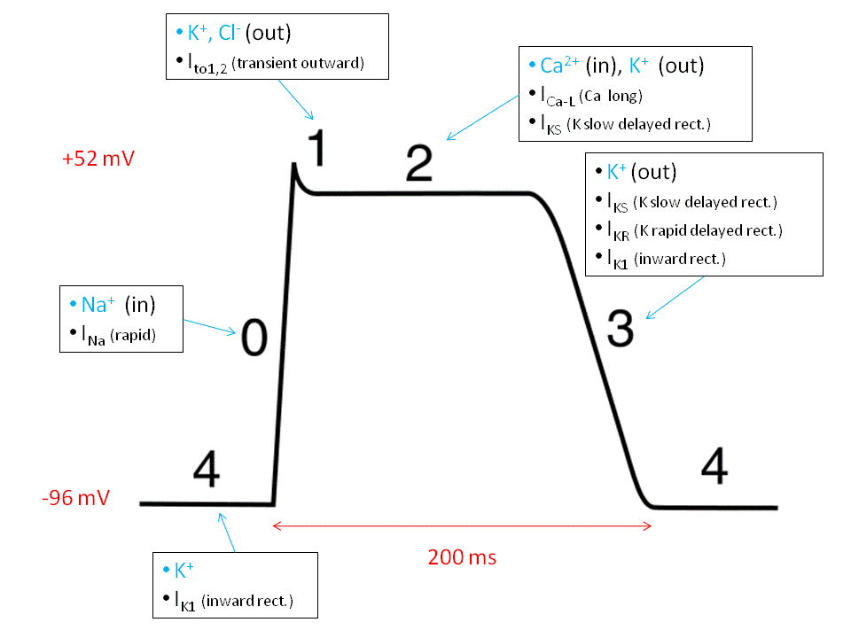
What is the peak potential of a skeletal muscle?
However, the peak potential achieved is approximately +30 mV. The Nernst potential is not achieved for two main reasons. First, just as the Na + channels are activated by membrane voltage changes, a process of inactivation is also initiated as the membrane potential becomes less negative. Inactivation is a slower mechanism than activation, so the Na + current continues to flow for a short period after the onset of inactivation, but not sufficiently to reach the Nernst potential. The second factor limiting the upstroke of the action potential is the voltage activation of rectifying potassium channels. Their activation is initiated also during the upstroke of the action potential but (in a similar way to Na + channel inactivation) there is a slight delay in channel opening. The resulting K + current, in addition to limiting the peak of the action potential, is also principally responsible for repolarization.
How many segments are in the dihydropyridine receptor?
The α1 subunit of the dihydropyridine receptor. Four transmembrane domains (I–IV) are each made up of six segments (1–6). The cytoplasmic loop between the sixth segment of domain II and the first segment of domain III interacts with the ryanodine receptor protein to cause Ca 2+ release from the sarcoplasmic reticulum.
How does action potential work in sarcolemma?
Once an action potential has been generated, it spreads as a wave over the sarcolemma. Skeletal muscle sarcolemma is characterized by invaginations called transverse- or t-tubules that run perpendicular to the surface of the cell deep into its body. By passing down the t-tubular membrane, the action potential is carried to the structures responsible for transducing an electrical into a chemical signal that will trigger activation of the contractile elements.
What are electrical events in muscle contraction?
Electrical events in muscle contraction. Muscle fibres are excitable cells. The cell membrane (sarcolemma) contains the ion channels and pumps necessary to maintain a very negative resting membrane potential and the voltage gated ion channels necessary for generation of an action potential .
How is smooth muscle contraction achieved?
Smooth and graded changes in force of contraction are achieved through summation of responses to successive stimuli and recruitment of motor units. Sustained muscle contraction requires de novo synthesis of ATP, which is principally aerobic or anaerobic depending on muscle fibre type.
What percentage of muscle mass is skeletal muscle?
Skeletal muscle constitutes 40% of muscle mass. Derangement of muscle function can have profound systemic effects. Physiological skeletal muscle contraction requires generation and spread of a membrane action potential, transduction of the electrical energy into an intracellular chemical signal that, in turn, triggers myofilament interaction.
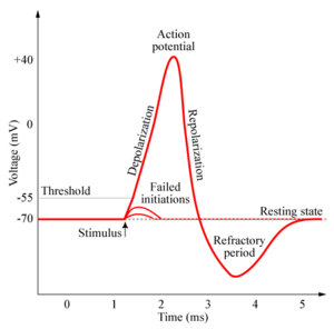
What happens to the membrane potential as the neuron repolarizes?
As the membrane continues to repolarize, the membrane potential becomes more negative than its resting level. This afterhyperpolarization is a result of K+ channels remaining open, allowing the continued efflux of K+ ions. Another way to think about afterhyperpolarization is that the membrane's permeability to K+ is higher than when the neuron is at rest. Consequently, the membrane potential is driven even more toward the K+ equilibrium potential (Fig. 3.5D).
What happens to the membrane potential of a nerve cell during the rising phase?
During the rising phase, the nerve cell membrane becomes more permeable to sodium ,- as a consequence, the membrane potential begins to shift more toward the equilibrium potential for sodium. However, before the membrane potential reaches ENa, sodium permeability begins to decrease and potassium permeability increases.
What happens at the peak of the action potential?
At the peak of the action potential, the sodium conductance begins to fall as an inactivation gate closes. Also, more K+ channels open, allowing more positively charged K+ ions to leave the neuron. The net effect of inactivating Na+ channels and opening additional K+ channels is the repolarization of the membrane (Fig. 3.5C).
What is the difference between the rising phase and the repolarization phase?
The rising phase of the action potential is the result of an increase in sodium conductance, while the repolarization phase is a result of a decrease in sodium conductance and a delayed increase in potassium conductance. The action potential may be recorded by placing a mi-croelectrode inside a nerve cell or its axon.
What are the characteristics of an action potential?
The action potential is a transient change in the membrane potential characterized by a gradual depolarization to threshold, a rapid rising phase, an overshoot, and a repolarization phase. The repolarization phase is followed by a brief afterhyperpolar-ization (undershoot) before the membrane potential again reaches resting level (Fig. 3.4A).
What is the action potential of sodium channels?
gated sodium channels initiates an action potential. The action potential then propagates to the axon terminal, where the associated depolarization causes the release of neuro-transmitter. The initial depolarization to start this process derives from synaptic inputs causing ligand-gated channels to open on the dendrites and somata of most neurons. For peripheral sensory neurons, the initial depolarization results from a generator potential initiated by a variety of sensory receptor mechanisms (see Chapter 4).
Where is action potential generated?
An Action Potential Is Generated at the Axon Hillock and Conducted Along the Axon. An action potential depends on the presence of voltage-gated sodium and potassium channels that open when the neuronal membrane is depolarized. These voltage-gated channels are restricted to the axon of most neurons.