
A retinaculum refers to any region on the body in which tendon groups from different muscles pass under one connective tissue band. Wrist retinacula include the flexor
Common flexor tendon
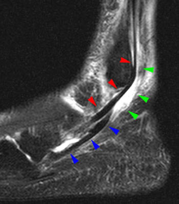
The common flexor tendon is a tendon that attaches to the medial epicondyle of the humerus.
What is another name for the retinaculum?
Related to retinaculum: lateral patellar retinaculum, extensor retinaculum. [ret″ĭ-nak´u-lum] a structure that retains an organ or tissue in place. flexor retinaculum of hand a fibrous band forming the carpal tunnel, through which pass the tendons of the flexor muscles of the hand and fingers.
What is the retinaculum of the wrist?
A retinaculum refers to any region on the body in which tendon groups from different muscles pass under one connective tissue band. Wrist retinacula include the flexor and the extensor retinacula of the hand.
What is the function of the flexor retinaculum?
A fibrous band, strap or ligament that holds another part in place. The flexor retinaculum prevents the flexor tendons from springing away from the front of the wrist when it is bent.
What is the function of the retinacula?
The retinacula, found in the hand, foot and knee, are covering muscle tendons and nerves as they cross vulnerable joints. There are two important tunnels that tendons and nerves pass through in the wrist and ankle. The carpal tunnel of the wrist, which most people have heard of, allows for the flow of the median nerve that innervates the hand.
What are the two parts of the wrist and ankle?
Why does my knee rupture after knee surgery?
What is retinaculum?
How long does it take for retinal amputation to manifest?
See 1 more
About this website
What is a retinaculum in anatomy?
The flexor retinaculum is a fibrous connective tissue band that forms the anterior roof of the carpal tunnel. Many experts consider the flexor retinaculum synonymous with the transverse carpal ligament and the annular ligament; for this discussion, they will be regarded as the same structure.
What is retinaculum and its function?
A retinaculum (plural retinacula) is a band of thickened deep fascia around tendons that holds them in place. It is not part of any muscle. Its function is mostly to stabilize a tendon. The term retinaculum is New Latin, derived from the Latin verb retinere (to retain).
What is the retinaculum made of?
Structurally, the retinaculum consists of three layers. The deepest layer, the gliding layer, consists of hyaluronic acid-secreting cells. The thick middle layer consists of interspersed elastin fibers, collagen bundles, and fibroblasts.
What is a retinaculum quizlet?
retinaculum. fibrous tissue for mechanical advantage.
Can torn retinaculum heal itself?
Superior peroneal retinaculum tears are often mistaken for lateral ankle instability. These tears often do not heal readily by themselves and must be identified so that proper treatment can begin.
What happens if you tear your retinaculum?
The peroneal tendons run behind the lateral malleolus to the foot. They are held in place by the (superior) peroneal retinaculum (SPR in the diagram opposite). If this retinaculum is torn as a result of injury, the tendons can pop out place, causing pain and swelling.
How is retinaculum treated?
Treatment: Rest, icing, and, compression socks to bring down swelling are generally the top recommended treatments for a strained flexor retinaculum.
Where is retinaculum located?
wristThe flexor retinaculum (transverse carpal ligament, or anterior annular ligament) is a fibrous band on the palmar side of the hand near the wrist. It arches over the carpal bones of the hands, covering them and forming the carpal tunnel.
Why is the retinaculum required?
Principal function of the flexor retinaculum is to serve as a pulley for the carpal flexor muscles and to stabilize the carpal system. In addition, The volar surface gives rise to muscles of the thenar and hypothenar eminences.
What is the function of retinaculum in foot?
A retinaculum is a band of thick deep fascia that holds the long tendons of your ankle (those that cross the ankle) in place. The retinaculum also acts as a pulley system increasing mechanical advantage. The retinaculum is a major source of neurological receptors involved in balance and proprioception.
What are the 3 retinaculum in the foot?
The ankle retinacula include the superior and inferior extensor and flexor retinacula and the superior and inferior peroneal retinac- ula.
What is the retinaculum of the foot?
Retinacula are thickenings of deep fascia in the region of joints, to keep the tendons in place as they cross the joints. Many retinacula are present in the vicinity of ankle joint. These retinacula are transverse bands across the ankle that holds down the tendons that cross from the leg to the foot.
What is the function of the retinaculum at the wrist?
Principal function of the flexor retinaculum is to serve as a pulley for the carpal flexor muscles and to stabilize the carpal system. In addition, The volar surface gives rise to muscles of the thenar and hypothenar eminences. It is Related to the tendon of the palmaris longus.
What is the function of retinaculum in foot?
A retinaculum is a band of thick deep fascia that holds the long tendons of your ankle (those that cross the ankle) in place. The retinaculum also acts as a pulley system increasing mechanical advantage. The retinaculum is a major source of neurological receptors involved in balance and proprioception.
What is the function of the ankle retinaculum?
The retinacula of the ankle are distinct structures defined as regions of localized thickening of the crural fascia covering the deep structures of the distal portion of the leg, ankle, and foot. Their role is to maintain the approximation of the tendons to the underlying bone.
Where is retinaculum located?
wristThe flexor retinaculum (transverse carpal ligament, or anterior annular ligament) is a fibrous band on the palmar side of the hand near the wrist. It arches over the carpal bones of the hands, covering them and forming the carpal tunnel.
Flexor Retinaculum of the Foot: Retinacula Injuries and Treatment
Dr. Justin is a LIFE SAVER! I have been dealing with low... back pain for the better part of the last 15 years. I've gone to over 20 chiropractors and therapists and I have never had experienced long lasting relief.
Inferior Extensor Retinaculum Anatomy, Function & Diagram - Healthline
A retinaculum refers to any region on the body where tendon groups from different muscles pass under one connective tissue band. The inferior extensor retinaculum is located at the front of the ...
Retinaculum Definition & Meaning - Merriam-Webster
retinaculum: [noun] any of several fibrous bands of fascia that pass over or under tendons (as at or near the ankle or wrist) and help to keep them in place.
Retinaculum - Differential Diagnosis of the Knee
Definition: Fibrous tissue on the lateral side of the patella that helps to control patellar tracking. If too tight may cause excessive lateral tracking which may lead to a lateral retinaculum release (Juhn).
Retinaculum - Wikipedia
A retinaculum (plural retinacula) is a band of thickened deep fascia around tendons that holds them in place. It is not part of any muscle.Its function is mostly to stabilize a tendon. The term retinaculum is New Latin, derived from the Latin verb retinere (to retain). Specific retinacula include: In the wrist: Flexor retinaculum of the hand
What is retinaculum?
Definition of retinaculum. : any of several fibrous bands of fascia that pass over or under tendons (as at or near the ankle or wrist) and help to keep them in place.
Where does the word "retinoculum" come from?
borrowed from New Latin retināculum, going back to Latin, "rope holding something in place, cable, hawser," from retinēre "to hold fast, detain, keep possession of" + -ā-, stem formative of verbs + -culum, suffix of instrument and place (going back to Indo-European *-tlom ), probably after gubernāculum "steering oar of a ship" (from gubernāre "to steer") or other derivatives of 1st-conjugation verbs — more at retain
What is the term for a band that holds another part in place?
A fibrous band, strap or ligament that holds another part in place. The flexor retinaculum prevents the flexor tendons from springing away from the front of the wrist when it is bent.
What is the term for a structure that retains an organ or tissue in place?
retinaculum. [ ret″ĭ-nak´u-lum] a structure that retains an organ or tissue in place. flexor retinaculum of hand a fibrous band forming the carpal tunnel, through which pass the tendons of the flexor muscles of the hand and fingers. retinaculum morga´gni a ridge formed by the coming together of segments of the ileocecal valve.
What is retinaculum morga?
retinaculum morga´gni a ridge formed by the coming together of segments of the ileocecal valve.
What is a frenulum?
A frenulum, or a retaining band or ligament.
What nerves pass through the wrist?
There are two important tunnels that tendons and nerves pass through in the wrist and ankle. The carpal tunnel of the wrist, which most people have heard of, allows for the flow of the median nerve that innervates the hand.
What is the retinaculum?
The retinaculum, part of the fascial system, is strong connective tissue found only in certain parts of the body. The retinaculum exists to provide stability and protection for tendons and nerves as they cross important joints. Fascia is a protective webbing that wraps the entire body. It works in different ways, connecting some things together, ...
Why are the retinacula of the knee so complex?
The retinacula of the knee are way more complex because of the structure of the knee itself. There are two retinacula of the knee, the medial and lateral and they sort of act ligamentously to balance and stabilize the kneecap, ideally sustaining an equal pull to the inside and outside.
Which part of the wrist is responsible for strapping nerves and tendons into place?
There are a number of tendons that move through these tunnels as well and the flexor retinacula of the wrist and ankle are essentially strapping these nerves and tendons into place. The extensor retinacula of the wrist and ankle play a similar role but don’t have nerves passing underneath them.
Where is the retinacula found?
The retinacula, found in the hand, foot and knee, are covering muscle tendons and nerves as they cross vulnerable joints.
How to use retinaculum in a sentence
This process is termed the retinaculum, and serves, in conjunction with the frenulum, to lock the wings together during flight.
Other words from retinaculum
The American Heritage® Stedman's Medical Dictionary Copyright © 2002, 2001, 1995 by Houghton Mifflin Company. Published by Houghton Mifflin Company.
What is the purpose of the fibrous capsule in the knee?
The fibrous capsule of the knee protects the delicate synovial membrane inside and seals the lubricating synovial fluid within the joint capsule. It also helps to maintain the proper position of the bones within the knee to prevent injury and premature wear . The medial patellar retinaculum fills in the gap between the patella, patellar ligament, ...
What connects the medial patellar retinaculum?
Several other important connections are formed between the medial patellar retinaculum and its surrounding structures. On its anterior edge, some collagen fibers extend to form connections to the patella and merge with the fibers of the patellar ligament. Posteriorly the fibers weave together with the fibers of the medial collateral ligament.
Which tendon of insertion forms the connection of the vastus medialis to the medial condyle?
Acting as a tendon of insertion, the medial patellar retinaculum forms the connection of the vastus medialis to the medial condyle of the tibia. Contraction of the vastus medialis extends the leg at the knee by pulling on the medial patellar retinaculum, which in turn pulls the tibia anteriorly and straightens the knee.
Which muscle is involved in the insertion of the vastus medialis?
This motion is assisted by the contraction of the other muscles of the quadriceps femoris group , which simultaneously pull on the tibia to extend the knee.
Where does the medial patellar retinaculum originate?
Most of the fibers of the medial patellar retinaculum originate in the medial femoral region from the vastus medialis muscle , just superior to the patella. Continue Scrolling To Read More Below... « Back Show on Map ». Anatomy Term.
What is the flexor retinaculum?
Flexor retinaculum is a strong fibrous band which bridges the anterior concavity of the carpal bones thus converts it into a tunnel, the carpal tunnel.
Which carpal bone gives attachments to both flexor and extensor retinacula?
These four bony points are all palpable in the living hand and it should be noted that pisiform is the only carpal bone that gives attachments to both flexor and extensor retinacula. On either side the retinaculum has a slip.
Which type of slip is attached to the pisiform bone?
Lateral deep slip - It is attached to the medial lip of the groove on the trapezium thus converts it into a fibro-osseous tunnel that transmits the tendon of the flexor carpi radialis and its synovial sheath. Medial superficial slip - It is attached to the pisiform bone and it forms a small canal (of Guyon).
Which side of the retina has a slip?
On either side the retinaculum has a slip.
Can you add videos to your watch history?
Videos you watch may be added to the TV's watch history and influence TV recommendations. To avoid this, cancel and sign in to YouTube on your computer.
How is lateral retinacular release done?
How is a lateral retinacular release done? This is an arthroscopic surgery ( a knee “scope” which is performed through 3 small incisions ( about ½ inch each) around the knee. From the inside of the knee the lateral retinaculum is incised, from the inside, allowing the kneecap to untilit itself.
Why does my knee tilt?
This occurs because of the chronic pull of the knee cap to the outside by the thigh muscles, creating a strain on the medical or inside tissues (the retinaculum).
What tissues are in the middle of the knee?
These tissues are call the medial (inside) and the lateral (outside) retinaculum.
How long does it take for a knee brace to be locked?
Partial weightbearing, with crutches, is recommended to minimize pain and swelling. The first office visit after surgery is between 7 and 14 days after surgery.
Where does the pain come from in the knee?
The pain comes from the tissue on the inside of the kneecap (the medial retinaculum).
What is retinal release?
It is a surgical release of the outside or lateral retinaculum.
Can lateral release be done?
Yes the arthroscopic lateral release is done to make the physical therapy work more efficiently. Failing to perform physical therapy will delay, and possibly prevent the eventual improvement from the surgery. This means more knee pain, fatigue, weakness and swelling for a longer time period.
What are the two parts of the wrist and ankle?
Wrist retinacula include the flexor and the extensor retinacula of the hand. Ankle retinacula include the peroneal retinacula and the flexor, superior extensor , and inferior extensor retinacula of the foot.
Why does my knee rupture after knee surgery?
During or following arthroscopic knee surgery, the quadriceps tendon may rupture due to the release of the lateral retinaculum.
What is retinaculum?
A retinaculum refers to any region on the body in which tendon groups from different muscles pass under one connective tissue band.
How long does it take for retinal amputation to manifest?
This complication is relatively rare and may manifest up to five weeks after the injury. To reduce the complications from retinaculum surgery, a procedure was developed that incorporates medial patellofemoral ligament overlap in addition to lateral retinaculum release.
