What are two structures form the hip joint?
iliofemoral ligament (y shaped, reinforces anterior aspect of hip joint) ischiofemoral ligament (spiral shaped, reinforces posterior aspect of hip joint) pubofemoral ligament (triangular thickening, supports inferior aspect) ligament of the head of the femur (small ligament, links head of femur with acetabulum)
Which pair of structures forms the hip joint?
The hip joint is a ball-and-socket type joint and is formed where the thigh bone (femur) meets the pelvis. The femur has a ball-shaped head on its end that fits into a socket formed in the pelvis, called the acetabulum. Large ligaments, tendons, and muscles around the hip joint hold the bones (ball and socket) in place and keep it from dislocating.
What are the components of the hip joint?
- Iliofemoral ligament
- Pubofemoral ligament
- Ischiofemoral ligament
- Transverse ligament of the acetabulum
- Ligament of the head of the femur (ligamentum teres capitis femoris)
How to strengthen the ligaments of the hip joint?
- Start by sitting down. ...
- Place the balance board between your feet.
- Place one foot on each side of the board. ...
- After you get used to the motion while sitting down, then try standing on the balance board. ...
- Try to keep your balance for as long as possible, but remember to step off if you're losing your footing. ...
What is the structure and function of the hip joint?
The hip joint connects the lower extremities with the axial skeleton. The hip joint allows for movement in three major axes, all of which are perpendicular to one another. The location of the center of the entire axis is at the femoral head. The transverse axis permits flexion and extension movement.
What holds the hip joint in place?
In a normal hip, the ball and socket are covered with a smooth layer of tissue called cartilage. The cartilage allows the ball to glide easily inside the socket and provides a cushion to your hip joint. Muscle and ligaments hold your hip joint in place.
What are the 3 main ligaments of the hip joint?
The hip joint capsule is formed by three major ligaments: the iliofemoral, pubofemoral, and ischiofemoral ligaments.
What muscles are attached to the hip joint?
The hip joint is surrounded by several muscles, including:Gluteal muscles, located on the back of the hip (buttocks);The adductor muscle on the inner thigh;The iliopsoas muscle, which extends from the lower back to upper femur;Quadriceps, a group of four muscles that comprise the front of the thigh; and.More items...
What tendon is in your hip?
There are two gluteus tendons that both attach at the outer aspect of the hip at the greater trochanter: the gluteus minimus and medius tendons. Both of these muscles abduct the hip, while the gluteus minimus also acts as the primary internal rotator of the hip.
What stabilizes the hip joint?
The stability of the hip joint depends on many ligaments including iliofemoral ligament, pubofemoral ligament, ischiofemoral ligament, ligamentum teres, zona orbicularis, and deep arcuate ligament, all of which work closely to reinforce the joint capsule2).
Which ligament of hip joint is the strongest?
The hip joint capsule and capsular ligaments The iliofemoral ligament is the strongest ligament in the body and attaches the anterior inferior iliac spine (AIIS) to the intertrochanteric crest of the femur.
What is the cartilage in the hip called?
The labrum is an additional, specialized piece of cartilage that runs along the rim of the socket to provide a suction seal and stability to the hip joint, absorbing shock and distributing pressure during hip motion. The hip labrum may become torn or even detached from the acetabular socket for a variety of reasons.
What stabilizes the hip joint?
The stability of the hip joint depends on many ligaments including iliofemoral ligament, pubofemoral ligament, ischiofemoral ligament, ligamentum teres, zona orbicularis, and deep arcuate ligament, all of which work closely to reinforce the joint capsule2).
What makes your hip joint hurt?
Osteoarthritis and rheumatoid arthritis are among the most common causes of hip pain, especially in older adults. Arthritis leads to inflammation of the hip joint and the breakdown of the cartilage that cushions your hip bones. The pain gradually gets worse.
What does a pulled hip muscle feel like?
Many people who experience hip flexor strain will have these symptoms as well: sudden, sharp pain in the hip or pelvis after trauma to the area. pain when lifting the leg. cramping, stiffness, and weakness in the muscles of the upper leg area.
What causes hip pain that radiates down the leg?
Sciatica. This one is often the most common cause of hip pain being funneled down your leg. Sciatica refers to the sciatic nerve that runs between your hip and down each leg. To be exact, it runs down the back or your hip and the front, back, and sides of your leg.
What are the muscles that attach to the hip joint?
Muscle Groups. The various muscles which attach to or cover the hip joint generate the hip’s movement. Gluteals: The gluteals are the muscles in your buttocks. The gluteals (gluteus maximus, gluteus minimus and gluteus medius) are the three muscles attached to back of the pelvis and insert into the greater trochanter of the femur.
What are the main ligaments in the hip?
As noted above, the stability of the hip joint is directly related to its muscles and ligaments. The most notable ligaments in the hip joint are: 1 Iliofemoral ligament, which connects the pelvis to the femur at the front of the joint. It keeps the hip from hyper-extension 2 Pubofemoral ligament, which attaches the most forward part of the pelvis known as the pubis to the femur 3 Ischiofemoral ligament, which attaches to the ischium (the lowest part of the pelvis) and between the two trochanters of the femur.
Why is the hip so strong?
The hip joint is normally very sturdy because of the fit between the femoral head and acetabulum as well as strong ligaments and muscles at the joint. All of the various components of the hip mechanism assist in the mobility of the joint. Damage to any single component can negatively affect range of motion and ability to bear weight on the joint.
What is the head of the femur?
At the top of the femur is a rounded protrusion which articulates with the pelvis. This portion is referred to as the head of the femur, or femoral head . There are two other protrusions near the top of the femur, known as the greater and lesser trochanters. The muscles involved in hip motion are attached to the joint at these trochanters.
What is the second largest joint in the body?
The hip is the body’s second largest weight-bearing joint (after the knee ). It is a ball and socket joint at the juncture of the leg and pelvis. The rounded head of the femur (thighbone) forms the ball, which fits into the acetabulum (a cup-shaped socket in the pelvis). Ligaments connect the ball to the socket and usually provide tremendous ...
Which ligaments attach to the femur?
The most notable ligaments in the hip joint are: Iliofemoral ligament, which connects the pelvis to the femur at the front of the joint. It keeps the hip from hyper-extension. Pubofemoral ligament, which attaches the most forward part of the pelvis known as the pubis to the femur. Ischiofemoral ligament, which attaches to ...
What is the outer layer of the acetabulum?
Labrum . The labrum is a circular layer of cartilage which surrounds the outer part of the acetabulum effectively making the socket deeper to provide more stability for the joint. Labrum tears are not an uncommon hip injury.
What is the hip joint?
The hip joint is a multiaxial joint and permits a wide range of motion; flexion, extension, abduction, adduction, external rotation, internal rotation and circumduction. Compared to the glenohumeral (shoulder) joint, however, this joint sacrifices mobility for stability as it is designed for weight bearing.
Where is the hip joint located?
The hip joint is the articulation between the ellipsoid head of the femur and the hemispherical concavity of the acetabulum located on the lateral aspect of the hip bone. The femoral head is covered with articular ( hyaline) cartilage with the exception of a rough central depression, the fovea capitis, which is a surface of attachment for the ligament of the femoral head (ligamentum teres capitis femoris).
How does internal rotation of the hip joint work?
During internal rotation, the femoral shaft moves anteriorly, causing the toes to point medially . The reverse occurs in external rotation where the femoral shaft moves posteriorly, causing the toes to point away from the midline. External rotation is much freer and more powerful than internal rotation. For example, the range of internal rotation with the hip extended is about 35o while external rotation is about 45o. Rotation at the hip joint is generally much freer with hip flexion rather than extension. Tightness in the lateral rotators and the ischiofemoral ligament limit internal rotation of the hip joint. Contrarily, external rotation is limited by tightness in the medial rotators of the thigh and the iliofemoral and pubofemoral ligaments.
What is the deep central nonarticular floor of the acetabulum?
The deep central nonarticular floor of the acetabulum is referred to as the acetabular fossa. This area is devoid of cartilage and is continuous with the acetabular notch. It contains loose connective tissue (fibroelastic fat pad) which is covered by synovial membrane. Attached to the margin of the acetabulum is a fibrocartilaginous collar called the acetabular labrum. This structure deepens the acetabulum by raising the rim of the acetabulum slightly, thereby increasing the acetabular articular area by about 10%. Inferiorly, the acetabular labrum continues as the transverse acetabular ligament, bridging the acetabular notch and transforming the notch into a foramen.
What is the acetabulum made of?
The acetabulum is formed by the fusion of the ilium, ischium and pubic bones. It plays a significant role in the stability of the hip joint as it almost entirely encompasses the head of the femur. The acetabulum bears a prominent semilunar region known as the lunate surface that is covered by articular cartilage.
Which ligaments are in the capsule of the hip joint?
The capsule of the hip joint is reinforced inferiorly by the pubofemoral ligament and posteriorly by the ischiofemoral ligament.
How does hip flexion affect knee extension?
Hip flexion is limited by the tension in the hamstrings when the knee is extended. Extension of the hip joint moves the thigh away from the trunk. Extension of the joint beyond the vertical is limited to about 30o by the tension of the capsular ligaments and the shape of the articular surfaces.
What is the hip joint?
The hip joint is a ball and socket synovial joint, formed by an articulation between the pelvic acetabulum and the head of the femur. It forms a connection from the lower limb to the pelvic girdle, and thus is designed for stability and weight-bearing – rather than a large range of movement.
Where is the capsule of the hip joint located?
The capsule of the hip joint attaches to the edge of the acetabulum proximally. Distally, it attaches to the intertrochanteric line anteriorly and the femoral neck posteriorly.
What is the ring around the acetabulum called?
There is a horseshoe shaped fibrocartilaginous ring around the acetabulum which increases its depth, known as the acetabular labrum. The increase in depth provides a larger articular surface, further improving the stability of the joint.
How many extracapsular ligaments are there?
There are three main extracapsular ligaments, continuous with the outer surface of the hip joint capsule:
Which ligaments are very strong?
The iliofemoral, pubofemoral and ischiofemoral ligaments are very strong, and along with the thickened joint capsule, provide a large degree of stability. These ligaments have a unique spiral orientation; this causes them to become tighter when the joint is extended.
Where is the acetabulum located?
The acetabulum is a cup-like depression located on the inferolateral aspect of the pelvis. Its cavity is deepened by the presence of a fibrocartilaginous collar – the acetabular labrum. The head of femur is hemispherical, and fits completely into the concavity of the acetabulum.
Which ligament runs from the acetabular fossa to the fovea of the femur?
The only intracapsular ligament is the ligament of head of femur. It is a relatively small structure, which runs from the acetabular fossa to the fovea of the femur.
What is the function of the hip joint?
Functionally, the hip joint enjoys a very high range of motion. The ball-and-socket structure of the joint allows the femur to circumduct freely through a 360-degree circle. The femur may also rotate around its axis about 90 degrees at the hip joint. Only the shoulder joint provides as high of a level of mobility as the hip joint. In addition to being flexible, each hip joint must be capable of supporting half of the body’s weight along with any other forces acting upon the body. During running and jumping, for example, the force of the body’s movements multiplies the force on the hip joint to many times the force exerted by the body’s weight. The hip joint must be able to accommodate these extreme forces repeatedly during intense physical activities.
How does the hip joint support the body?
In addition to being flexible, each hip joint must be capable of supporting half of the body’s weight along with any other forces acting upon the body. During running and jumping, for example, the force of the body’s movements multiplies the force on the hip joint to many times the force exerted by the body’s weight.
What is the joint between the femur and the os coxa?
The hip joint is a ball-and-socket synovial joint formed between the os coxa (hip bone) and the femur. A round, cup-shaped structure on the os coxa, known as the acetabulum, forms the socket for the hip joint. The rounded head of the femur
What is the function of hyaline cartilage?
Hyaline cartilage also acts as a flexible shock absorber to prevent the collision of the bones during movement. Between the layers of hyaline cartilage, synovial membranes secrete watery synovial fluid to lubricate the joint capsule.
What is the most important joint in the human body?
The hip joint is one of the most important joints in the human body. It allows us to walk, run, and jump. It bears our body’s weight and the force of the strong muscles of the hip and leg. Yet the hip joint is also one of our most flexible joints and allows a greater range of motion than all other joints in the body except for the shoulder.
Why was hip replacement impossible?
Hip replacement was once impossible because, although joints could easily be produced in a laboratory, the human body rejected the materials. Sometimes the pins that held the artificial joint to other bones worked loose and required further surgery.
Which joint has the highest range of motion?
Functionally, the hip joint enjoys a very high range of motion. The ball-and-socket structure of the joint allows the femur to circumduct freely through a 360-degree circle. The femur may also rotate around its axis about 90 degrees at the hip joint. Only the shoulder joint provides as high of a level of mobility as the hip joint.
What is the hip joint?
The hip joints are among the joints in the lower body that have to work hard. They are the joints that support our entire body weight when we are walking and standing. If the load is too high or if the muscles are exposed to repeated loads for a long period, the muscles can suffer from excess strain. On a microscopic level, the muscles can suffer micro-ruptures which cause pain.
What is the anatomy of the hip?
The anatomy of the hip is complicated in the form of several muscles, ligaments and a tight joint capsule that hold the joint together. This design means that we can move the joint.
What are the bones that connect the hips to the lower body?
The hips consist of two major skeletal parts: the pelvis and the femur. In turn, the pelvis consists of four different symmetrical parts on the right and left side. These skeletal parts in the pelvis are called the pubis, the ischium, the ilium and the sacrum. These bones are all flat and strong, they connect the upper body to the lower body. The pelvis also protects our internal organs.
What is the largest joint in the body?
The hip, or more specifically the hip joint, is one of the largest joints in the body. It consists of what is known as a ball-and-socket type joint, which means that the head of the joint looks like a ball. This allows the joint to move in all directions, even if the hip is not as mobile as the shoulder joint, for example, which is also a ball-and-socket joint. The hip anatomy also includes the acetabulum, which is located in the pelvis. The ball of the hip joint fits into this. Both joint surfaces are also covered with a layer of cartilage. This articular cartilage allows smooth movements without friction and protects the joints. In addition, the ligaments, joint capsule and muscles hold the hip joint in place. They stabilize the hip joint and enable it to support a large part of the body’s weight.
What is the hip joint?
The hip joint consists of two main parts: Femoral head – a ball-shaped piece of bone located at the top of your thigh bone, or femur. Acetabulum – a socket in your pelvis into which the femoral head fits. Bands of tissue, called ligaments, connect the ball to the socket, stabilizing the hip and forming the joint capsule.
What muscles are involved in hip joint movement?
The hip is surrounded by large muscles that support the joint and enable movement. They include: 1 Gluteals – muscles of the buttocks, located on the back of the hip 2 Adductor muscles – muscles of the inner thigh, which pull the leg inward toward the opposite leg 3 Iliopsoas muscle – a muscle that begins in the lower back and connects to the upper femur 4 Quadriceps – four muscles on the front of the thigh that run from the hip to the knee 5 Hamstrings – muscles on the back of the thigh, which run from the hip to just below the knee
What are the sacs in the hip called?
Fluid-filled sacs called bursae provide cushioning where there is friction between muscle, tendons and bones. The hip is surrounded by large muscles that support the joint and enable movement. They include: Gluteals – muscles of the buttocks, located on the back of the hip.
Which muscle pulls the leg inward toward the opposite leg?
Adductor muscles – muscles of the inner thigh, which pull the leg inward toward the opposite leg
What is the membrane that connects the ball to the socket called?
The joint capsule is lined with a thin membrane called synovium, which produces a viscous fluid to lubricate the joint.
What is the anatomy of the hip?
Anatomy of the Hip. One of the two ball and socket joints in the human body, the pelvis and femur form the Hip Joint. The femoral head locks into the acetabulum, forming a joint capable of weight-bearing with adequate range of motion. Due to the weight-bearing nature of the joint, the hip joint often suffers from athrtitis.
What is the ligament of the hip?
Ligaments of the hip. Within the hip joint, only one ligament resides; the ligamentum teres, or ligament of head of femur. A triangle shaped ligament, the ligamentum teres supplies blood to the femoral head with help from the obturator artery, and branches from the acetabular fossa to the fovea of the femur.
What part of the femur is responsible for stabilizing the hip joint?
All parts of the proximal femur play a role in creating and stabilizing the hip joint. The femoral head locks into the acetabulum and suctioned in with a piece of fibrocartilage called the labrum. The hip joint should be considered a saucer rather than a cup, and the labrum deepens this saucer, allowing the femur to securely move.
Which ligament is the strongest?
Of the three ligaments outside of the hip joint, the iliofemoral ligament boasts the strongest. Like the ligamentum teres, the pubofemoral ligament has a triangular shape. The pubofemoral helps prevent excessive abduction and extension. Abduction occurs when an individual moves their leg out to the side. Extension occurs when an individual moves ...
Which ligaments help stabilize the hip joint?
Outside of the hip joint, three ligaments help stabilize the joint from the outside. The iliofemoral ligament, pubofemoral ligament, and ischiofemoral ligament. The iliofemoral ligament has a ‘Y’ shape and prevents hyperextension of the hip. Of the three ligaments outside of the hip joint, the iliofemoral ligament boasts the strongest.
What are the bones that make up the pelvis?
The Pelvis. The sacrum and coccyx compose the bony framework for the pelvis, or pelvic girdle. The ilium, ischium, and pubis fuse together to create the hip bone.
Which ligament prevents hyperextension?
Lastly, the ischofemoral ligament prevents hyperextension and has a spiral shape. In addition, the ischiofemoral ligament helps hold the femoral head into the acetabulum.
What is the bone structure of the hip?
Bony Structure of the Hip Joint. The hip joint is a multi-axial joint that connects the pelvis to the lower extremities. As compared with the glenohumeral joint (shoulder), the hip has less range of motion and is designed primarily for weight bearing and stability. Type: ball-and-socket diarthrodial joint.
Which muscle group attaches to the hip joint?
The muscles that attach to the hip joint include those of the gluteal region and thigh. Last update: January 26, 2021.
What is the acetabulum of the hip bone?
Acetabulum of the hip bone (acetabular labrum: a ring of cartilage that increases the depth and stability of the articular surface) Supporting structures: Fibrous capsule. Intra-articular and extra-articular ligaments. Iliopectineal, trochanteric, and ischial bursae.
Which nerve passes through the greater sciatic foramen to the gluteal region?
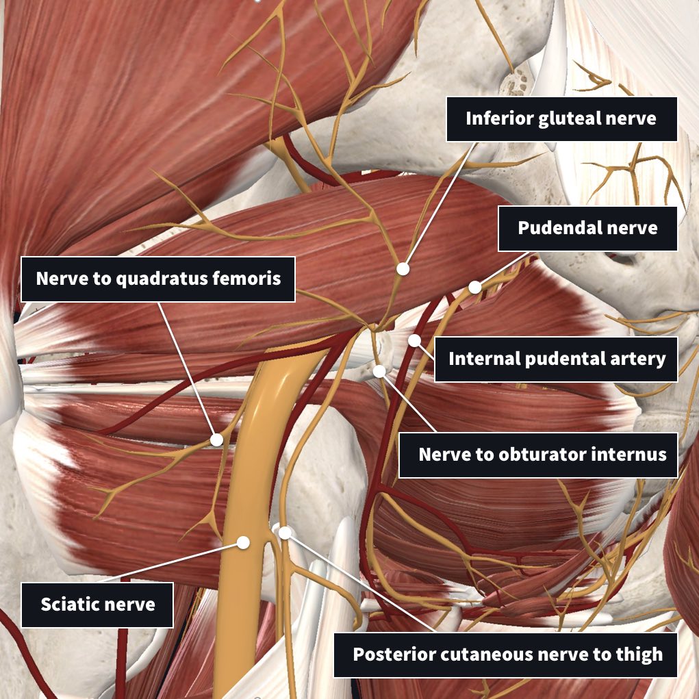
Sciatic nerve (L4–S3): passes through the greater sciatic foramen to the gluteal region; the sciatic nerve is the longest and widest nerve in the body. Superior gluteal nerve (L4–S1): innervates the gluteus medius, gluteus minimus, and tensor fascia latae muscles, and the superior aspect of the joint.
What is the name of the membrane that forms a collar around the femoral neck?
Has deep circular fibers that form a collar around the femoral neck, called the zona orbicularis (or annular ligament) Synovial membrane: The internal layer of the capsule. Produces synovial fluid, a viscous substance that lubricates and circulates nutrients to the joint.
Where does the femoral head originate?
Originates at the margin of the articular surface of the fe moral head, covers a portion of the femoral neck, is reflected on the internal surface of the capsule, reaches the fat tissue contained in the acetabular fossa, and encloses the ligament of the head of the femur.
Which artery divides into a posterior trunk and the anterior trunk?
Internal iliac artery— divides into a posterior trunk and the anterior trunk, which has multiple branches: Gluteal arteries: superior and inferior branches. Obturator artery: gives rise to the artery of the head of the femur within the ligament of the head of the femur.