What are the three layers of the cerebrum?
The six layers of the cerebral cortex are:
- I. Molecular layer (lamina molecularis) - consists only a few nerve cells
- II. External granular layer (lamina granularis externa) – relatively thin layer consisting of numerous small, densely packed neurons
- III. Pyramidal layer or external pyramidal layer (lamina pyramidalis externa) - is composed of medium-sized pyramidal nerve cells
- IV. ...
- V. ...
- VI. ...
What are the lobes of the cerebral cortex?
- Structure Lobes of the cerebral cortex Histology of the cerebral cortex Columnar organization of the cerebral cortex Functional areas of the cerebral cortex Phylogenetic types of cortex Brodmann areas
- Principal gyri of the cerebral cortex Frontal lobe Parietal lobe Temporal lobe Occipital lobe Insular lobe Limbic lobe
- Blood supply
- Function
- Sources
What is the difference between the cerebrum and the cortex?
What are the Similarities Between Cerebrum and Cerebral Cortex?
- Cerebrum and cerebral cortex are components of the forebrain.
- Specifically, cerebral cortex is the outer layer of the gray matter of cerebrum. Hence, the cerebral cortex is a part of the cerebrum.
- Thus, both are located in the uppermost region of the central nervous system.
- Furthermore, they are important in the coordination of functions of the body.
What is the cerebral cortex responsible for?
The cerebral cortex is responsible for the processes of thought, perception and memory and serves as the seat of advanced motor function, social abilities, language, and problem solving. It is composed of gray matter, consisting mainly of cell bodies and capillaries. It contrasts with the underlying white matter, consisting mainly of the white ...

What is cerebral cortex and its function?
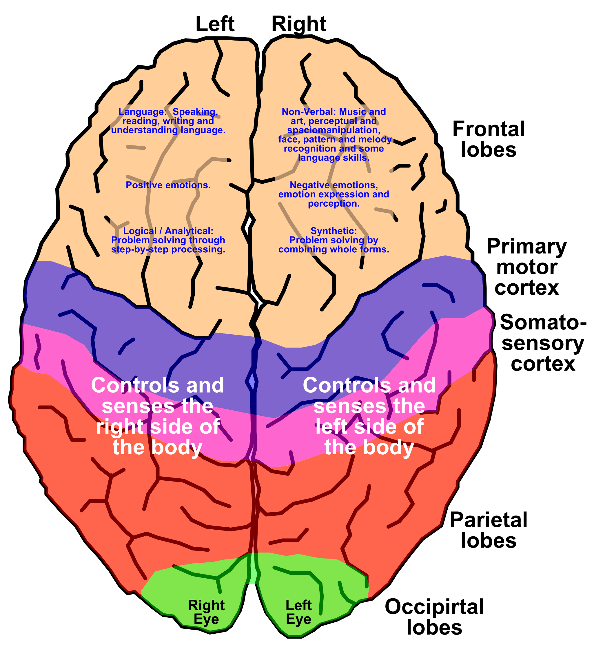
The cerebral cortex, which is the outer surface of the brain, is associated with higher level processes such as consciousness, thought, emotion, reasoning, language, and memory. Each cerebral hemisphere can be subdivided into four lobes, each associated with different functions.
What are the 3 main functions of the cerebral cortex?
The cerebral cortex is involved in several functions of the body including:Determining intelligence.Determining personality.Motor function.Planning and organization.Touch sensation.Processing sensory information.Language processing.
What best describes the cerebral cortex?
Terms in this set (123) Which best describes the cerebral cortex? Surface layer of gray matter on the cerebellum.
What are the 4 parts of the cerebral cortex?
There are four lobes in the cortex, the frontal lobe, parietal lobe, temporal lobe, occipital lobe.
What is the function of the cerebral cortex quizlet?
The coiled outer layer of the brain's cerebral hemispheres that is involved with information-processing activities such as perception, language, learning, memory, thinking, and problem solving, as well as the planning and control of voluntary bodily movements.
What are the 4 lobes of the cerebrum and their functions?
The four lobes include the occipital, temporal, frontal, and parietal lobes. Each lobe is responsible for a specific task. The frontal lobe functions in solving problems, controlling body movements, sentence formation, and personality traits. The occipital lobe functions in processing visual images.
What are three features that characterize the structure of cerebral cortex?
As a means of simplification, the cerebral cortex is often characterized as being made up of three types of areas: sensory, motor, and association areas. Sensory areas receive information related to sensation, and different areas of the cortex specialize in processing information from different sense modalities.
What is the cerebral cortex simple definition?
The cerebral cortex is the outer layer of the cerebrum, and it is the largest part of the human brain. The cerebral cortex consists of many gyri (r...
What are the 4 cerebral cortices?
The four cerebral cortices (also known as lobes) include the frontal lobe, parietal lobe, temporal lobe, and parietal lobe. Each of these lobes has...
What are the 3 functional areas of the cerebral cortex?
The three functional areas of the cerebral cortex include the sensory area, motor area, and association area. The sensory area processes informatio...
What Is the Cerebral Cortex?
Cerebral cortex definition: The cerebral cortex (sometimes known as the brain cortex) is the outer layer of the cerebrum, and it is the largest part of the brain. The cerebral cortex consists of many gyri and sulci which give it a wrinkled appearance.
Cerebral Cortex Location and Structure
The cerebral cortex forms the outer layer of the brain, and it is a few millimeters thick. Even though it is only a few millimeters thick, the cerebral cortex makes up roughly half of the brain's weight. The cerebral cortex is made up of grey matter which is a type of brain tissue that consists of neurons.
Cerebral Cortex Functions
What does the cerebral cortex do? In addition to being divided into four lobes, the cerebral cortex is also divided into three functional areas as well. These three functional areas include:
How many layers does the cerebral cortex have?
On the basis of light microscopic preparations stained by methods in which the cell bodies are displayed (e.g., Nissl method) and those where myelinated fibres are stained (e.g., Weigert method) the cerebral cortex is described as having six layers or laminae (Fig. 15.2). From the superficial surface downwards these laminae are as follows.
How much of the cerebral cortex is on the surface of the brain?
Because of the presence of a large number of sulci, only about one third of the total area of cerebral cortex is seen on the surface of the brain. The total area of the cerebral cortex has been estimated to be about 2000 cm2.
What are the similarities between the regions of the cortex?
The similarities between different regions of the cortex, in terms of the total neuronal population per unit volume of cortex, and in terms of the ratios of pyramidal and stellate (granular) neurons, are more striking than differences in laminar structure. Counts of neurons in delimited zones of cortex have shown that the total number of neurons is remarkably constant over different areas. However, the striate area (area 17) is an important exception, the neurons being much more numerous (about two and a half times) here as compared to other parts of cortex. In a given area of cortex about two thirds of neurons are pyramidal, and one third non-pyramidal.
What are the most abundant types of cortical neurons?
1. The most abundant type of cortical neurons are the pyramidal cells. (In contrast all other neurons in the cortex are referred to as non-pyramidal neurons). About two thirds of all cortical neurons are pyramidal. Their cell bodies are triangular, with the apex generally directed towards the surface of the cortex. A large dendrite arises from the apex. Other dendrites arise from basal angles. The axon arises from the base of the pyramid. The processes of pyramidal cells extend vertically through the entire thickness of cortex and establish numerous synapses. The axon of a pyramidal cell may terminate in one of the following ways.
Which type of cortex is closest to the agranular cortex?
The frontal type is nearest to the agranular cortex, the pyramidal cells being prominent; while the polar type is nearest to the granular cortex. The terms frontal and parietal are unfortunate as these types are not confined to the regions suggested by their names. The approximate distribution of the five types of cortex described above, on the superolateral surface of the cerebral hemisphere, is shown in Fig. 15.3
Which layer of the brain contains neurons?
The plexiform layer is made up predominantly of fibres although a few cells are present. All the remaining layers contain both stellate and pyramidal neurons as well as other types of neurons. The external and internal granular layers are made up predominantly of stellate (granular) cells. The predominant neurons in the pyramidal layer and in the ganglionic layer are pyramidal. The largest pyramidal cells (giant pyramidal cells of Betz) are found in the ganglionic layer. The multiform layer contains cells of various sizes andshapes.
Which type of neurons are found in the cortex?
In addition to the stellate and pyramidal neurons the cortex contains numerous other cell types some of which are illustrated in Fig. 15.1.
What is the cerebral cortex?
The cerebral cortex is required for voluntary activities, language, speech, and multiple brain functions, such as thinking and memory. In addition to the neuron bodies, the cortex also contains endings of neurons that reach it from other parts of the brain as well as a rich network of blood vessels. It is thanks to this content that the cortex has ...
What is the largest part of the brain?
The largest part of the cortex, as much as 90%, consists of a phylogenetically newer structure - a new cortex, consisting of six layers of stacked nerve cell bodies. The phylogenetically older structure of the cortex consists of the limbic part, which is part of the limbic system and the olfactory zone ( 1 ).
Why is the cerebral cortex wrinkled?
Since space in the skull is very limited, the cerebral cortex is wrinkled, as we have already said, which causes a much larger area of the cerebral cortex to fit into the same volume. The cerebral cortex is the most developed part of the human brain - four times the size of a gorilla.
What is the role of the basal ganglia in the cerebral hemisphere?
There is a thin but extensive cerebral cortex on the surface of our brain. The basal ganglia play an important role in initiating and controlling movement.
What is the function of the frontal lobe?
The frontal lobe is responsible for thinking, planning, performing actions, voluntary movements, speech production, and emotional control. The anterior portion of this lobe is called the prefrontal cortex and it represents the highest part of the CNS.
What is the damage to the upper layer of the brain?
Damage to the upper layer of the brain, i.e. it's surface or the cerebral cortex usually impairs a person's ability to think, manage emotions, and behave in a regular, usual way ( 2 ). As specific areas of the cerebral cortex are generally responsible for specific types of behavior, the type of damage determines the exact location and extent of the injury.
Where is the primary somatosensory cortex located?
The primary somatosensory cortex is. located in postcentral whorls and corresponds to areas 3, 2, and 1. It receives. information from the opposite side of the body for touch, pain, temperature, position, and vibration. The primary visual cortex.
Which brain lobe encompasses the cerebral cortex and brain lobes?
Forebrain - encompasses the cerebral cortex and brain lobes.
Why is the cerebral cortex gray?
The cortex (thin layer of tissue) is gray because nerves in this area lack the insulation that makes most other parts of the brain appear to be white. The cortex covers the outer portion (1.5mm to 5mm) of the cerebrum and cerebellum .
What is the outer part of the brain?
The cortex covers the outer portion (1.5mm to 5mm) of the cerebrum and cerebellum . The cerebral cortex is divided into four lobes. Each of these lobes is found in both the right and left hemispheres of the brain. The cortex encompasses about two-thirds of the brain mass and lies over and around most of the structures of the brain.
What is the most developed part of the brain?
The cortex encompasses about two-thirds of the brain mass and lies over and around most of the structures of the brain. It is the most highly developed part of the human brain and is responsible for thinking, perceiving, producing and understanding language. The cerebral cortex is also the most recent structure in the history of brain evolution.
Which lobes of the brain are responsible for processing and interpreting input from various sources?
Structures of the limbic system, including the olfactory cortex, amygdala, and the hippocampus are located within the temporal lobes. In summary, the cerebral cortex is divided into four lobes that are responsible for processing and interpreting input from various sources and maintaining cognitive function. Sensory functions interpreted by the ...
Which lobes are involved in receiving and processing sensory information?
Parietal Lobes: These lobes are positioned posteriorly to the frontal lobes and above the occipital lobes. They are involved in receiving and processing of sensory information. The somatosensory cortex is found within the parietal lobes and is essential for processing touch sensations.
Which lobe of the brain controls the left side of the body?
Frontal Lobes: These lobes are positioned at the front-most region of the cerebral cortex. They are involved with movement, decision-making, problem-solving, and planning. The right frontal lobe controls activity on the left side of the body and the left frontal lobe controls activity on the right side.
What is the grey matter of the brain?
The grey matter of the cerebral cortex is a convoluted, layered sheet of tissue, 2–3 millimetres thick in man but with a surface area of several hundred square centimetres. This is not an adaptation to promote gaseous exchange, or heat loss — rather, if the grey matter is compact in at least one dimension, it is outgoing axons that may readily escape it; once outside, they club together and form the cortical white matter. If grey and white were intermixed, the average separation of neurons would be greater, creating extra neural ‘wiring’. The speed of cortical computation would suffer accordingly.
Which layer of the descending pathway is a unique stratum almost entirely devoid of neuronal cell bodies,?
As mentioned above, both the origins and terminations of the descending pathway tend to avoid the middle layers. Layer 1 , often the principal target of this pathway, is a unique stratum almost entirely devoid of neuronal cell bodies, apart from a few inhibitory neurons. It is composed largely of incoming axonal ramifications and the branching apical dendrites of pyramidal cells, mostly those of the upper layers but some sited as deep as layer 6. The remaining component is a rich contingent of glial cells, perhaps signifying a distinct local blend of neurochemistry, but one whose physiological significance remains wholly mysterious.
What are the two types of neurons?
Neurons come in two main forms: excitatory (pyramidal) and inhibitory. Pyramidal neurons are named for their prominent apical dendrite, which typically points superficially. Customarily, a neuron ‘belongs’ to the layer in which its cell body is sited — even if the apical and basal dendrites, between them, span several more layers, picking up a broader range of signals. Pyramidal neuron dendrites are covered in spines (specialised postsynaptic structures) and the density of spines, coupled to the degree of dendritic branching within a layer, indicate the parent neuron's commitment to sample signals from that layer.
What are the lateral connections between pyramidal cells?
Typically these are patchy in appearance, as if nests or clusters of cells with certain properties in common — such as similar orientation tuning, in visual cortex — selectively connect with each other. There is, in addition, the bewildering diversity of selective connections made by inhibitory neurons to ponder. It is reasonable to suspect that certain basic neural operations, known in shorthand as a ‘canonical microcircuit’, are repeated over and again in the serial chain of areas — ultimately resulting in the synthesis of such exemplars as face selective neurons — but the details are little understood.
Which layer of the pyramidal cell is a minority of inhibitory types?
The granule cells of layer 4 include a minority of inhibitory types, whose postsynaptic structures can be recognised by electron microscopy, forming about 10–20% of all contacts made by the ascending axon terminals. Most of these terminals contact the dendritic spines of excitatory cells; however, fewer than 5% of excitatory synapses with spines in layer 4 are actually formed by the ascending input. The remainder are intrinsic connections between layer 4 pyramidal cells, and this may be a means of amplifying the incoming signal.
Is cortical circuitry a fiendish problem?
The complexities of cortical circuitry are nothing short of fiendish, and the problem of integrating genetic, morphological and physiological details from diverse cortical areas and across diverse species is a worthy challenge to the burgeoning science of neuroinformatics. Though inconsistencies abound, the fact that some trans-areal, trans-specific generalisations are possible, and justified, is a quite remarkable observation. Following the strategy of ‘know thine enemy’, it appears that the cortical fiend has some interesting habits, which we can usefully begin to tag with some shorthand, functional labels.
Which lobe of the brain has the primary motor cortex?
The frontal lobe has the motor cortex divided into two regions: the primary motor area located posterior to the precentral sulcus and non-primary motor areas, including the premotor cortex, supplementary motor area, and cingulate motor areas. The exact function of each structure and its role in the movement is still an active research area. [6]
How many areas are there in the visual cortex?
Typically based on the function and structure, the visual cortex is divided into five areas (v1-v5). The primary visual cortex (v1, BA 17) is the first area that receives the visual information from the thalamus, and its located around the calcarine sulcus.
What is the occipital lobe?
The occipital lobe is the smallest lobe in the cerebrum cortex. It is located in the most posterior region of the brain, posterior to the parietal lobe and temporal lobe. The role of this lobe is visual processing and interpretation. Typically based on the function and structure, the visual cortex is divided into five areas (v1-v5). The primary visual cortex (v1, BA 17) is the first area that receives the visual information from the thalamus, and its located around the calcarine sulcus. The visual cortex receive, process, interpret the visual information, then this processed information is sent to the other regions of the brain to be further analyzed (example: inferior temporal lobe). This visual information helps us to determine, recognize, and compare the objects to each other. [15]
Why does the cerebral cortex fail?
Cerebral cortex dysfunction can occur due to various causes (lesions) like tumors, trauma, infections, autoimmune diseases, cerebrovascular accident. The clinical features for each cause will depend on which lobe is affected. I will review some of the clinical features and their relation to each lobe.
What are the three main divisions of the brain?
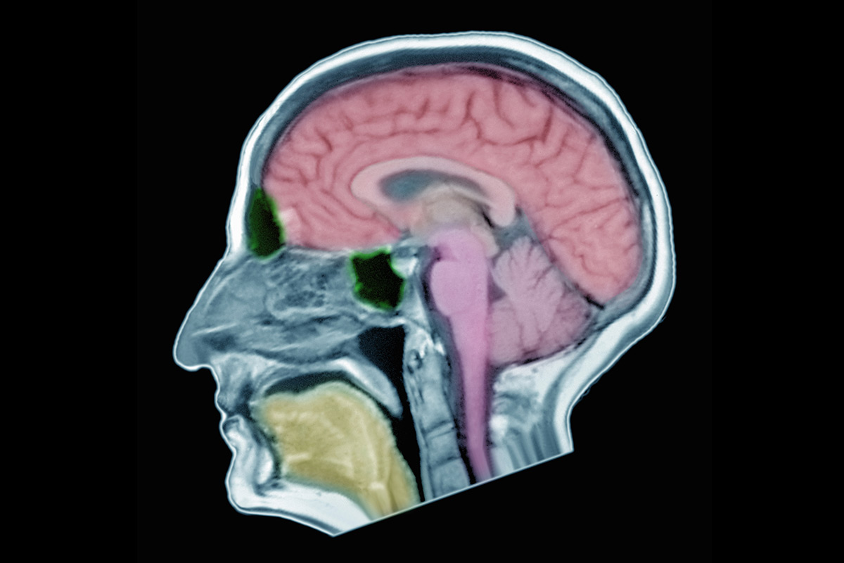
There are three main divisions cerebrum, cerebellum, brain stem. The cerebrum consists of two cerebral hemispheres the outer layer called the cortex (gray matter) and the inner layer (white matter). There are four lobes in the cortex, the frontal lobe, parietal lobe, temporal lobe, occipital lobe. The brain is one of the largest ...
How many grams does the brain weigh?
The brain weight is different between men and women; The male brain weighs about 1336 grams, ...
How much does the brain weigh?
The brain weight is different between men and women; The male brain weighs about 1336 grams, and the female comes in at about 1198 grams, ...
What is the Cerebral Cortex?
The cerebral cortex is the outermost layer of the brain that is associated with our highest mental capabilities. The cerebral cortex is primarily constructed of grey matter (neural tissue that is made up of neurons), with between 14 and 16 billion neurons being found here.
Where are the lobes of the cerebral cortex located?
The largest lobes of the cerebral cortex are the frontal lobes. These are located at the front of the brain behind the forehead.
Why do the folds and wrinkles of the cerebral cortex allow for a wider surface area for an increased number of?
The many folds and wrinkles of the cerebral cortex allow for a wider surface area for an increased number of neurons to live there , permitting large amounts of information to be processed.
What causes frontal lobe injury?
Frontal lobe injury symptoms can include one or more of the following: memory issues, personality changes, issues with problem-solving, difficulties with working memory, inattentiveness, emotional deficiencies, socially inappropriate behavior, behavioral changes, aphasia, weakness, and paralysis. Common causes of damage to this area of the cortex include traumatic brain injuries or neurogenerative diseases such as dementia. A literature review investigated the frontal lobe’s association with schizophrenia and found that many patients had differences in grey matter volumes and functional activity in their frontal lobes, compared to those without the disorder (Mubarik & Tohid, 2016).
What is the thickness of the cerebral cortex?
Although the cerebral cortex is only a few millimeters in thickness, it consists of approximately half the weight of the total brain mass. The cerebral cortex has a wrinkled appearance, consisting of bulges, also known as gyri, and deep furrows, known as sulci. The many folds and wrinkles of the cerebral cortex allow for a wider surface area ...
Which lobes of the brain are located between the frontal and occipital lobes?
Parietal Lobes. The parietal lobes of the cerebral cortex are situated between the frontal and occipital lobes, above the temporal lobes. This region is especially important for integrating the body’s sensory information, so we can build a picture of the world around us.
Which lobes of the brain are controlled by the gyri?
These lobes are called the frontal lobes, temporal lobes, parietal lobes, and occipital lobes.

Structure of The Cerebral Cortex
- Because of the presence of a large number of sulci, only about one third of the total area of cerebral cortex is seen on the surface of the brain. The total area of the cerebral cortex has been estimated to be about 2000 cm2. Like other masses of grey matter the cerebral cortex contains the cell bodies of an innumerable number of neurons along with...
Neurons in The Cerebral Cortex
- Cortical neurons vary in size, in the shape of their cell bodies, and in the lengths, branching patterns and orientation of their processes. Some of these are described below (Fig. 15.1). 1. The most abundant type of cortical neurons are the pyramidal cells. (In contrast all other neurons in the cortex are referred to as non-pyramidal neurons). About two thirds of all cortical neurons ar…
Laminae of Cerebral Cortex
- On the basis of light microscopic preparations stained by methods in which the cell bodies are displayed (e.g., Nissl method) and those where myelinated fibres are stained (e.g., Weigert method) the cerebral cortex is described as having six layers or laminae (Fig. 15.2). From the superficial surface downwards these laminae are as follows. 1. Plexiform or molecular layer.2. E…