What is transitional epithelium?
January 12, 2017. Transitional epithelium is a stratified tissue made of multiple cell layers, where the cells constituting the tissue can change shape depending on the distention in the organ. When the organ is filled with fluid, cells on the topmost layer of this epithelium can stretch and appear flattened.
What is the shape of transitional epithelium in bladder?
The bladder for example has a need for great distension. The appearance of transitional epithelium differs according to its cell layer. Cells of the basal layer are cuboidal (cube-shaped), or columnar (column-shaped), while the cells of the superficial layer vary in appearance depending on the degree of distension.
Why do transitional epithelium cells stretch?
The transitional epithelium cells stretch readily in order to accommodate fluctuation of volume of the liquid in an organ (the distal part of the urethra becomes non-keratinized stratified squamous epithelium in females; the part that lines the bottom of the tissue is called the basement membrane ).
What is the structure and function of transitional epithelium?
Transitional epithelium: A transitional epithelium (also known as urothelium) is made up of several layers of cells that become flattened when stretched. It lines most of your urinary tract and allows your bladder to expand.
What is the structure of transitional?
Structure of Transitional Epithelium Transitional epithelia are made of 3-4 layers of cells with the lowermost or basal layer staying in contact with the basement membrane. The cells of this basal layer are attached to the lamina propria through tonofilaments and hemi-desmosomes.
What is the shape of transitional epithelium?
Transitional epithelium is a stratified tissue in which the cells are all have a fairly round shape when the organ it lines is not distended (stretched out).
What structures are lined by transitional epithelium?
Where is transitional epithelium found? Transitional epithelium is found lining the structures of the urinary system. These structures include the ureters (transport urine from kidneys to bladder), urinary bladder (holds urine), and urethra (transports urine form the bladder to outside the body).
Which description is best for transitional epithelium?
Which description is best for transitional epithelium? Cells that are more cuboidal in a relaxed state but more squamous when stretched.
How do you identify transitional epithelium in histology?
1:136:09Transitional Epithelium - YouTubeYouTubeStart of suggested clipEnd of suggested clipSo you can see the flat nuclei in the umbrella shaped cells or the dome shaped cells on the surfaceMoreSo you can see the flat nuclei in the umbrella shaped cells or the dome shaped cells on the surface view of the transitional epithelium.
Why is it called transitional epithelium?
The bladder is lined by epithelial cells that are somewhere in between the thick layers of squamous cells and the single layer of tall cells of glandular epithelia. Logically, these cells are called transitional cells because they represent a transition between these two disparate epithelial cell types.
What is a unique feature of transitional epithelium quizlet?
Transitional epithelium is unique in that it is composed of differing cell shapes in a stratified or layered, epithelial sheet. Bone-destroying cells are called osteoblasts.
Where are transitional epithelial cells found?
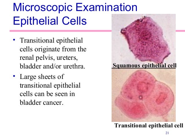
Transitional epithelial cells can occur in spherical, polyhedral, and caudate (bottom image) forms. They originate from the bladder, ureters, and renal pelvis.
Where is transitional epithelium found in the body quizlet?
Transitional epithelium is found in the urinary system. It lines the ureters, bladder, and proximal part of the urethra--organs that are subjected to distention or stretching as urine passes through or fills them.
How is transitional epithelium adapted for its function?
To help achieve this, the cells of transitional epithelium are connected by tight junctions, or virtually impenetrable junctions that seal together to the cellular membranes of neighboring cells. This barrier prevents re-absorption of toxic wastes and pathogens by the bloodstream.
How does transitional epithelium differ from stratified squamous?
However there are two main differences between these two epithelia. With regard to the epithelial cells themselves, the stretched transitional epithelium has considerably fewer cell layers than the stratified squamous epithelium and the immediately underlying cells tend to be cuboidal rather than squamous.
What is a transitional piece?
A sheet metal device shaped to form a transition from one shape of duct to different shape or size of duct.
What does transitional mean in fashion?
Transitional wear is typically used to describe clothing that can interchange between seasons and carry you through that time of year when Mother Nature is being most fickle.
What is the main function of transitional epithelium?
The main function of transitional epithelium is to allow tissue to expand and contract. For example, the transitional epithelium lines the inner wa...
What are the features of transitional epithelium?
There are several features of transitional epithelium. This tissue has several layers of cells and the cells contain gap junctions and microvilli....
Where is transitional epithelium found?
Transitional epithelium is found lining the structures of the urinary system. These structures include the ureters (transport urine from kidneys to...
Why do transitional epithelium cells stretch?
The transitional epithelium cells stretch readily in order to accommodate fluctuation of volume of the liquid in an organ (the distal part of the urethra becomes non-keratinized stratified squamous epithelium in females; the part that lines the bottom of the tissue is called the basement membrane ).
What are the layers of the epithelium?
Transitional epithelium is made up of three types of cell layers: basal, intermediate, and superficial. The basal layer fosters the epithelial stem cells in order to provide constant renewal of the epithelium. These cells' cytoplasm is rich in tonofilaments and mitochondria; however, they contain few rough endoplasmic reticulum. The tonofilaments play a role in the attachment of the basal layer to the basement membrane via desmosomes. The intermediate cell layer is highly proliferative and, therefore, provides for rapid cell regeneration in response to injury or infection of the organ or tube in which it resides. These cells contain a prominent Golgi apparatus and an array of membrane-bound vesicles. These function in the packaging and transport of proteins, such as keratin, to the superficial cell layer. The cells of the superficial cell layer that lines the lumen are known as facet cells or umbrella cells. This layer is the only fully differentiated layer of the epithelium. It provides an impenetrable barrier between the lumen and the bloodstream, so as not to allow the bloodstream to reabsorb harmful wastes or pathogens. All transitional epithelial cells are covered in microvilli and a fibrillar mucous coat.
How does transitional cell carcinoma spread?
It can spread to the tissues and fat surrounding the kidney, the fat surrounding the ureter , or, more progressively, lymph nodes and other organs, including bone. Common risk factors of transitional cell carcinoma include long-term misuse of pain medication, smoking, and exposure to chemicals used in the making of leather, plastic, textiles, and rubber.
Why is the transitional epithelium impermeable to water?
Because of its importance in acting as an osmotic barrier between the contents of the urinary tract and the surrounding organs and tissues, transitional epithelium is relatively impermeable to water and salts. This impermeability is due to a highly keratinized cellular membrane synthesized in the Golgi apparatus. The membrane is made up of a hexagonal lattice put together in the Golgi apparatus and implanted into the surface of the cell by reverse pinocytosis, a type of exocytosis. The cells in the superficial layer of the transitional epithelium are highly differentiated, allowing for maintenance of this barrier membrane. The basal layer of the epithelium is much less differentiated; however, it does act as a replacement source for more superficial layer. While the Golgi complex is much less prominent in the cells of the basal layer, these cells are rich in cytoplasmic proteins that bundle together to form tonofibrils. These tonofibrils converge at hemidesmosomes to attach the cells at the basement membrane.
How to treat transitional cell carcinoma?
Transitional cell carcinoma patients have a variety of treatment options. These include nephroureterectomy, or the removal of kidney, ureter, and bladder cuff, and segmental resection of the ureter. This is an option only when the cancer is superficial and infects only the bottom third of the ureter. The procedure entails removing the segment of cancerous ureter and reattaching the end. Patients with advanced bladder cancer or disease, also often look to bladder reconstruction as a treatment. Current methods of bladder reconstruction include the use of gastrointestinal tissue. However, while this method is effective in improving the function of the bladder, it can actually increases the risk of cancer, and can cause other complications, such as infections, urinary stones, and electrolyte imbalance. Therefore, other methods loom in the future. For example, current research paves the way for use of pluripotent stem cells to derive urothelium, as they are highly and indefinitely proliferative in vitro (i.e. outside of the body).
What is the transitional epithelium of the urinary bladder?
Transitional epithelium of the urinary bladder, known as urothelium. The rounded surface of the apical cells is a distinguishing characteristic of this type of epithelium. Transitional epithelium animation, highlighting the epithelial layer, then underlying connective tissue.
What is the role of tonofilaments in the cell cycle?
The tonofilaments play a role in the attachment of the basal layer to the basement membrane via desmosomes. The intermediate cell layer is highly proliferative and, therefore, provides for rapid cell regeneration in response to injury or infection of the organ or tube in which it resides.
What is the transitional epithelium?
Transitional epithelium is a type of layered epithelium that consists of several layers of cells, with the shape of the cell changing depending on the function of the organ. The epithelium is variable and appears cubic or round when relaxed, except for the apical layer, which appears to flatten when stretched. This epithelium is almost exclusively limited to the urinary tract, which is why it is also called “urothelium”.
Where is the epithelium located?
This epithelium is found in the bladder, ureters, and urethra, as well as in the ducts of the prostate.
How are cells connected to the epithelium?
All cells of this epithelium are deeply connected by connecting complexes. Junction complexes are symmetrical connections between two cells, usually made up of three components: a band of tight junctions on the apical surface, followed by an intermediate series of adherent junctions and desmosomes on the base. These multiprotein complexes hold the epithelial cells together and form an uninterrupted surface in the lumen of the organ.
Why is the bladder a permeability barrier?
Because of its location in the excretory system, particularly the ureters and bladder , one of the main functions of this tissue is to be a highly effective permeability barrier that is impermeable to water and most small molecules. The cells of this tissue are probably among the most resistant to osmotic pressure. Urine is hypertonic with a much higher concentration of many solutes than the cytoplasm of epithelial cells. However, these cells are protected from dehydration even when the epithelium is fully expanded. Toxic waste is also prevented from getting back into the bloodstream.
What is a simple transitional cell?
Simple transitional cell hyperplasia is a sessile or uniformly thickened urothelium lining the pelvis without any prominent outward or inward growth ( Figure 10.12a ). Papillary transitional hyperplasia represents an exophytic proliferation often with a fibrovascular core whereas nodular transitional hyperplasia is a solid nodule-like growth projecting into the lumen or into the underlying renal parenchyma. In atypical hyperplasia there is disturbance of the normal regular urothelial growth pattern, nuclear hyperchromasia and cellular pleomorphism ( Figure 10.12b ).
Which epithelium is responsible for ectopic osteogenesis?
Transitional epithelium of the urinary bladder can induce ectopic osteogenesis from inducible osteogenic progenitor cells from a variety of tissues in postnatal mammals (see preceding text), demonstrating that epithelial–connective tissue interactions can elicit skeletogenesis outside the normal bounds of the skeleton from cells that would normally become neither chondroblasts nor osteoblasts. The site of the epithelium dictates the location of the ectopic skeletal nodule.
What is the epithelium of the urinary bladder?
The urinary bladder has a transitional epithelium, which appears smooth on a normal cystogram. The urinary bladder epithelium is capable of metaplastic, neoplastic, and nonneoplastic proliferation.38 Mucosal proliferation appears as an irregular outline along the inside urinary bladder surface and may be accentuated with inadequate urinary bladder distention. The irregular mucosa is usually focally distributed but may be diffuse; it may vary in severity from a slightly irregular brush-type surface to a severe cobblestone appearance ( Fig. 42.16 ). Mild mucosal irregularity may be obliterated on a cystogram if the urinary bladder is maximally distended. 36 Ulcers may be present with mucosal proliferation and can be identified where contrast medium adheres to the mucosal surface.
What are the effects of irritants on the respiratory epithelium?
A common superficial, and often reversible, effect of irritants on respiratory epithelium involves attenuation and/or loss of cilia along the luminal surface in the proximal nasal cavity. This effect was frequently seen in mice and rats exposed to chlorine gas (Jiang et al. 1986) and was a common alteration observed in monkeys exposed to 0.15 and 0.30 ppm ozone for 6 or 90 days (8 h day −1) ( Harkema et al. 1987c ). Ciliated cell necrosis, mucous cell hyperplasia, and inflammatory cell influx (after 6 days of exposure only) were additional features in the nasal respiratory epithelium of monkeys exposed to ozone.
What are the tissues that are independent of the inducer?
Skeletal tissues that form part of the normal skeleton become independent of the action of the inducer soon after induction. This has been established for the apical ectodermal ridge (AER) and limb chondrogenesis, notochord/spinal cord and somitic chondrogenesis, mandibular epithelium and mandibular osteogenesis, otic capsular cartilages and otic epithelium, and so forth. Formation of epithelially induced ectopic bone, however, continues only while the epithelium is present. This is also the case for somitic cartilage induced in vitro ( Chapter 41 ), and may be a general feature of induced skeletal tissues; see Keilisborok et al. (1982) for induced bone, and Hall (*1977a) for induced cartilage.
What is ectopic bone?
Ectopic bone forms in the bladder in situ; when transplanted subcutaneously, pelvic transitional epithelium of the pelvis induces ectopic bone ( Tavassoli and Crosby, 1971 ). Tumours of the urinary bladder evoke ectopic osteoid, bone and cartilage, and cells within some tumours contain or acquire skeletogenic ability themselves. Meningiomas are one example ( Ball et al., 1975 ). Skeletogenesis in association with breast and prostate cancers is discussed in Box 23.2.
Does the transitional epithelium repair bones?
Transitional epithelium can act on osteogenic progenitor cells located intra skeletally, as demonstrated in two studies on repair of fractured bones. Pieces of bladder dispersed in polyurethane sponge or in protein-free bone matrix accelerate repair of fractured cranial bones in guinea pigs and rats, while urinary bladder mucosa accelerates fracture repair in dogs ( Beresford and Hancox, 1967; Gilbert and Gorman, 1971 ). Whether the epithelium commits more cells to osteogenesis or accelerates the rate of bone formation of already committed cells is unclear. Friedenstein’s work suggests the latter.
What is the epithelium?
The epithelium is a type of body tissue that forms the covering on all internal and external surfaces of your body, lines body cavities and hollow organs and is the major tissue in glands . Epithelial tissue has a variety of functions depending on where it’s located in your body, including protection, secretion and absorption.
What are epithelial cells?
Epithelial tissue is made up of epithelial cells. The cells can be different shapes and can be arranged in a single layer or multiple layers depending on where they are in your body and what kind of functions they have.
What is the difference between epithelium, endothelium and mesothelium?
Epithelium, endothelium and mesothelium are three types of epithelial cell layers that line your internal organs, body cavities and form the outer layer of your skin.
What are the different kinds of epithelial cell tests?
Since epithelial cells exist in several important parts of your body, several types of tests examine epithelial cells to check for certain medical conditions. In medicine, pathology is the laboratory examination of cells in samples of body tissue or fluids for diagnostic purposes. A scientist called a pathologist examines the cells.
Structure of The Transitional Epithelium
- Transitional epithelium is an epithelial tissue that, when at rest, resembles stratified cuboidal epithelium.
- The pear-or round-shaped cells of the transitional epithelium flatten down as the tissue is stretched, generating the look of stratified squamous epithelium.
- According to the extent of extension, the cells in the apical layer flatten, whereas cells in the …
- Transitional epithelium is an epithelial tissue that, when at rest, resembles stratified cuboidal epithelium.
- The pear-or round-shaped cells of the transitional epithelium flatten down as the tissue is stretched, generating the look of stratified squamous epithelium.
- According to the extent of extension, the cells in the apical layer flatten, whereas cells in the base layer have a cuboidal or columnar appearance.
- Three groups of cells make up the layers of the epithelium.
Functions of The Transitional Epithelium
- The transitional epithelium serves two basic purposes, which are determined by the composition and structure of the cell: 1. Permeability barrier 1. The tissue offers high impermeability to water and other molecules because of the significant keratin deposits present in the cells. 2. Even when the cells are fully stretched, desiccation is prevented by the tissue cells’ great resistance to osm…
Location and Examples
- The urothelium is the most well-known type of transitional epithelium.
- The transitional epithelium, also known as urothelium, lines the urethra, ureters, and urine bladder.
- Similar to this, the transitional epithelium that lines the prostatic urethra in the male reproductive system is likewise continuous with the urothelium of the urine bladder.
References and Sources
- Mescher AL (2016). Basic Histology. Fourteenth Edition. McGraw-Hill Education.
- Tortora GJ and Derrickson B (2017). Principles of Physiology and Anatomy. Fifteenth Edition. John Wiley & Sons, Inc.
- Waugh A and Grant A. (2004) Anatomy and Physiology. Ninth Edition. Churchill Livingstone.
- 4% – https://microbenotes.com/simple-squamous-epithelium/
Transitional Epithelium Definition
- Transitional epitheliumis a type of stratified epithelium consisting of multiple layers of cells where the shape of the cell changes according to the function of the organ. The epithelium has a varying appearance as they appear cubical or round when in a relaxed state, except the apical layer which seems to be flattened when stretched. This epithelium is almost limited to the urinary system, w…
Structure of The Transitional Epithelium
- Transitional epithelium is an epithelial tissue which in a relaxed state appears as a stratified cuboidal epithelium.
- The cells in the transitional epithelium are pear-shaped or round, but as tissue is stretched, cells become flattened, giving the appearance of stratified squamous epithelium.
- The cells in the basal layer appear cuboidal or columnar, but the cells in the apical layer beco…
- Transitional epithelium is an epithelial tissue which in a relaxed state appears as a stratified cuboidal epithelium.
- The cells in the transitional epithelium are pear-shaped or round, but as tissue is stretched, cells become flattened, giving the appearance of stratified squamous epithelium.
- The cells in the basal layer appear cuboidal or columnar, but the cells in the apical layer become flattened depending on the degree of extension.
- The layers of cells in the epithelium are divided into three groups.
Functions of The Transitional Epithelium
- Based on the structure of the cell and its composition, the transitional epithelium performs two main functions, which are:
Location and Examples
- The most prominent example of transitional epithelium is the urothelium.
- As the urothelium, the transitional epithelium lines the urinary bladder, ureters, and parts of the urethra.
- Similarly, the lining of the prostatic urethra in the male reproductive system is also lined with the transitional epithelium, which is continuous with the urothelium of the urinary bladder.
References and Sources
- Mescher AL (2016). Basic Histology. Fourteenth Edition. McGraw-Hill Education.
- Tortora GJ and Derrickson B (2017). Principles of Physiology and Anatomy. Fifteenth Edition. John Wiley & Sons, Inc.
- Waugh A and Grant A. (2004) Anatomy and Physiology. Ninth Edition. Churchill Livingstone.
- 4% – https://microbenotes.com/simple-squamous-epithelium/
Overview
Transitional epithelium also known as urothelium is a type of stratified epithelium. Transitional epithelium is a type of tissue that changes shape in response to stretching (stretchable epithelium). The transitional epithelium usually appears cuboidal when relaxed and squamous when stretched. This tissue consists of multiple layers of epithelial cells which can contract and exp…
Structure
The appearance of transitional epithelium differs according to its cell layer. Cells of the basal layer are cuboidal (cube-shaped), or columnar (column-shaped), while the cells of the superficial layer vary in appearance depending on the degree of distension. These cells appear to be cuboidal with a domed apex when the organ or the tube in which they reside is not stretched. When the organ or tube is stretched (such as when the bladder is filled with urine), the tissue compresses …
Function
The transitional epithelium cells stretch readily in order to accommodate fluctuation of volume of the liquid in an organ (the distal part of the urethra becomes non-keratinized stratified squamous epithelium in females; the part that lines the bottom of the tissue is called the basement membrane). Transitional epithelium also functions as a barrier between the lumen, or inside hollow space of the tract that it lines and the bloodstream. To help achieve this, the cells of tran…
Clinical significance
Urothelium is susceptible to carcinoma. Because the bladder is in contact with urine for extended periods, chemicals that become concentrated in the urine can cause bladder cancer. For example, cigarette smoking leads to the concentration of carcinogens in the urine and is a leading cause of bladder cancer. Aristolochic acid, a compound found in plants of the family Aristolochiaceae, also causes DNA mutations and is a cause of liver, urothelial and bladder cancers. Occupational expo…
Gallery
• Types of epithelium
• Schematic view of transitional epithelium
• Vertical section of bladder wall.
• Transverse section of ureter.
External links
• Histology at utmb.edu
• Histology image: 36_02 at the University of Oklahoma Health Sciences Center - "ureter"
• Histology image: 37_02 at the University of Oklahoma Health Sciences Center - "urinary bladder"