Terms in this set (3)
- 1. T tubules propagate the action potential from the plasma membrane into the interior of the muscle cell via voltage-gated Na and K channels
- 2. An action potential carried by a T tubule regulates the opening and closing of Calcium channels in the sarcoplasmic reticulum. The resulting change in the cytosolic Calcium concentration triggers the contraction of the myofibrils
Full Answer
What is the role of T-tubules in muscle contraction?
During muscle contraction, T-tubules allow the depolarization impulse to rapidly propagate through the interior of the muscle fiber. This ensures that calcium release from the sarcoplasmic reticulum occurs uniformly throughout the fiber allowing for synchronized contraction of myofibrils in each muscle cell.
What is the function of the T-tubule in the heart?
T-tubule. With membranes that contain large concentrations of ion channels, transporters, and pumps, T-tubules permit rapid transmission of the action potential into the cell, and also play an important role in regulating cellular calcium concentration. Through these mechanisms, T-tubules allow heart muscle cells to contract more forcefully by...
What is the structure of T tubules in skeletal muscle?
Structure. In skeletal muscle cells, T-tubules are between 20 and 40 nm in diameter and are typically located either side of the myosin strip, at the junction of overlap between the A and I bands. T-tubules in skeletal muscle are associated with two terminal cisternae, known as a triad.
What is the function of T-tubules in sarcolemma?
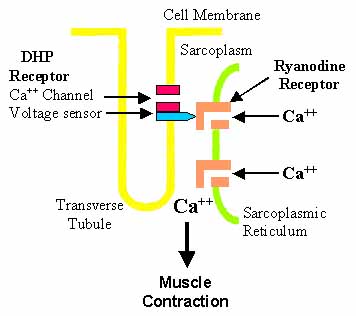
T-tubules contain a higher concentration of L-type calcium channels than the rest of the sarcolemma and therefore the majority of the calcium that enters the cell occurs via T-tubules. This calcium binds to and activates a receptor, known as a ryanodine receptor, located on the cell's own internal calcium store, the sarcoplasmic reticulum.

Why is there no change in ICa density?
This can be due to a number of factors, including upregulation of the remaining channels, consistent with studies showing an increase in the activity of single channels from failing myocytes. This upregulation may result from channel phosphorylation, which may partly explain the blunted response of failing hearts to β-adrenergic stimulation. The ability of ICa to trigger CICR from the SR is reduced in myocytes from failing rat hearts ( Gomez et al., 1997 ). Possible changes in the colocalization of LTCC with RyR 2, increased physical separation of the T-tubule from the SR, and T-tubule remodeling processes have been suggested as underlying causes for the lack of LTCC/RyR2 integration. A porcine postischemic cardiomyopathy model suggests that T-tubule dysfunction in the failing heart is related to contractile abnormalities ( Heinzel et al., 2008 ). In this model, impaired contractility was associated with reduced Ca 2 + release synchronicity, a longer time to reach peak Ca 2 + release, and a lower peak concentration of Ca 2 + ( Heinzel et al., 2008 ). Furthermore, the authors observed a significant reduction in T-tubule density, while the LTCC current and SR Ca 2 + content remained unaffected ( Heinzel et al., 2008 ). From their data, the authors proposed that the dysfunction was due to a gain in the CICR process, implicating T-tubule structural disorganization in the pathophysiology of the failing myocyte. Follow-up studies also demonstrated that failing myocytes have increased Ca 2 + spark frequency, consistent with an uncoupling of the Ca 2 + release machinery and disruption of the T-tubules. It has also been argued that such a mechanism would produce only transient/short-lived changes in the size of the Ca 2 + transient and that it is more likely that a decrease in SR Ca 2 + content underlies the decrease in Ca 2 + transients. However, a reduction in T-tubule density could desynchronize Ca 2 + following electric stimulation, reducing peak Ca 2 + and slowing the time course. Although the loss of T-tubules cannot be completely responsible for the changes in Ca 2 + handling observed in heart failure, it is important to note that many of the functional changes exhibited in heart failure are observed in detubulation experiments, suggesting that the loss of T-tubules in heart failure may contribute to or exacerbate the phenotype observed in models of heart failure.
What is the role of T tubules in muscle relaxation?
During muscle relaxation, ATPase sends some Ca++ back to the sarcoplasmic reticulum. Stimulation of the sympathetic heart nerves (b-adrenoceptor) causes an increased contraction and an increase in the second messenger cAMP. ATP makes cAMP and creates pathways to assist certain hormones such as adrenaline and glucose, which normally have trouble entering cells, to enter and activate energy within a cell. Phosphodiesterase is an enzyme in cAMP, which degrades signaling effects over time. Caffeine and other stimulants can prolong the effect of cAMP.
What are junctional domains of the SR?
The junctional domains of the SR (jSR, oblique arrows) are wide, contain calsequestrin and directly face the T-tubules. The triads are the sites of calcium release during muscle activation and thus they are also called calcium release units (CRUs).
Where do T tubules extend?
T tubules extend from the free surface throughout the cells, in a generally transverse direction, coursing around the myofibrils at the levels of the Z bands;
What is the SR in a frog muscle?
The SR is composed of elements that repeat longitudinally with a period equal to that of the sarcomere. In the case of frog muscle, shown in Figure 53.2, for example, the SR shows a network of longitudinal elements opposite the A band, connected to the two enlarged cisternae on either side of the Z-line. The T-tubules are located in the space between the two SR cisternae ( Figure 53.2B) and the assembly of two SR and one T-tubule is called a triad. The SR, like the ER, is a totally internal membrane system that creates a segregated space: its lumen is not connected to either the cytoplasm or the extracellular space.
What is a T tubule?
T-tubules Are Surface Invaginations; the SR Is an Internal Membrane System. T-tubules have a random, mostly longitudinal, disposition between the myofibrils during their initial formation but even at this stage they immediately form junctions with SR elements.
What are transverse tubules?
Transverse tubules, or T-tubules, invaginations of the sarcolemma in ventricular, but not at rial, myocardial cells have been mentioned in the discussion of myocardial performance. They allow transmission of the action potential, with its attendant ion shifts, to all parts of the cell, which allows rapid activation of the entire cell. T-tubules are thus a required component of larger cells ( Gotoh, 1983 ). Maturation of T-tubules is associated with the increased size of mature myocardial cells, with the sarcolemmal calcium channels further from the contractile apparatus. Species that have a relatively mature myocardium at birth have well-developed T-tubules at birth, whereas animals with immature myocardium at birth do not. T-tubules first appear at about 30 weeks gestation in humans ( Kim et al., 1992 ).
Which action potential regulates the opening and closing of Calcium channels in the sarcoplasmic reticulum?
2. An action potential carried by a T tubule regulates the opening and closing of Calcium channels in the sarcoplasmic reticulum. The resulting change in the cytosolic Calcium concentration triggers the contraction of the myofibrils
Which membrane extends deep into the muscle cell?
Invaginations of the muscle cell plasma membrane that extend deep in to the muscle cell and are in close contact with (but not continuous with) the sarcoplasmic reticulum.