Thoracic wall. The thoracic wall or chest wall is the boundary of the thoracic cavity. Body cavities. A transverse section of the thorax, showing the contents of the middle and the posterior mediastinum. [edit on Wikidata] The bony skeletal part of the thoracic wall is the rib cage, and the rest is made up of muscle, skin, and fasciae.
Full Answer

What thoracic wall is lined with?
The thoracic wall consists of a bony framework that is held together by twelve thoracic vertebrae posteriorly which give rise to ribs that encircle the lateral and anterior thoracic cavity. The first nine ribs curve around the lateral thoracic wall and connect to the manubrium and sternum. Ribs 10-12 are relatively short and attach to the costal margins of the ribs just above them.
What does thoracic wall mean?
The thoracic wall actually encloses a cavity, or space, that is filled with various anatomical structures. Since there are so many of them, the thoracic cavity is divided into several compartments to aid their localization. There is a mediastinum located centrally bordered by two pleural cavities laterally.
What are thoracic landmarks?
- The posterior iliac crest.
- The greater trochanter.
- The groin.
What is the thorax wall?
The thoracic, or chest wall, consists of a skeletal framework, fascia, muscles, and neurovasculature – all connected together to form a strong and protective yet flexible cage. The thorax has two major openings: the superior thoracic aperture found superiorly and the inferior thoracic aperture located inferiorly.

What is the function of the thoracic wall?
Structure and Function The structures of the thoracic wall protect the heart, lungs, and great vessels as well as some abdominal organs. Additionally, the bony structures provide attachment points for muscles and allow for the mechanical function of ventilation.
Is the thoracic wall the same as the chest wall?
The thoracic, or chest wall, consists of a skeletal framework, fascia, muscles, and neurovasculature – all connected together to form a strong and protective yet flexible cage. The thorax has two major openings: the superior thoracic aperture found superiorly and the inferior thoracic aperture located inferiorly.
What does thoracic wall mean?
The thoracic wall or chest wall is the boundary of the thoracic cavity. Thoracic wall. Body cavities. A transverse section of the thorax, showing the contents of the middle and the posterior mediastinum.
What is the thoracic wall made up of?
The thoracic wall is made up of five muscles: the external intercostal muscles, internal intercostal muscles, innermost intercostal muscles, subcostalis, and transversus thoracis. These muscles are primarily responsible for changing the volume of the thoracic cavity during respiration.
Where is thoracic wall located?
The thoracic wall consists of a bony framework that is held together by twelve thoracic vertebrae posteriorly which give rise to ribs that encircle the lateral and anterior thoracic cavity. The first nine ribs curve around the lateral thoracic wall and connect to the manubrium and sternum.
What does a chest wall tumor feel like?
Symptoms of a Chest Wall Tumor Pain or soreness in the chest area. Swelling. Impaired movement. A lump or bump protruding from the chest.
What organs are located in the thoracic cavity?
[2] The thoracic cavity contains organs and tissues that function in the respiratory (lungs, bronchi, trachea, pleura), cardiovascular (heart, pericardium, great vessels, lymphatics), nervous (vagus nerve, sympathetic chain, phrenic nerve, recurrent laryngeal nerve), immune (thymus) and digestive (esophagus) systems.
Is the chest wall your ribs?
The bones in the chest wall include the ribs, sternum (breastbone), and spine. The chest wall also helps support breathing and movement of the upper arms and shoulders.
What helps chest wall pain?
Treatment for chest wall pain depends on the cause of the pain. Minor chest wall pain is treated with rest, ice or heat applied to the area, and non-steroidal anti-inflammatory drugs (NSAIDs) or acetaminophen. If the chest wall pain is the result of coughing, the pain should improve as the cough improves.
How the thoracic wall is formed?
The thoracic cage is formed by the 12 pairs of ribs with their costal cartilages and the sternum. The ribs are attached posteriorly to the 12 thoracic vertebrae and most are anchored anteriorly either directly or indirectly to the sternum. The thoracic cage functions to protect the heart and lungs.
Which arteries supply blood to the thoracic wall?
The thoracic wall is richly supplied with blood arising from three sources. These are the thoracic aorta, subclavian artery and axillary artery.
Is the heart in the thoracic cavity?
Overview. The heart and lungs are located in the thorax, or chest cavity.
Where is the intercostal chest wall?
The intercostal muscles are the muscles between the ribs. During breathing, these muscles normally tighten and pull the rib cage up. Your chest expands and the lungs fill with air.
What are the boundaries of the thoracic wall?
The boundaries of the Thoracic Cavity are the Ribs (and Sternum), Vertebral Column, and the Diaphragm. The Diaphragm seperates the Thoracic Cavity from the Abdominal Cavity.
What is the anatomy of the chest?
thoracic cavity, also called chest cavity, the second largest hollow space of the body. It is enclosed by the ribs, the vertebral column, and the sternum, or breastbone, and is separated from the abdominal cavity (the body's largest hollow space) by a muscular and membranous partition, the diaphragm.
What is a chest wall injury?
What is a chest wall injury? Injuries to the chest wall include fractured ribs, fractured sternum (breastbone) and bruising to the lungs. They normally occur following a high impact trauma such as falling from a height, a road traffic accident or high impact sports. Signs and symptoms.
What is the thoracic wall?
Thoracic wall. The first step in understanding thorax anatomy is to find out its boundaries. The thoracic, or chest wall, consists of a skeletal framework, fascia, muscles, and neurovasculature – all connected together to form a strong and protective yet flexible cage.
Which muscles are involved in the thoracic wall?
These include the transversus thoracis, subcostals, levatores costarum, serratus posterior superior, and serratus posterior inferior muscles. Broadly speaking, they attach to the ribs, their cartilages, or thoracic vertebrae–ultimately depressing or elevating the ribs. In addition, all of the thoracic muscles provide further support and strength for the thorax.
What are the arteries that supply breasts?
Anatomy of the female breast (lateral view) They are supplied by several arteries of the thoracic wall, namely branches of the internal thoracic, axillary, lateral thoracic, thoracoacromial, and posterior intercostal arteries.
What is the chest?
The chest, properly called the thorax, is the superior part of the trunk located between the neck and abdomen. It consists of several components: 1 Thoracic wall 2 Several cavities 3 Neurovasculature and lymphatics 4 Internal organs 5 Breasts
How does the thoracic cavity communicate with the neck?
The thoracic cavity communicates with the neck via the superior thoracic aperture and with the abdominal cavity via the inferior thoracic aperture through anatomical spaces piercing the diaphragm. Cavities of the body Explore study unit.
What is the inferior thoracic aperture?
The inferior thoracic aperture is almost completely covered by the diaphragm, separating it from the abdominal cavity. Moving forward with the skeletal scaffold of the thorax, we have the thoracic skeleton. It is made up of the sternum, twelve pairs of ribs, twelve thoracic vertebrae, and interconnecting joints.
What muscles support the thorax?
In addition, all of the thoracic muscles provide further support and strength for the thorax. If you want to learn more about the muscles of the thoracic wall and get one step closer to mastering chest anatomy, take a look at our muscle anatomy charts!
What is the most superior portion of the sternum?
The most superior portion of the sternum is the manubrium , and it is also the first to form during embryogenesis. The sternal body and xiphoid process soon follow the manubrium in development. Anatomically, the manubrium is located at the level of thoracic vertebral bodies T3 and T4. The manubrium is also the widest and thickest segment of the sternum. During a physical exam of the chest, one noticeable feature of the manubrium is the presence of the suprasternal notch. On either side of this notch, one will feel the thick attachment from the clavicles. For access to the superior mediastinum, suprasternal goiter or thymus, some thoracic surgeons will only make a midline incision in the manubrium .
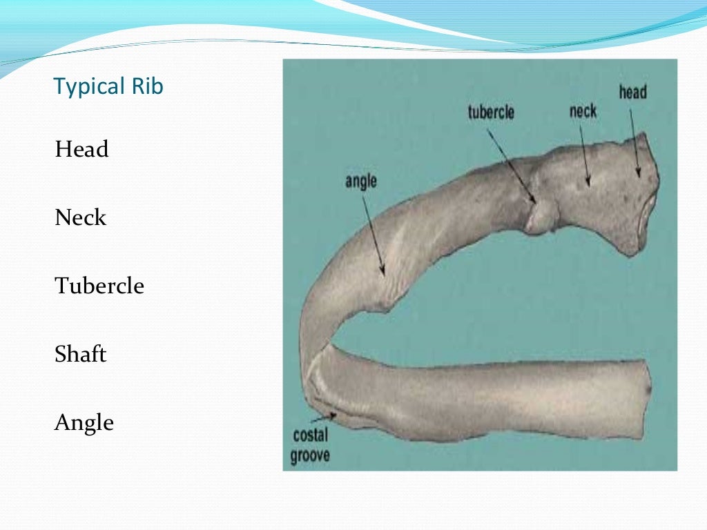
What are the ribs attached to?
The first seven ribs are termed true ribs and attach to the manubrium and directly attach to the body of the sternum. Ribs eight to ten only attach to the inferior part of sternum via the costal cartilages. Ribs 11-12 are termed floating ribs because they do not attach directly to the sternum. Ribs eight to ten are known as false ribs because they lack direct attachment to the sternum. At the level of the spine, the ribs articulate with the costal facet of two opposing vertebrae. An articular capsule surrounds the head of each rib, and the attachment to the transverse process is made with the help of the radiate ligament. Once the ribs leave the vertebrae, they gently curve around the lateral thoracic wall and approach the anterior wall of the thoracic cavity. [1]
How many ribs are in the thoracic wall?
The thoracic wall consists of a bony framework that is held together by twelve thoracic vertebrae posteriorly which give rise to ribs that encircle the lateral and anterior thoracic cavity. The first nine ribs curve around the lateral thoracic wall and connect to the manubrium and sternum. Ribs 10-12 are relatively short and attach to the costal margins of the ribs just above them. Ribs 10-12, due to their short course, they do not reach the sternum.
Why is the sternum used for bone marrow aspiration?
The sternum is a commonly used site for bone marrow aspiration because it possesses hematopoietic marrow throughout life. However, the surgeon needs to exercise great care because if the sternal puncture is improperly executed, the needle can pierce the structures related to the posterior surface of the manubrium such as the left brachiocephalic vein in the upper part and aortic arch in the lower part. [9]
What is the vertical bone of the chest?
The vertical bone of the chest, the sternum , defines the anterior chest wall. The three separate bone segments of different size and shape that make up the sternum include 1) the thick manubrium, 2) long body of the sternum , and 3) the xiphoid process. It develops independently of the ribs. In sporadic cases, the sternum may not fully form, and the underlying heart may be exposed.
How many arteries are there in the intercostal space?
Three arteries supply each intercostal space; the posterior intercostal artery and two branches of anterior intercostal arteries. These intercostal blood vessels run along with the nerves between the internal intercostal muscle and innermost intercostal muscles in the costal groove. They are arranged in order from superior to inferior: vein, artery, and nerve.
Where does the posterior intercostal artery originate?
The posterior intercostal artery for first two intercostal spaces is fed from the superior (supreme) intercostal artery. This artery arises from the costocervical trunk of the subclavian artery. The remaining pair of posterior intercostal arteries from 3rd - 11th intercostal spaces and a pair of subcostal arteries emerge directly descending thoracic aorta. [1]
What is the chest wall?
chest wall. in respiratory physiology, the total system of structures outside the lungs that move as a part of breathing; it includes the rib cage, diaphragm, abdominal wall, and abdominal contents.
What causes TB in the thoracic wall?
TB of the thoracic wall is usually caused by reactivation of some latent foci of primary tuberculosis formed during hematogenous or lymphatic dissemination but direct extension from contiguous mediastinal lymph nodes can also occur (9).
What flap is used to repair thoracic wall defects in dogs?
Repair of thoracic wall defects in the dog with an omental pedicle flap.
What is the meaning of chest wall?
chest wall. ( chest wawl) respiratory physiology the total system of structures outside the lungs that move as a part of breathing; it comprises the rib cage, diaphragm, abdominal wall, and abdominal contents. Synonym (s): thoracic wall. Medical Dictionary for the Health Professions and Nursing © Farlex 2012.
Where do thoracic tumors grow?
Thoracic wall tumors generally grow towards the thoracic cavity and may reach a large size when the diagnosis is made.
Is a metallic foreign body in the thoracic wall rare?
Metallic foreign bodies in the thoracic wall are rare and are usually related to a history of trauma. Metallic Foreign Bodies in the Thoracic Wall in Three Cases. Thoracic wall reconstruction after tumor resection.
What is the most superior portion of the sternum?
The most superior portion of the sternum is the manubrium , and it is also the first to form during embryogenesis. The sternal body and xiphoid process soon follow the manubrium in development. Anatomically, the manubrium is located at the level of thoracic vertebral bodies T3 and T4. The manubrium is also the widest and thickest segment of the sternum. During a physical exam of the chest, one noticeable feature of the manubrium is the presence of the suprasternal notch. On either side of this notch, one will feel the thick attachment from the clavicles. For access to the superior mediastinum, suprasternal goiter or thymus, some thoracic surgeons will only make a midline incision in the manubrium .
How many vertebrae are in the thoracic wall?
The thoracic wall consists of a bony framework that is held together by twelve thoracic vertebrae posteriorly which give rise to ribs that encircle the lateral and anterior thoracic cavity. The first nine ribs curve around the lateral thoracic wall and connect to the manubrium and sternum. Ribs 10-1 …
What are the ribs attached to?
The first seven ribs are termed true ribs and attach to the manubrium and directly attach to the body of the sternum. Ribs eight to ten only attach to the inferior part of sternum via the costal cartilages. Ribs 11-12 are termed floating ribs because they do not attach directly to the sternum. Ribs eight to ten are known as false ribs because they lack direct attachment to the sternum. At the level of the spine, the ribs articulate with the costal facet of two opposing vertebrae. An articular capsule surrounds the head of each rib, and the attachment to the transverse process is made with the help of the radiate ligament. Once the ribs leave the vertebrae, they gently curve around the lateral thoracic wall and approach the anterior wall of the thoracic cavity.
What is the vertical bone of the chest?
The vertical bone of the chest, the sternum , defines the anterior chest wall. The three separate bone segments of different size and shape that make up the sternum include 1) the thick manubrium, 2) long body of the sternum , and 3) the xiphoid process. It develops independently of the ribs. In sporadic cases, the sternum may not fully form, and the underlying heart may be exposed.
Where is the sternal body located?
The sternal body is located at the level of vertebral bodies T5-T9. It covers a significant portion of the mid-chest and is very strong. To access the chest cavity, surgeons usually cut through the sternum with a mechanical saw.
Is the xiphoid process calcified?
The xiphoid may appear bifid, oval or be curved inwards/outwards. In younger individuals, the xiphoid is mostly cartilaginous but is nearly wholly ossified by age 40. By the age of 60 and over, the xiphoid is almost certainly completely calcified. To perform pericardiocentesis safely the needle has to be placed directly underneath the xiphoid because the heart is just a few fingerbreadths below.
What muscles are on each side of the spine?
Let’s start with the two serratus posterior muscles. There’s a serratus posterior superior muscle and serratus posterior inferior muscle on each side. Serratus posterior superior muscles have their proximal attachment at the nuchal ligament and the spinous processes of C7 through T3 vertebrae and their distal attachment at the superior borders ...
What is the intercostal membrane?
Between the ribs and medial to the angles, the internal intercostal muscles are replaced by fascia called the internal intercostal membranes. The inferior internal intercostal muscles are continuous with the internal oblique muscles in the anterior abdominal wall. The internal intercostal muscles mostly play a role during expiration.
What is the thoracic wall?
The thoracic wall surrounds the thoracic cavity, which is the anatomical region where viscera like the heart and lungs can be found. The thoracic wall contains a variety of muscles, where many of the muscles that cover and attach to the thoracic wall have a primary action ...
How many pairs of intercostal muscles are there?
Then there are the 11 pairs of internal intercostal muscles, which run deep to and at right angles to the external intercostals.
Which muscles are continuous anteriorly with the external oblique muscles in the abdominal wall?
The lower external intercostals are continuous anteriorly with the external oblique muscles in the abdominal wall. The external intercostal muscles elevate the ribs during forced inspiration. Then there are the 11 pairs of internal intercostal muscles, which run deep to and at right angles to the external intercostals.
Which muscles are involved in the upper limb?
For example, the pectoralis major and pectoralis minor muscles overlay the thoracic wall but they are primarily involved with movements of the upper limb and their secondary action is to act on the thoracic wall as accessory muscles of respiration.
Where do the external intercostals run?
The lower external intercostals are continuous anteriorly with the external oblique muscles in the abdominal wall.
What is the purpose of the chest wall?
The chest wall’s main job is to surround and protect your vital organs, including your heart, lungs, and liver.
What landmarks help doctors locate thoracic artery?
These landmarks, as well as specific muscles and bones that are identifiable from the exterior, such as your pecs, ribs, clavicle, and sternum, help doctors locate your thoracic artery, aid them in placing a thoracostomy tube, and other similar procedures.
What is the Rockland Thoracic and Vascular Associates?
But at Rockland Thoracic & Vascular Associates, we think about it a lot. Our experts specialize in keeping your chest wall and all its contents healthy and thriving. That’s why we’re taking some time to take a deeper look into the chest wall and give you a glimpse of its daily duties.
What is the chest wall made of?
You can think of it as a box made of muscles, fat, skin, cartilage, and bones. The muscles and flexible cartilage give the chest wall a dynamic quality that allows for expansion when you inhale.
What is the term for fluid build up in the inner chest wall?
Empyema occurs when fluid builds up in your inner chest wall lining called the pleural space. You may have a dry cough, sweating, fever and chills, and chest pain. This condition often stems from lung infections, trauma, surgery, and pneumonia.
Is a tumor in the chest benign?
Tumors in your chest wall are often benign (noncancerous) and may be one of three types:
Can you add chest wall?
On life’s long list of things we take for granted, you can add your chest wall. If you are like most people, you never give a second thought to this structural part of your anatomy that defines your upper torso.
What is the thoracic wall?
The thoracic wall forms part of the axial skeleton and is comprised of bone, muscle, and connective tissue. It develops from the mesoderm where blocks become somites from which the sclerotome separates. The sclerotome is the origin of the vertebrae and transverse elements which begin elongating into ribs during the fifth week of gestation. Ossification begins during the fetal period but will not be complete until adulthood.[4] The myotome gives rise to the muscles of the thoracic wall. The sternum forms from sternal bars that meet in the midline and begin to fuse during the seventh week of gestation. Fusion starts superiorly and ends inferiorly by the tenth week. [5]
What are the considerations for a thoracotomy?
Some essential surgical considerations regarding the thorax pertain to needle decompression and chest tube placement. As discussed, the blood vessels and nerves of the anterior thorax follow the costal groove on the inferior aspect of each rib.[11] Therefore, needle thoracostomy should be completed by piercing over the superior aspect of the rib on the affected side. Several sites have been studied to include the second intercostal space at the midclavicular line, as well as the fourth and fifth intercostal spaces at the anterior axillary line.[18] Similarly, chest tube placement (tube thoracostomy) should be performed in the fourth or fifth intercostal space and should also track above the rib. Chest tubes should be placed between the mid to anterior axillary line.[19] Lastly, while performing a left-sided thoracotomy, providers must remember that the left phrenic nerve runs along the pericardium and should be avoided to reduce the risk of phrenic nerve palsy. [20]
What are the parts of the sternum?
The sternum is composed of 3 parts: the manubrium, body , and xiphoid process.[3] The manubrium is the widest portion and contains the jugular, or sternal notch as well as the clavicular notch. The sternal angle, or angle of Louis, is where the manubrium joins the body of the sternum. The second rib attaches at this point. The xiphoid process is attached inferiorly to the body of the sternum and provides an attachment point for the diaphragm and rectus abdominis, but no ribs.
Which artery supplies the anterior intercostal arteries?
The internal thoracic artery is another branch of the subclavian artery that supplies the anterior intercostal arteries of spaces 1 through 6 before dividing into the superior epigastric and musculophrenic arteries.[8] The musculophrenic artery supplies the anterior intercostal arteries of spaces 7 through 9. The 2 most inferior intercostal spaces are supplied from the posterior intercostal arteries and their collateral branches and do not have anterior intercostal arteries.
Where does the blood supply come from in the intercostal space?
Each intercostal space receives its blood supply from 3 arteries, a posterior intercostal artery and a pair of anterior intercostal arteries. The posterior arteries of the first 2 intercostal spaces are fed from the superior (supreme) intercostal artery which comes off the subclavian artery.[6] The remaining pairs of posterior intercostal arteries and a pair of subcostal arteries emerge directly from the thoracic aorta.[7] These posterior arteries enter the costal groove near the angle of each rib where they travel between the intercostal vein and nerve. The posterior and anterior intercostal arteries anastomose laterally.
Which organ elevates the ribs?
Beyond the diaphragm compressing the abdominal cavity and the external intercostals lifting the ribs, the serratus posterior superior attaches to ribs 2 through 5 and elevates them during inhalation. When the neck is fixed, the scalene helps to elevate the first and second ribs. Likewise, the sternocleidomastoid can assist in raising the sternum.[13]. When needed, the pectoralis minor assists in lifting third, fourth, and fifth ribs.
Where do the intercostal veins run?
The intercostal veins run most superiorly in the costal grooves. There are 11 pairs of posterior intercostal veins and one pair of subcostal veins. Like the posterior arteries, the posterior veins also anastomose with the anterior intercostal veins. Several of the superior posterior intercostal veins merge to form the left and right superior intercostal veins at the level of T3 or T4 which can then empty into the brachiocephalic vein or the superior vena cava (SVC).[9] The remaining posterior intercostal veins end at the azygous vein before emptying into the SVC. The internal thoracic veins accompany the internal thoracic arteries before emptying into the brachiocephalic vein.
What is the pleura of the rib cage?
Human rib cage. Encyclopædia Britannica, Inc. The pleura is a continuous sheet of endothelial, or lining, cells supported by a thin base of loose connective tissue. The membrane is well supplied with blood vessels, nerves, and lymph channels.
What happens when air enters the pleural cavity?
The penetration of air into the pleural cavity from outside, as from a penetrating wound of the chest, or from within, by rupture of dilated alveoli (air sacs of the lung) or of a cyst, will produce a pneumothorax, converting this cavity into a positive pressure chamber and collapsing the lung , which in turn will lead to decreased oxygenation of the venous blood. The collapse may also have a deleterious effect on the heart.
What is the term for fluid accumulation in the pleural cavity?
Subscribe Now. Accumulation of fluid in the pleural cavity is called hydrothorax. If the fluid is bloody, the condition is described as hemothorax; if it contains pus, pyothorax. The accumulation of fluid may or may not be accompanied by air.
How long does it take for Bornholm disease to go away?
That pain is usually increased by respiration and cough, and pain in other muscles is often present. The condition subsides in two to five days but sometimes may take weeks to disappear.
What is the thoracic cavity?
The thoracic cavity also contains the esophagus, the channel through which food is passed from the throat to the stomach. The chest cavity is lined with a serous membrane, which exudes a thin fluid.
What is the name of the condition in which the pleura is irritated?
The collapse may also have a deleterious effect on the heart. Inflammation of the pleura, usually diffuse, affecting one or both sides, is called pleurisy. Two forms are distinguished: (1) simple, dry, or fibrinous pleurisy; and (2) exudative pleurisy, in which the membrane gives off excessive fluid.
Which part of the pleura is closely related to the lungs?
The vessels of the visceral part of the pleura are intimately related with those of the lungs and bronchi; its arteries are branches of the bronchial arteries, and its veins mingle with the pulmonary network of capillaries.