The transverse plane or axial plane (also called the horizontal plane or transaxial plane) is an imaginary plane that divides the body into superior and inferior parts. It is perpendicular to the coronal plane and sagittal plane.
What is the thoracic plane?
Dr Craig Hacking ◉ ◈ and Dr Henry Knipe ◉ ◈ et al. The thoracic plane, also known as the transthoracic plane or the plane of Ludwig is an artificial horizontal plane used to divide the mediastinum into the superior mediastinum and the inferior mediastinum.
What is the anatomy of the transversus thoracis?
Anatomical terms of muscle. The transversus thoracis muscle (/trænzˈvɜːrsəs θəˈreɪsɪs/) lies internal to the thoracic cage, anteriorly. It is a thin plane of muscular and tendinous fibers, situated upon the inner surface of the front wall of the chest. It is in the same layer as the subcostal muscles and the innermost intercostal muscles.
What is the transverse plane also known as?
*The transverse plane is also called the "horizontal" plane. Movements in the transverse plane are rotational-based movements, or movements across the body. *The transverse plane is also called the "horizontal" plane, as movements align with a horizontal direction. What exact movements can be performed in the transverse plane?
What are movements in the transverse plane?
Movements in the transverse plane are rotational-based movements, or movements across the body. *The transverse plane is also called the "horizontal" plane, as movements align with a horizontal direction. What exact movements can be performed in the transverse plane?

What happens at the transverse thoracic plane?
The thoracic plane, also known as the transthoracic plane or the plane of Ludwig is an artificial horizontal plane used to divide the mediastinum into the superior mediastinum and the inferior mediastinum.
What level is the transverse thoracic plane?
T4-T5It is at the level of the T4-T5 intervertebral disc. It marks the level of the transverse thoracic plane which divides the mediastinum into the superior and inferior mediastinum. It overlies the aortic arch on the left and the superior vena cava on the right.
How do you remember the thoracic plane?
A handy mnemonic to remember the structures found at the level of the thoracic plane (also known as the plane of Ludwig) is:CLAPTRAP.RAT PLANT.
Why is it called the transverse plane?
It is perpendicular to the coronal plane and sagittal plane. It is one of the planes of the body used to describe the location of body parts in relation to each other.
What is transverse plane in anatomy?
Axial Plane (Transverse Plane) - A horizontal plane; divides the body or any of its parts into upper and lower parts. Median plane - Sagittal plane through the midline of the body; divides the body or any of its parts into right and left halves.
What does the transverse plane do?
The transverse plane (axial or X-Z plane) divides the body into superior and inferior (head and tail) portions. It is typically a horizontal plane through the center of the body and is parallel to the ground.
Why sternal angle is important?
The sternal angle is the angle formed between the manubrium of the sternum and the body of the sternum (manubriosternal junction), and is an important anatomical landmark. It marks the level of the 2nd pair of costal cartilages which lies at the level of the intervertebral disc between thoracic vertebrae 4 and 5.
Why is sternal angle called angle of Louis?
History. The sternal angle is also called the angle of Louis, but the reason for that name was lost. Once thought to be after Antoine Louis or Wilhelm Friedrich von Ludwig, it is now believed to be after Pierre Charles Alexandre Louis.
Where is thoracic duct?
The thoracic duct ascends through the aortic hiatus of the diaphragm entering the posterior mediastinum, still to the right of the vertebral column. It courses posterior to the esophagus at the T7 level and crosses over the midline to the left side of the thorax around the T5 vertebral level.
What movements occur in the transverse plane?
Transverse plane movements include: Rotation: Moving the torso or a limb around its vertical axis. Pronation: Rotating the forearm or foot to a palm-side or foot-side down position. Supination: Rotating the forearm or foot to a palm-side or foot-side up position.
What exercises are in the transverse plane?
Transverse Plane Exercises: Standing Clamshell. Twisting Lunges. Side Plank with Rotation. Forward Plank Knee to Opposite Elbow.
How many transverse planes exist in the human body?
A transverse plane (right) divides the body, or part of it, into top and bottom portions. Note that each of the three anatomical planes can be moved and still retain the name associated with its direction of orientation. Think of standing in the shallow end of a swimming pool with the water at about navel level.
Where is your transverse plane?
The transverse plane is an imaginary line dividing the body into superior and inferior parts (or top and bottom). Along with the frontal and sagittal sections, it's one of the three anatomical planes of the body, which describe how each body part moves on its axis.
What is the level of the sternal angle?
Anatomy. The sternal angle, which varies around 162 degrees in males, marks the approximate level of the 2nd pair of costal cartilages, which attach to the second ribs, and the level of the intervertebral disc between T4 and T5. In clinical applications, the sternal angle can be palpated at the T4 vertebral level.
What are the 3 planes of the body?
What are the 3 planes of motion in the body?Sagittal Plane: Cuts the body into left and right halves. Forward and backward movements.Coronal (or Frontal Plane): Cuts the body into front and back halves. Side-to-side movements.Transverse Plane: Cuts the body into top and bottom halves. Twisting movements.
Where is 2nd intercostal space?
From the angle of Louis, move your fingers to the right and you will feel a gap between the ribs. This gap is the 2nd Intercostal space.
What is the function of transversus thoracis?
Like all these muscles, transversus thoracis helps to move the ribs during forced breathing and support the thoracic cage during the breathing process. This article will discuss ...
Where does the transversus thoracis muscle originate?
The transversus thoracis muscle originates from three points; inferior third of the posterior surface of the body of sternum, posterior surface of the xiphoid process and sternal ends of the costal cartilages of ribs 4-7. Its fibers diverge and course superolaterally, forming 4-5 slips on each side of the sternum.
What muscle pulls ribs 2-6?
Functions. Transversus thoracis is an accessory respiratory muscle that is active during forced expiration. It pulls ribs 2-6 towards the sternum during forced expiration, which results in depression of those ribs. This action consequently decreases the anteroposterior diameter of the thoracic cavity.
How long does it take to read a transversus thoracis?
Reading time: 4 minutes. Transversus thoracis muscle (Musculus transversus thoracis) Transversus thoracis (triangularis sternae, sternocostalis) is a muscle found on the inner surface of the anterior chest wall. It belongs to the intrinsic muscles of the chest wall, along with the intercostals, subcostal, levatores costarum ...
Which nerve innervates transversus thoracis?
Transversus thoracis is innervated by the second to fifth thoracic intercostal nerves. These nerves are the anterior rami of spinal nerves T2-T6 .
Where to harvest a thoracic artery?
When harvesting the artery, surgeons prefer to start between the first rib and the highest insertion tendon of the transversus thoracis, as this is usually the easiest place to detect and dissect the vessel. More caudally, the internal thoracic artery is increasingly covered by fibers of the transversus thoracis muscle.
Which artery separates the pleura from the intercostal nerves?
It separates the pleura from the intercostal nerves. The upper border of transversus abdominis is in direct contact with the inferior border of transversus thoracis. The superior epigastric artery and vein pass anterior to both muscles.
What is the thoracic plane?
The thoracic plane, also known as the transthoracic plane or the plane of Ludwig is an artificial horizontal plane used to divide the mediastinum into the superior mediastinum and the inferior mediastinum. It is defined as a horizontal line that runs from the manubriosternal joint (sternal angle or angle of Louis) to the inferior endplate of T4 1.
What is the line that runs from the manubriosternal joint to the inferior endplate of T4?
It is defined as a horizontal line that runs from the manubriosternal joint (sternal angle or angle of Louis) to the inferior endplate of T4 1. Above the thoracic plane is the superior mediastinum and below the thoracic plane is the inferior mediastinum.
What is the ISBN for Lasts Anatomy Regional and Applied?
1. Mcminn. Lasts Anatomy Regional and Applied. Churchill Livingstone. (2003) ISBN:0729537528. Read it at Google Books - Find it at Amazon
Which nerve loops around the aortic arch?
left recurrent laryngeal nerve loops around the aortic arch. thoracic duct moves from right to left hand side posterior to the esophagus. ligamentum arteriosum. cardiac plexus (superficial and deep parts) termination of the prevertebral fascia and pretracheal fascia. thoracic plane (mnemonic)
What muscle is located on the posterior surface of the sternum?
Posterior surface of sternum and costal cartilages, showing transversus thoracis. The transversus thoracis muscle ( / trænzˈvɜːrsəs θəˈreɪsɪs / ), also known as triangularis sterni, lies internal to the thoracic cage, anteriorly. It is usually a thin plane of muscular and tendinous fibers, however on athletic individuals it can be a thick 'slab ...
How does contraction help with exertional expiration?
Contraction of this muscle aids in exertional expiration by decreasing the transverse diameter of the thoracic cage.
Where is the transversus thoracis muscle located?
The transversus thoracis muscle ( / trænzˈvɜːrsəs θəˈreɪsɪs / ), also known as triangularis sterni, lies internal to the thoracic cage, anterior ly. It is usually a thin plane of muscular and tendinous fibers, however on athletic individuals it can be a thick 'slab of meat', situated upon the inner surface of the front wall of the chest.
Which muscle is horizontal?
The lowest fibers of this muscle are horizontal in their direction, and are continuous with those of the transversus abdominis; the intermediate fibers are oblique, while the highest are almost vertical. This muscle varies in its attachments, not only in different subjects, but on opposite sides of the same subject.
Where does the xiphoid process come from?
It arises on either side from the lower third of the posterior surface of the body of the sternum, from the posterior surface of the xiphoid process, and from the sternal ends of the costal cartilages of the lower three or four true ribs.
Does the symlink muscle vary?
This muscle varies in its attachments, not only in different subjects, but on opposite sides of the same subject.

Patient Positioning and Equipment Selection
- Needle
Use a 21-gauge, 5- or 10-cm blunt tip echogenic needle. Determine the needle length by clinical judgement based on patients’ body habitus and relevant anatomy.
Medication Selection
- Local Anesthetic
Use bupivacaine 0.25% or ropivacaine 0.25% (10–20 mL on each side). A patient’s weight, type of surgery, location of artery, and risk of vascular puncture should be considered when determining both volume and concentration of local anesthetic. - Adjuvants
Preservative-free dexamethasone (1–3 mg on each side) has anti-inflammatory properties and limits ectopic discharge in neural membranes. Clonidine (0.5 mcg/kg with a maximum dose of 150 mcg) allows for prolongation of block via vasoconstriction secondary to hyperpolarization of gate…
Description of Technique
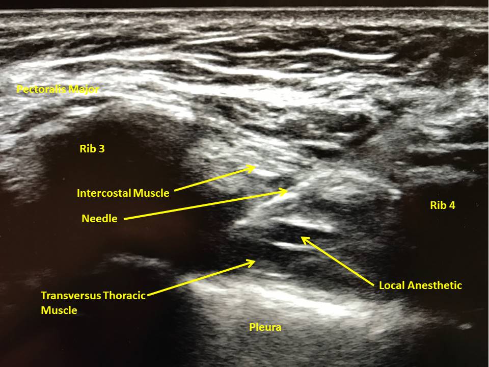
- The goal is to achieve blockade of the anterior cutaneous branches of intercostal nerves 2–6, which innervate the sternum as originally described by Ueshiema and Otake.The chest is disinfected and sterile ultrasound gel is applied. A 50-mm, high-frequency linear ultrasound probe is placed on the chest in a parasagittal plane over the third and fourth ribs at the midclavicular lin…
Conclusion
- As reliance on opioids as the sole analgesic option wanes, new regional anesthesia techniques that focus on injection of local anesthetics into various fascial planes have gained prominence as critical components of multimodal analgesic regimens. Patients experiencing medial chest wall pain had not previously benefited from regional anesthesia techniques and have largely depend…