What is close ventral mesogastrium?
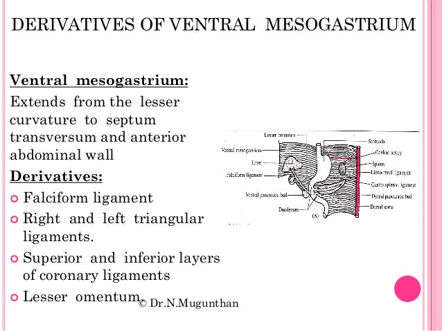
Close ventral mesogastrium ven·tral mes·o·gas·tri·um the primitive midline mesentery extending between future stomach and proximal duodenum and the anterior abdominal wall superior to the umbilicus (umbilical vein). The liver develops within it; consequently, the lesser omentum, coronary and falciform ligaments are derivatives of it.
What is the meaning of mesogastrium?
Medical Definition of mesogastrium 1 : a ventral mesentery of the embryonic stomach that persists as the falciform ligament and the lesser omentum — called also ventral mesogastrium 2 : a dorsal mesentery of the embryonic stomach that gives rise to ligaments between the stomach and spleen and the spleen and kidney
What is ventral mesentery 1?
1 : a ventral mesentery of the embryonic stomach that persists as the falciform ligament and the lesser omentum — called also ventral mesogastrium.
What are the two attachments of the mesogastrium?
Its two attachments are the dorsal mesogastrium behind, and the ventral mesogastrium in front. As the foregut rotates, the dorsal and ventral mesogastrium rotate with it. The line of attachment of the ventral mesogastrium swings round to the right as the foregut develops.

Where is the ventral Mesogastrium?
The part of the ventral mesentery that attaches to the stomach is known as the ventral mesogastrium. The lesser omentum is formed, by a thinning of the mesoderm or ventral mesogastrium, which attaches the stomach and duodenum to the anterior abdominal wall.
What does the ventral Mesogastrium become?
The ventral mesogastrium of the foregut is carried rightwards with the liver. The border of the mesogastrium between the liver and the ventral wall of the embryo becomes the falciform ligament. The portion between the stomach and liver becomes the lesser omentum.
What does the dorsal Mesogastrium become?
The portion of the dorsal mesogastrium that lies between the stomach and spleen becomes the gastrosplenic ligament, whilst the part lying between the spleen and the dorsal wall of the embryo becomes the lienorenal ligament (Fig. 7.5). Lying across the dorsal wall of the embryo is the pancreas (see later).
Where is the dorsal Mesogastrium?
the stomachThe portion of mesentery attached to the greater curvature of the stomach is named the dorsal mesentery (or dorsal mesogastrium, when referring to the portion at the stomach), and the part which suspends the colon is termed the mesocolon. The dorsal mesogastrium develops into the greater omentum.
What are the derivatives of dorsal Mesogastrium?
The spleen and body of the pancreas develop within it, and thus the splenorenal and gastrosplenic ligaments are derivatives of the (dorsal) mesogastrium.
What is the origin of gut tube?
The primitive gut tube is derived from the dorsal part of the yolk sac, which is incorporated into the body of the embryo during folding of the embryo during the fourth week. The primitive gut tube is divided into three sections.
What is dorsal and ventral Mesogastrium?
- Dorsal mesogastrium anchors the stomach to the posterior body wall. - Visceral peritoneum covers the stomach. - Ventral mesogastrium connects the stomach to the liver. - Falciform ligament anchors the liver to the ventral body wall.
What is ventral mesentery?
The ventral mesentery is a double-layered membrane that gives rise to the falciform ligament, connecting liver to anterior abdominal wall, the visceral peritoneum of the liver, and the lesser omentum. The ventral mesentery originally stretches between the stomach and the anterior abdominal wall.
Does omentum grow back?
No. This misunderstanding comes from the fact that your omentum is part of your peritoneum, the tissue that lines your abdominal cavity and wraps around your organs. Peritoneal tissue is known to have rapid healing properties. But no, your omentum won't grow back after being removed.
What is dorsal and ventral Mesogastrium?
- Dorsal mesogastrium anchors the stomach to the posterior body wall. - Visceral peritoneum covers the stomach. - Ventral mesogastrium connects the stomach to the liver. - Falciform ligament anchors the liver to the ventral body wall.
How does the greater omentum develop?
The stomach then rotates a second time, and here the cranial end moves inferiorly and the caudal end moves superiorly, giving the stomach its final position in the abdomen. This second rotation causes the double folded dorsal mesentery to move downwards, becoming the greater omentum.
What is the epiploic foramen?
Anatomical Parts The epiploic foramen (foramen epiploicum; foramen of Winslow) is the passage of communication between the general cavity and the omental bursa.
Does omentum grow back?
No. This misunderstanding comes from the fact that your omentum is part of your peritoneum, the tissue that lines your abdominal cavity and wraps around your organs. Peritoneal tissue is known to have rapid healing properties. But no, your omentum won't grow back after being removed.
What is the ventral mesentery?
The ventral mesentery forms the gastrohepatic ligament16 and the fibrous visceral peritoneum of the liver. This was first described by Glisson in 1659,17 as a peritoneal sheath that envelops the organ, except for a “bare area” on the superoposterior surface of the right lobe where the organ is in direct contact with the inferior vena cava, diaphragm, and superior aspect of the right adrenal gland. Glisson’s capsule involutes into the parenchyma as intrahepatic septa or trabeculae that support vascular structures and serve as surgical landmarks.18,19
What is the diaphragm?
The diaphragm, which separates the thoracic from the abdominal cavity in adults, is a composite structure derived from several embryonic components (see Fig. 15.38 ). The large ventral component of the diaphragm arises from the septum transversum, which fuses with the ventral part of the esophageal mesentery.
What are the pleural cavities?
The pleural cavities become divided from the abdominal cavities by the pulmonary folds (Plate 128 ), each of which arises medially from a lung bud and extend laterally, left or right ( Text-Figure 60; Plate 65 ). (In mammals the corresponding folds arise laterally and extend medially.) Each pulmonary fold fuses with the peritoneal lining of the body wall to form the pleuro-peritoneal membrane, with the pericardium to form the pleuro-pericardial membrane, and with the peritoneum covering the liver to form eventually, the lateral hepatic ligament ( Plate 204 ). During development the lateral thoracic air sacs on each side invade the pulmonary fold ( Text-Figure 60; Plate 204 ), splitting apart the two layers so that two separate septa are formed, the horizontal septum (also known as pulmonary aponeurosis, sacco-pleural membrane ( Plate 204), or pulmonary diaphragm) and the oblique septum (also known as thoraco-abdominal diaphragm or sacco-peritoneal membrane). (Note: these are not true diaphragms, as found in mammals, so this term is best avoided.) The ventral mesentery persists as the falciform ligament (Plate 204), a transverse sheet connecting the ventral body wall (inner face of the sternum) with the ventral surface of the liver. It thus corresponds with the falciform ligament of mammals. The post-hepatic septum is an extension of the mesentery arising at the level of the gizzard.
What is the shape of the stomach?
Very early in the formation of the digestive tract, the stomach is recognizable as a dilated region with a shape remarkably similar to that of the adult stomach (see Figure 3). The early stomach is suspended from the dorsal body wall by a portion of the dorsal mesentery called the dorsal mesogastrium. It is connected to the ventral body wall by a ventral mesentery that also encloses the developing liver (Figure 6 ).
Where is the tail of the pancreas located?
At the point on the dorsal wall of the embryo, where the lienorenal ligament is reflected on to the wall, the tail of the pancreas comes to lie between the two layers of the double layer of that ligament. As the liver develops in the ventral mesogastrium and enlarges it is carried towards the right.
Which gland is the largest in the body?
The liver. This is the largest gland in the body weighing approximately 1500 g which forms from an outgrowth of foregut. It develops in the septum transversum and protrudes into the abdomen dividing the ventral mesentery into two: anteriorly the falciform ligament is produced, posteriorly the lesser omentum.
Where is ventral mesentery located?
A ventral mesentery only exists in the terminal part of the oesophagus, stomach and the proximal part of the duodenum.
What is the midline mesentery?
the primitive midline mesentery extending between future stomach and proximal duodenum and the anterior abdominal wall superior to the umbilicus (umbilical vein). The liver develops within it; consequently, the lesser omentum, coronary and falciform ligaments are derivatives of it.
Which direction does the ventral mesogastrium attach?
The line of attachment of the ventral mesogastrium swings round to the right as the foregut develops. It ends up running along the lesser curve of the stomach, and the top of the proximal duodenum. On the back, the attachment of the dorsal mesogastrium swings round to the left.
Where is the dorsal mesogastium?
The dorsal mesogastium hangs down in front of the transverse colon. To follow its growth we'll look at a sagittal section made in this line. Here's the lesser omentum, between the liver and the stomach. Here's the greater omentum, hanging down in a double fold.
What are the attachments of the hindgut and midgut?
The foregut is attached also at the front. Its two attachments are the dorsal mesogastrium behind, and the ventral mesogastrium in front. As the foregut rotates, the dorsal and ventral mesogastrium rotate with it.
Which organ grows downward in front of the transverse colon?
This is the pancreas. The greater omentum grows downward in front of the transverse colon. The greater omentum and the transverse mesocolon come together, and the duplicated layers are absorbed, so that we're left with the greater omentum stuck to the transverse colon, and hanging down below it.