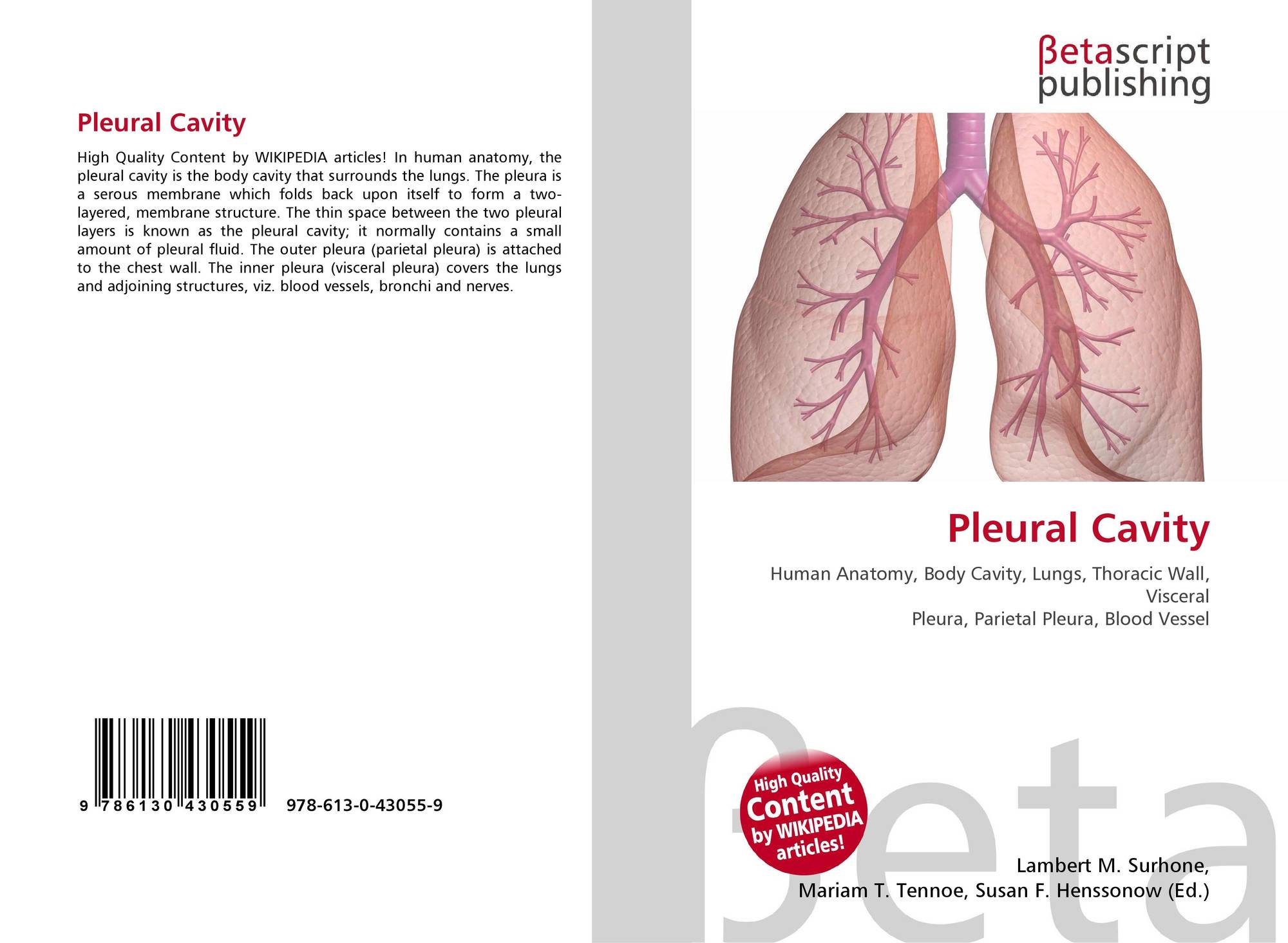
Visceral pleura. The pleura that covers the lungs and enters into and lines the interlobar fissures. It is loose at the base and at the sternal and vertebral borders to allow for lung expansion. The thin, double-layered membrane that separated the lungs from the inside of the chest wall.
What are the roles of the pleura?
What is the role of pleural fluid?
- Function. The pleural cavity, with its associated pleurae, aids optimal functioning of the lungs during breathing.
- Pleural fluid. The space containing the fluid is referred to as the pleural cavity or pleural space. ...
- pleural. Consequently, what is the color of pleural fluid? ...
- Normally. What is the most common cause of pleural effusion? ...
What is the structure and function of the pleura?
Pleura function & Structure - Knowledgist . The visceral and parietal pleurae connect to each other at the hilum. The pleural cavity is the space between the visceral and parietal layers. The pleurae perform two major functions: They produce pleural fluid (serous fluid) and create cavities that separate the lungs from each other and other ...
How serious is a pleural effusion?
The seriousness of the condition depends on the primary cause of pleural effusion, whether breathing is affected, and whether it can be treated effectively. Causes of pleural effusion that can be effectively treated or controlled include an infection due to a virus, pneumonia or heart failure.
What are the causes of visceral pain?
Visceral Pain
- Causes of True Visceral Pain. Any stimulus that excites pain nerve endings in diffuse areas of the viscera can cause visceral pain.
- “Parietal Pain” Caused by Visceral Disease. ...
- Localization of Visceral Pain— “Visceral” and the “Parietal” Pain Transmission Pathways. ...

What does visceral pleura mean?
A pleura is a serous membrane that folds back on itself to form a two-layered membranous pleural sac. The outer layer is called the parietal pleura and attaches to the chest wall. The inner layer is called the visceral pleura and covers the lungs, blood vessels, nerves, and bronchi.
What is the pleura and its function?
(PLOOR-uh) A thin layer of tissue that covers the lungs and lines the interior wall of the chest cavity. It protects and cushions the lungs. This tissue secretes a small amount of fluid that acts as a lubricant, allowing the lungs to move smoothly in the chest cavity while breathing.
What keeps the visceral and parietal pleura?
The parietal pleura lines the thoracic wall and superior surface of the diaphragm. It continues around the heart forming the lateral walls of the mediastinum. The pleura extends over the surface of the lungs as the visceral pleura. The surface tension of the fluid in the pleural cavity secures the pleura together.
What is the visceral lining?
Serous membrane lining the wall of a serous cavity is designated parietal while that covering viscera is called visceral. Connecting serous membrane runs between parietal and visceral components. The serous membranes are: Peritoneum — the peritoneal cavity is found within the abdominal & pelvic body cavities.
Is the visceral pleura sensitive to pain?
The parietal pleurae are highly sensitive to pain, while the visceral pleura are not, due to its lack of sensory innervation.
Can you live without pleura?
Once the pleural lining is removed, one can live a normal life but will lack physical endurance. Removal of the pleural is a major surgical undertaking, and is usually only an option for those who are healthy enough to undergo such an invasive procedure.
What is the other name for visceral pleura?
The inner pleura, called the visceral pleura, covers the surface of each lung and dips between the lobes of the lung as fissures, and is formed by the invagination of lung buds into each thoracic sac during embryonic development....Pulmonary pleuraeLatinpleurae pulmonariusMeSHD010994TA98A07.1.02.001TA233229 more rows
What is the difference between the visceral and parietal?
Parietal serosa line the body cavities and visceral serosa line the outer part of the organs within the body cavity. Therefore, parietal serous membranes are the outer membranes lining a body cavity and visceral serous membranes are the inner membranes lining a body cavity.
Why is visceral pleura clinically important?
The layer of pleura that covers the lung parenchyma is named the visceral pleura. By invaginating and folding back on itself to form the fissures of the lungs, it is responsible for creating the different lung lobes.
What is the function of visceral layer?
The visceral layer of the serous pericardium is also known as the epicardium. The main purpose of all of the aforementioned layers are the same: to protect the heart and assist in contractration.
What is the purpose of visceral?
It is called visceral fat because it is located close to the viscera. The major physical function of the visceral fat is protection of the internal organs. It also provides a reserve source for energy if needed by the animal.
What does the visceral layer do?
The inner (visceral) layer of the serous pericardium lines the outer surface of the heart itself. Between the two layers of the serous pericardium is the pericardial cavity, which contains pericardial fluid. It is this fluid that provides lubrication between the two layers, and allows the heart to expand and contract.
What is the pleura and its function quizlet?
Describe the pleura and its function. - Thin, slippery pleura form an envelope between the lungs and chest wall. - Outside layer visceral; inside layer parietal. - Function: form cushion for lungs. - Pleural cavity has negative pressure or vacuum which holds the lungs tightly against chest wall.
What is the main function of pleura Brainly?
Answer. Answer: Pleural membranes :Thin layers that reduce friction between the lungs and the inside of the chest wall during breathing. The function of the pleura is to allow optimal expansion and contraction of the lungs during breathing.
What is the function of the Pleurae quizlet?
The pleural cavity, with its associated pleurae, aids optimal functioning of the lungs during breathing. The pleural cavity also contains pleural fluid, which acts as a lubricant and allows the pleurae to slide effortlessly against each other during respiratory movements.
What is the purpose of the pleura that surrounds the lungs?
The function of the pleura is to allow optimal expansion and contraction of the lungs during breathing. The pleural fluid acts as a lubricant, allowing the parietal and visceral pleura to glide over each other friction free. This fluid is produced by the pleural layers themselves.
What is the visceral pleura?
Visceral pleura is composed of outer mesothelial layer (which is easily denuded by mechanical manipulation and autolysis) with underlying connective tissue layered between 2 elastic lamina layers, and connective tissue layered at interface with alveolated parenchyma
What nerves are involved in the visceral pleura?
The visceral pleura is innervated by autonomic nerves containing fibers that are sensitive to stretch but not to pain. On the contrary, sensory nerve endings recognizing and conducting pain sensation are present in the parietal pleura. Costal pleura and the peripheral parts of the diaphragmatic pleura are supplied by the sensory fibers of the intercostal nerves arising from the thoracic spinal nerves. Thus, the irritation of costal pleura is perceived as pain in the corresponding dermatome of the chest or the medial part of the upper limb (Th1 dermatome). The cupula, mediastinal and a central portion of the diaphragmatic pleura are innervated by nerve fibers originating from the neck (C3–C5 spinal nerves). Stimulation of these pleura regions may cause the pain to be felt in the ipsilateral shoulder (C4 dermatome) (Yalcin et al., 2013 ).
What is the double layer of the parietal pleura?
Several structures (e.g., the hilae and sometimes the great veins) within the thoracic cavity acquire a double layer of parietal pleura during embryological development. Such a double layer is pulled into the thorax by the developing lung, and extends from the lung hilum vertically downward to the diaphragm on both sides – these form the pulmonary ligaments. They are of importance surgically as they may contain lymphatics, tumor, or vessels. Their presence may prevent torsion of the lower lobes.
How much fluid can a pleura produce?
The pleura can produce up to 100 mL of fluid in an hour, and the absorption capacity of the pleural surface is approximately 300 mL per hour. The parietal pleura is supplied by systemic capillary vessels and drains into the right atrium by way of the azygos, hemiazygos, and internal mammary veins.
What is the pleural cavity?
Between these two delicate membranes lies the pleural cavity, a sealed space maintained ∼10–20 μm across.
Which pleura lines the chest wall?
As the costal pleura, the parietal pleura lines the chest wall, as the mediastinal pleura it lines the lateral surface of the mediastinum, and as the diaphragmatic pleura it lines the diaphragm. The parietal pleura is significantly more firmly fixed to its surroundings than the visceral pleura because of the demands made on it by mechanical traction. The costal pleura is firmly attached to the endothoracic fascia and the diaphragmatic pleura to the phrenicopleural fascia.
Where is the costal pleura innervated?
Thus, the irritation of costal pleura is perceived as pain in the corresponding dermatome of the chest or the medial part of the upper limb (Th1 dermatome). The cupula, mediastinal and a central portion of the diaphragmatic pleura are innervated by nerve fibers originating from the neck (C3–C5 spinal nerves).
Which nerves innervate the visceral pleura?
Innervation. The visceral pleura is innervated by autonomic nerves containing fibers that are sensitive to stretch but not to pain. On the contrary, sensory nerve endings recognizing and conducting pain sensation are present in the parietal pleura.
Which pleura has a covering of connective tissue?
(c) The parietal pleura also has a covering of mesothelium and there is generally more underlying connective tissue here than in the visceral pleura.
What are the two components of the deep network of pulmonary lymphatics?
The deep network of pulmonary lymphatics consists of two separate components: lymphatic vessels that connect interacinar regions and a rich plexus of peribronchiolar lymphatic vessels. The collagen and elastic fibers of the visceral pleura merge with the fibroelastic framework of the lung parenchyma.
How many layers are there in the pleural membrane?
Layers of the pleural membrane. Both visceral and parietal pleura in humans are approximately 40 μm thick. Between pleural surface and underlying tissue, five layers are identified histologically, consisting of a single-cellular layer and four subcellular layers, as follows: 1. a monolayer of mesothelial cells;
What is the connective tissue of the pleura?
The connective tissue of the human visceral pleura contains a network of lymphatic vessels that prevent the accumulation of fluid in the pleural space, the tiny area between the visceral and parietal pleurae. This network, known as the superficial lymphatic network, is drained by lymphatic vessels that run through the interlobular septa. These lymphatic vessels, together with the lymphatic vessels that follow the bronchial tree and the pulmonary vessels, form the deep lymphatic network. The superficial lymphatic network in rodent lungs is not as well characterized. The deep network of pulmonary lymphatics consists of two separate components: lymphatic vessels that connect interacinar regions and a rich plexus of peribronchiolar lymphatic vessels.
Why do ventilatory muscles affect the pleural space?
Because the visceral and parietal pleurae are maintained in close apposition, the lung and the thorax interact mechanically. The work performed by the ventilatory muscles induces changes in the pressure of the intrapleural space. During inspiration, the pleural pressure (which, at rest and in quiet conditions, is subatmospheric) decreases, and at the end of inspiration, when the air flow returns to 0, the pleural pressure increases slightly. During expiration, the pleural pressure increases toward its value at functional residual capacity, that is, lung volume before inspiration (see Figure 9-15 ).
How many layers does the pleura have?
Pleura. The visceral pleura has five layers. A single layer of mesothelial cells without a basement membrane rests on a submesothelial layer of loose connective tissue approximately as thick as the mesothelial cell layer.
Which pleura envelopes all surfaces of the lungs?
The lungs are lined by the visceral pleura, which at the level of the hilum folds back upon itself to form the parietal pleura, with the cavity between these two layers being defined as pleural space. Pleural effusion: diagnosis and management. The visceral pleura envelopes all surfaces of the lungs.
Where are cysts attached in pleural fissures?
Cysts in pleural fissures were indeed attached by a thin pedicle to the visceral pleura (1, 2).
What is tension pneumothorax?
Tension pneumothorax occurs when an increasing amount of air accumulates between the parietal and visceral pleura, collapsing the ipsilateral lung and displacing the mediastinum. Malposition of thoracostomy tubes leading to missed haemothorax and tension pneumothorax.
Which membrane invests in the lungs?
the serous membrane investing the lungs and dipping into the fissures between the lobes of the lungs.
What is EPP surgery?
EPP is a radical surgical procedure involving complete removal of the ipsilateral lung along with the parietal and visceral pleura, pericardium with portions of the phrenic nerve, and the majority of the hemidiaphragm (10).
What is the pleural cavity?
The pleural cavity, also known as the intrapleural space, contains pleural fluid secreted by the mesothelial cells.
What is the name of the fluid that separates the pleura?
The layers are separated by a small amount of viscous lubricant known as pleural fluid. 1 . There are a number of medical conditions that can affect the pleura, including pleural effusions, a collapsed lung, and cancer.
How many layers are there in the pleura?
Anatomy. There are two pleurae, one for each lung, and each pleura is a single membrane that folds back on itself to form two layers. The space between the membranes (called the pleural cavity) is filled with a thin, lubricating liquid (called pleural fluid ). The pleura is comprised of two distinct layers: 1 .
What is pleural effusion?
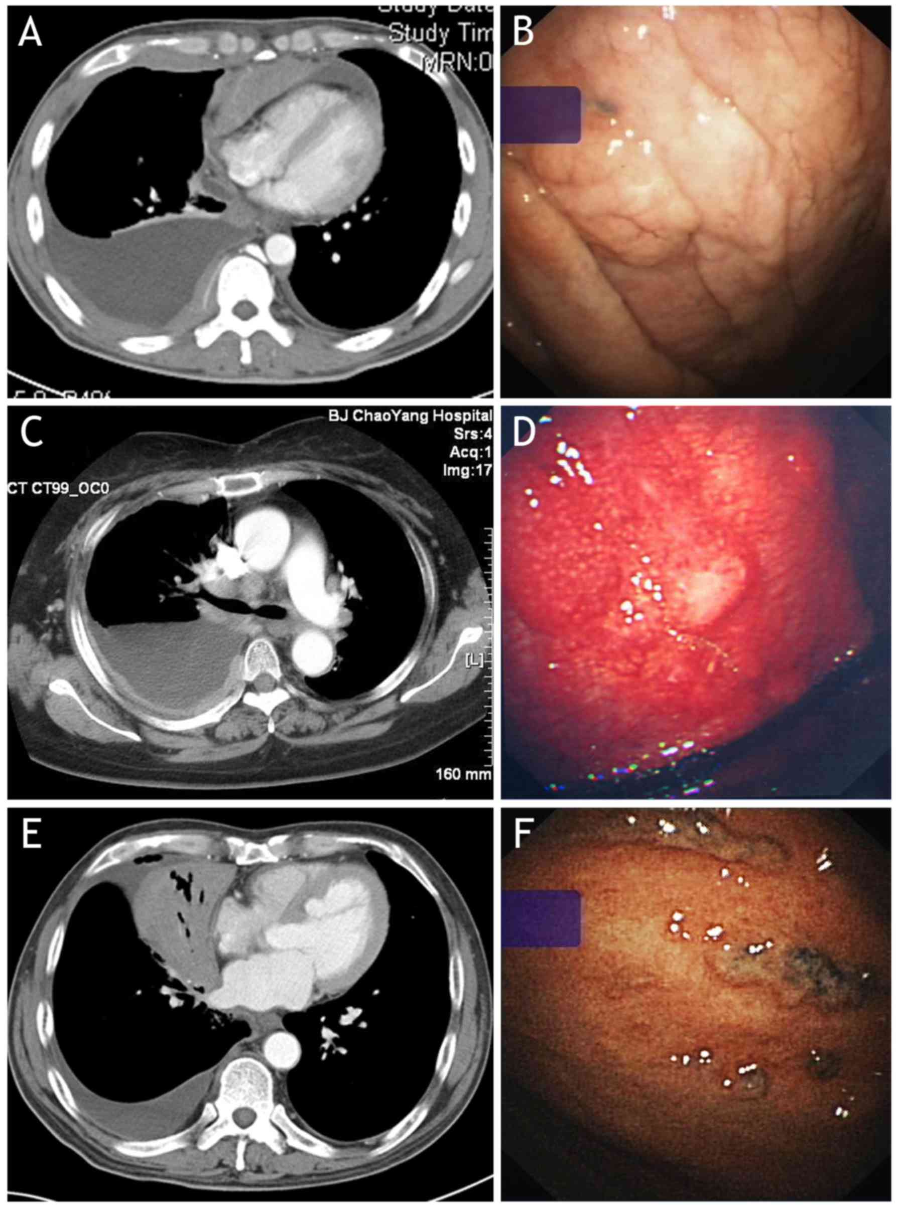
A pleural effusion is the accumulation of excess fluid in the pleural space. When this happens, breathing can be impaired, sometimes significantly.
How many ccs of pleural fluid are in the lungs?
The intrapleural space contains roughly 4 cubic centimeters (ccs) to 5 ccs of pleural fluid which reduces friction whenever the lungs expand or contract. 1 . The pleura fluid itself has a slightly adhesive quality that helps draw the lungs outward during inhalation rather than slipping round in the chest cavity.
What is malignant pleural effusion?
A malignant pleural effusion refers to an effusion that contains cancer cells. It's most commonly associated with lung cancer or breast cancer that has metastasized (spread) to the lungs. 5 .
How to tell if pleural effusion is small?
A pleural effusion can be very small (detectable only by a chest X-ray or CT scan) or be large and contain several pints of fluid. 4 Common symptoms include chest pain, dry cough, shortness of breath, difficulty taking deep breaths, and persistent hiccups. Common Disorders of the Pleural Fluid.
Where is the visceral pleura located?
The visceral pleura covers the outer surface of the lungs, and extends into the interlobar fissures. It is continuous with the parietal pleura at the hilum of each lung (this is where structures enter and leave the lung).
Which part of the lungs is covered by the visceral pleura?
Visceral pleura – covers the lungs. Parietal pleura – covers the internal surface of the thoracic cavity. These two parts are continuous with each other at the hilum of each lung. There is a potential space between the viscera and parietal pleura, known as the pleural cavity.
What fluid pulls the parietal and visceral pleura together?
It lubricates the surfaces of the pleurae, allowing them to slide over each other. The serous fluid also produces a surface tension, pulling the parietal and visceral pleura together. This ensures that when the thorax expands, the lung also expands, filling with air.
How many pleurae are there in the human body?
There are two pleurae in the body: one associated with each lung. They consist of a serous membrane – a layer of simple squamous cells supported by connective tissue. This simple squamous epithelial layer is also known as the mesothelium. Each pleura can be divided into two parts: Visceral pleura – covers the lungs.
What is the name of the line that lines the extension of the pleural cavity into the neck?
Cervical pleura – Lines the extension of the pleural cavity into the neck.
What is the term for a collapsed lung?
A pneumothorax (commonly referred to a collapsed lung) occurs when air or gas is present within the pleural space. This removes the surface tension of the serous fluid present in the space, reducing lung extension.
How to treat a pneumothorax?
Treatment depends on identifying the underlying cause. Primary pneumothoraces tend to be small and generally require minimal intervention, whereas secondary and traumatic pneumothoraces may require decompression to remove the extra air/gas in order for the lung to reinflate (this is achieved via the insertion of a chest drain ).
What is the Difference Between Parietal and Visceral Pleura?
The parietal pleura is the outer layer of the pleural membrane, while the visceral pleura is the inner layer of the pleural membrane. Thus, this is the key difference between parietal and visceral pleura. Moreover, the parietal pleura lines the inner surfaces of the thoracic cavity on each side of the mediastinum, while the visceral pleura lines the lungs, blood vessels, nerves, and bronchi.
What is Parietal Pleura?
Parietal pleura is the outer layer of pleural membrane. Normally, the parietal pleura is attached to the chest wall. It also lines the inner surfaces of the thoracic cavity on each side of the mediastinum. The parietal pleura is set apart from the thoracic wall by the endothoracic fascia. The parietal pleura is further subdivided into mediastinal, diaphragmatic, costal, and cervical pleurae.
