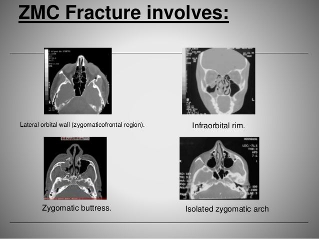
zygomatic buttress the structural pillar of the mid-face that extends superiorly from the maxillary
Maxillary sinus
The pyramid-shaped maxillary sinus is the largest of the paranasal sinuses, and drains into the middle meatus of the nose.
Zygomatic bone
In the human skull, the zygomatic bone is a paired irregular bone which articulates with the maxilla, the temporal bone, the sphenoid bone and the frontal bone. It is situated at the upper and lateral part of the face and forms the prominence of the cheek, part of the lateral wall and floor of the orbit, and parts of the temporal fossa and the infratemporal fossa. It presents a malar and a temporal surface; fou…
How to minimize the morbidity of zygomaticomaxillary buttress surgery?
fixation of the zygomaticomaxillary buttress. In addition, it is possible to install a miniplate when necessary. These advan - tages minimize morbidity and reduce surgical time 39. 10. Fixation techniques
How to fix Zygomatic buttress block bone graft?
A 5 inch long mini titanium plate was adapted and fixed by passing it below the arch through lateral eye brow incision and brining it behind the zygomatic buttress to the front of maxilla. A clinical study of the outcomes and complications associated with zygomatic buttress block bone graft for limited preimplant augmentation procedures.
What does zygomatic mean in medical terms?
The term zygomatic derives from the Greek Ζυγόμα zygoma meaning "yoke". The zygomatic bone is occasionally referred to as the zygoma, but this term may also refer to the zygomatic arch .
Why is the zygomatic bone important for facial symmetry?
The lateral prominence and convexity of the zygomatic bone makes it the most important bone for providing the aesthetic facial look and sets up the facial width but at the same time this prominence and convexity makes this bone more vulnerable to injury.
What is a buttress in bone?
Bony Buttresses of the Facial Skeleton These buttresses define the vertical height of the face and provide the bony support required for mastication. Masticatory forces imparted to the midface are transferred to the skull base through the ZM, NM and PM buttresses.
What does the zygomatic arch protect?
The function of the zygomatic arch is protection of the eye, origin for the masseter and part of the temporal muscles, and to provide an articulation for the mandible. The zygomatic arch is approached by an incision made along its ventral border.
How many facial buttresses are there?
Anatomy of the Facial Buttresses There are three paired vertical and three transverse buttresses (Figure 1).
What is the zygomatic arch known as?
Zygomatic arch: The part of the temporal bone of the skull that forms the prominence of the cheek. The zygomatic arch is also known as the zygomatic bone, the zygoma, the malar bone, the cheek bone and the yoke bone.
What is the main function of the zygomatic bone?
Definition. The zygomatic bone is a paired facial bone. Both zygoma or cheek bones are irregular and articulate with other bones of the cranium and face. They are important contributors to mastication or chewing, providing an attachment point for the masseter muscle – a jaw adductor that closes the jaw.
Is the zygomatic arch the cheekbone?
The facial area includes the zygomatic, or malar, bones (cheekbones), which join with the temporal and maxillary bones to form the zygomatic arch below the eye socket; the palatine bone; and the maxillary, or upper jaw, bones.
What is a buttress plate?
Buttress plates are osteosynthetic implants commonly used in the metaphyseal area for internal fixation of articular fractures to support intraarticular fragments.
What happens if the zygomatic bone is damaged?
Fractures of the ZMC or zygomatic arch can often lead to unsightly malar depression, which should be corrected to restore a normal facial contour. ZMC fractures can also cause significant functional issues, including trismus, enophthalmos and/or diplopia, and paresthesias of the infraorbital nerve.
What is the 3rd most common facial fracture?
The most common isolated fracture site was the nasal bone (37.7%), followed by the mandible (30%), orbital bones (7.6%), zygoma (5.7%), maxilla (1.3%) and the frontal bone (0.3%).
What is the zygomatic arch composed of?
The zygomatic arch (cheek bone) is formed by the zygomatic process of temporal bone and the temporal process of the zygomatic bone, the two being united by an oblique suture (zygomaticotemporal suture).
What are the parts of the zygomatic bone?
The zygomatic bone has three surfaces: lateral, posteromedial and orbital. The lateral (facial) surface faces towards the outside. It is smooth and convex, and it features a small opening called the zygomaticofacial foramen.
What is the most common condition associated with the zygomatic bone?
The most common condition associated with the zygomatic bone is a fracture. 3 A fracture to the orbital floor, the portion of the zygomatic bone which is attached to the eye, also has an impact on the function of the zygomatic bone. This type of fracture is called a blowout and can cause a fracture to the zygomatic bone, displace the upper portion of the zygomatic bone which articulates with the skull, and can cause a deeper fracture to the eye socket. Jaw fractures can also impact the lower portion of the zygomatic bone, causing difficulty chewing, speaking, and other functions associated with the mouth.
What bone connects to the upper portion of the skull?
This allows the zygomatic bone and other facial bones to connect with the upper portion of the skull.
Why is the front part of the zygomatic bone thick?
The front portion of the bone is thick and jagged to allow for its joining with other bones of the face. This thickness also allows the bone to remain strong and sturdy to protect the more delicate features of the face. Other portions of the zygomatic bone include joints near the jaw, near the ears, and near the forehead and skull.
What is the function of the zygomatic bone?
The zygomatic bone functions as a structure which joins the bones of the face while protecting the arteries, nerves, veins, and organs which lie below the surface. The arches of the zygomatic bone provide a person’s cheeks with the structure to fill out the face. The zygomatic bone itself has no ability to move, ...
What bones join together in the face?
The zygomatic bones join with several other bones of the face, including the nose, jaw, portions of the eye, and bones just in front of the ears. The zygomatic bone consists of cartilage when a fetus is in utero, with bone-forming immediately after birth. Due to its size and function in joining many facial bones together, ...
Which bone allows for the passage of integral veins and arteries through the face?
There is also a tunnel within the zygomatic bone called the zygomaticofacial foramen which allows for the passage of integral veins and arteries through the face. Sebastian Kaulitzki / Science Photo Library / Getty Images.
Which bone joins the jaw bone?
However, the lower portion of the zygomatic bone which joins with the jaw bone assists in providing movement to the jaw bone. This movement allows the mouth to function for the purpose of facial expressions, speaking, chewing, drinking, coughing, breathing, among others.
What is partial maxillectomy?
Partial maxillectomy techniques involve removal of various segments of the incisive, nasal, maxilla, palatine, vomer, lacrimal and zygomatic bones with adjacent neoplastic and normal soft tissues and teeth. The term premaxillectomy is often used in the veterinary literature to denote an excision confined to the incisive bone. However, the term premaxilla is not accepted veterinary anatomical nomenclature; 1 the term incisivectomy is therefore more appropriate. The term hemimaxillectomy is equally confusing and incorrect if used to describe the surgical excision of one maxilla. Removing most of one maxillary bone (typically combined with the excision of all or parts of the incisive and palatine bones) is a complete or total, unilateral maxillectomy. The term hemimaxillectomy could be used to denote the excision of half of one maxillary bone, but the term partial maxillectomy is more appropriately used for this. An orbitectomy involves removal of portions of bones that comprise the orbit including the maxilla, palatine, zygomatic, lacrimal and frontal bones. 2
What is the maxilla?
The term ‘maxilla’ is used here to refer to the incisive, palatine, zygomatic, lacrimal, frontal and nasal bones, in addition to the maxillary bone proper. By definition, the forces exerted on the mandible also are exerted on the maxilla. However, the distribution of these forces is much different, and it is generally accepted that the maxilla is subject to much less strain. The maxillofacial area can most easily be thought of as an ‘outer facial frame,’ which acts as a link between the base of the skull and the occlusal surfaces. 31 The support of the facial region is provided by a series of anatomic buttresses that distribute the masticatory forces to the head. These buttresses exist in the horizontal, vertical and coronal planes. 31–34 There are three primary buttresses: rostral (medial), lateral, and caudal ( Fig. 25.11 ). 31–34 These buttresses also can be defined anatomically: the rostral (medial) as the nasomaxillary buttress, the lateral as the zygomaticomaxillary buttress, and the caudal as the pterygomaxillary buttress. The anatomic definitions mirror the bones of the skull that compose these buttresses. The caudal buttress (which is not readily accessible) is composed of the lacrimal, palatine and pterygoid bones. In the presence of a fracture, the facial frame can be adequately reconstructed with two of the three buttresses: medial and lateral (see below). Therefore, the palatine and lacrimal bones are more likely to be dealt with secondarily, i.e., with primary stabilization of other fractures of the skull. The incisive bones are not part of the buttresses, and therefore may not need to be stabilized as this area generally does not provide essential support to the skull, or can effectively be treated by other means (see Ch. 28 ). The nasal bones also may be fractured without disturbing the medial buttress. Fixation of these areas still may be useful in these areas in either providing the support for the incisors or reestablishing the cosmetic appearance of the nasal area when depressed fractures occur. 5 Similarly, maxillary fractures often may not require stabilization unless the buttresses are compromised. 4 In the latter case, occlusion or support of the orbit is compromised, and fixation (most often plate) is ideally suited to stabilize these fractures and reestablish the medial buttress. Likewise, if the lateral buttress is compromised, which often will affect the orbit, plate fixation again is ideally suited for stabilization and to reestablish this buttress ( Ch. 31 ). 4,5
What bone is in the rostral part of the dog's orbit?
In the dog, the rostral part of the bony orbit is formed by the zygomatic bone, lacrimal bone, maxillary bone, sphenoid bone, palate bone, and frontal bone. 9 On the medial (nasal) side the orbit is formed by the frontal bone. This continues laterally (temporally) as the zygomatic process and connects to the frontal process of the zygomatic bone by a bridge of connective tissue. The bony parts are inspected and palpated for symmetry.
What is the floor of the orbit?
The floor of the orbit is formed by the zygomatic bone, the orbital surface of the maxilla, and the orbital process of the palatine bone (Figure 6 ). It is the shortest of the orbital walls (∼40 mm). Similar to the roof, it is triangular in shape. Posteriorly, the floor is separated from the lateral wall by the inferior orbital fissure. It continues anteriorly as the infraorbital groove and canal. The canal runs approximately in the center of the floor from posterior to anterior and carries the maxillary division of the trigeminal nerve and associated infraorbital artery. This bundle exits approximately 4–10 mm inferior to the orbital rim through the infraorbital foramen. The visualization of this groove during orbital floor surgery is important in preventing inadvertent trigeminal hypoesthesia. Medial to the infraorbital canal is the thinnest portion of the orbit where most blowout fractures occur. It is also where the floor is decompressed for surgical treatment of patients with thyroid eye disease.
What is a zygomatic fracture?
Fractures of the zygomatic bone are usually associated with trauma to the eye and orbit. This fracture may result in exophthalmos if the bone fragments have been displaced medially, or alternatively, either a caudal or ventral displacement of the eye if the lateral support provided by this bone is lost. A direct approach to the zygomatic arch is performed. The arch may be reconstructed either with a single plate that spans the arch or, in highly comminuted fractures, pieced together with smaller plates across the individual fracture lines and then additionally spanned over the entire distance (Fig. 31.11 ). Caution must be exercised to protect the zygomatic branch of the facial nerve, which courses over the caudodorsal aspect of the zygomatic arch. Repair of fractures at the caudal aspect of the zygomatic arch must be approached with great caution due to the proximity of the facial nerve and maxillary artery. It may be more prudent to follow a conservative, noninterventional approach, for fractures at this location.
Where does the zygomaticotemporal nerve travel?
The zygomaticotemporal nerve travels in a groove or canal in the zygomatic bone as it passes along the inferolateral angle of the orbit anteriorly. It gives off a communicating branch (carrying postganglionic parasympathetic fibers from the PPG to the lacrimal gland) to the lacrimal nerve of CN V1 before it exits the orbit through a zygomatico-orbital foramen and emerges into the temporal fossa through the zygomaticotemporal foramen. The zygomaticotemporal nerve then ascends between bone and the temporalis muscle to supply the skin of the temple ( Totonchi, Pashmini, & Guyuron, 2005 ). In this region, the nerve also sends communicating branches to the auriculotemporal nerve of CN V 3 and the facial nerve (CN VII) ( Standring, 2008 ).
What are the bones of sheep and goats?
Sheep and goats have an enclosed orbit, typical of most grazing animals. Both species have lacrimal, zygomatic, frontal, sphenoid, and palatine bones comprising the bony fossa of the orbit. In addition, sheep have a maxillary bone and goats have an ethmoid bone that forms part of the orbit. The size, shape, and position of the orbit are closely associated with visual activity and feeding behavior. 1 In general, prey species such as sheep and goats have eyes that are located more laterally on the skull and have mostly monocular vision. 1 Nerves and blood vessels travel into the orbital region by the rostral alar, ethmoidal, lacrimal, orbital, ovale, optic, rotundum, and supraorbital foramina or fissures. The pterygopalatine region has nerves and vessels associated with the orbit as well, including the caudal palatine, maxillary, and sphenopalatine foramina. Glands of the infraorbital sinus are present only in sheep and are better developed in rams than in ewes. These are specialized cutaneous glands that produce pheromones; their secretions exit from the infraorbital sinus in a depression just rostral to the eye. 2
What is zygomatic complex?
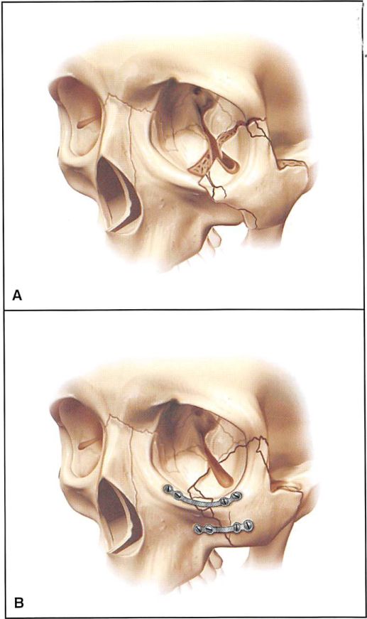
Introduction: The zygomatic complex is integral to the facial contour, protection of the eye and other facial structures, and dental occlusion. Its importance in facial function and aesthetics requires high quality outcomes of the treatment. Case presentation: This paper reports the case of a 46-year-old man who had an occupational accident resulting in extensive facial trauma and zygomatic fractures. The patient presented with hyposphagma, palpable step in the area of the infraorbital rim, paresthesia of the right infraorbital nerve, flattening of zygomatic prominence, abrasion of the chin and nose, a 7-cm laceration in the midface region, ecchymosis in the palate, and alteration in the dental occlusion without limitation of mouth opening. Computed tomography (CT) confirmed the zygomatic complex fractures. The treatment was reduction and fixation with plates and screws. CT was used throughout the treatment period as an essential diagnostic tool for accurate fracture assessment and classification, formulation of the surgical plan, and postoperative evaluation. Conclusion: This case study illustrated the correct use of CT for improved and efficient treatment of traumatic injury of the zygoma, an anatomical area where restoration of function and aesthetics is challenging. The patient signed a written informed consent statement for publication.
What is a zygomatic fracture?
Zygomatic fractures are the second most common fractures of the facial skeleton, after nasal bone fractures. Due to its uniqueness, the malar bone plays a very important role in maintaining appropriate facial contours. Zygomatic fractures can cause ocular and mandibular functional impairment, along with cosmetic defects. With the help of advanced imaging techniques and various treatment options, the management of zygomatic fractures has become more sophisticated and less invasive. This article discusses zygomatic fractures in detail: their clinical and radiographic features, and the various treatment options available.
What is the zygomatic bone?
In the human skull, the zygomatic bone ( cheekbone or malar bone) is a paired irregular bone which articulates with the maxilla, the temporal bone, the sphenoid bone and the frontal bone. It is situated at the upper and lateral part of the face and forms the prominence of the cheek, ...
What is the jugal bone?
In non- mammalian vertebrates, the zygomatic bone is referred to as the jugal bone, since these animals have no zygomatic arch. It is found in most reptiles, amphibians, and birds. It is connected to the quadratojugal and maxilla, as well as other bones, which may vary by species.
What are high cheekbones called?
Society and culture. Pronounced zygomatic arches, commonly called " high cheekbones ", are considered a beauty trait in some cultures, in both males and females. Ancient Chinese sculptures of goddesses typically have a "broad forehead, raised eyebrows, high cheekbones, and large, sensuous mouth".
What is the division of the zygomatic bone?
After birth, the bone is sometimes divided by a horizontal suture into an upper larger, and a lower smaller division. In some quadrumana the zygomatic bone consisted of two parts, an orbital and a malar.
What are the three processes of the zygomatic bone?
Each zygomatic bone is diamond-shaped and composed of three processes with similarly named associated bony articulations: frontal, temporal, and maxillary. Each process of the zygomatic bone forms important structures of the skull.
What is the orbital surface?
The orbital surface forms the lateral part and some of the inferior part of the bony orbit. The zygomatic nerve passes through the zygomatic-orbital foramen on this surface. The lateral palpebral ligament attaches to a small protuberance called the orbital tubercle.
Where does the word "zygomatic" come from?
The term zygomatic derives from the Greek Ζυγόμα zygoma meaning "yoke". The zygomatic bone is occasionally referred to as the zygoma, but this term may also refer to the zygomatic arch .