Explore
Where does the thoracic duct originate? The thoracic duct originates in the abdomen from the confluence of the right and left lumbar trunks and the intestinal trunk, forming a significant pathway upward called the cisterna chyli.
Where does the thoracic duct begin?
Thoracic-duct. definition. (0) Meanings. The main duct of the lymphatic system, ascending through the thoracic cavity in front of the spinal column and discharging lymph and chyle into the blood through the left subclavian vein. noun. 0. 0.
What does thoracic duct mean?
The thoracic duct continues superiorly to empty into the junction of the left subclavian and internal jugular veins. The right lymphatic duct drains the right side of the thorax, right upper extremity, and right side of the neck and head.
What does the thoracic duct empty into?
The thoracic duct delivers lymph into junction between left subclavian and left internal jugular veins. Right lymphatic duct delivers lymph into junction between right subclavian and right internal jugular veins. What is a lymphatic duct?
Where does thoracic duct and right lymphatic duct lead?
See 4 key topics from this page & related content
What's the thoracic duct?
In human anatomy, the thoracic duct is the larger of the two lymph ducts of the lymphatic system. It is also known as the left lymphatic duct, alimentary duct, chyliferous duct, and Van Hoorne's canal. The other duct is the right lymphatic duct.
Where does the thoracic duct drains into?
The cisterna chyli and other major lymphatic trunks join to form the thoracic duct, which passes through the aortic hiatus to enter the mediastinum. After picking up additional lymphatic trunks within the thorax, the thoracic duct empties into the left subclavian or innominate vein.
What happens when there is blockage in the main thoracic duct?
Obstruction of the thoracic duct(s) causes chronic upper extremity lymphedema. Lymphatics have bicuspid valves like the venous system. Metaplastic fibrosis resulting from obstruction of lymph drainage in the upper extremities impedes vertebral venous plexus / Batson's plexus circulation.
Where do the thoracic duct and right lymphatic duct drain into quizlet?
Right lymphatic duct drains lymph from the right upper limb, right side of thorax and right halves of head and neck. The thoracic duct drains lymph into the circulatory system at the left brachiocephalic vein between the left subclavian and left internal jugular veins.
Where does the thoracic duct terminate quizlet?
Q5: Exactly where does the Thoracic Duct terminate at the venous system? A5: The TD usually terminates at the jugulovenous angle (at the junction of the Lt internal jugular vein & the Lt Subclavian vein.
What is the thoracic duct?
Anatomical terminology. In human anatomy, the thoracic duct is the larger of the two lymph ducts of the lymphatic system. It is also known as the left lymphatic duct, alimentary duct, chyliferous duct, and Van Hoorne's canal. The other duct is the right lymphatic duct. The thoracic duct carries chyle, a liquid containing both lymph ...
Where does the thoracic duct drain?
It drains into the systemic (blood) circulation at the junction of the left subclavian and internal jugular veins, at the commencement of the brachiocephalic vein. When the duct ruptures, ...
What is the lymph transport in the thoracic duct?
The lymph transport, in the thoracic duct, is mainly caused by the action of breathing, aided by the duct's smooth muscle and by internal valves which prevent the lymph from flowing back down again. There are also two valves at the junction of the duct with the left subclavian vein, to prevent the flow of venous blood into the duct.
Which lymphatic duct is drained by the right lymphatic duct?
The other duct is the right lymphatic duct. The thoracic duct carries chyle, a liquid containing both lymph and emulsified fats, rather than pure lymph. It also collects most of the lymph in the body other than from the right thorax, arm, head, and neck (which are drained by the right lymphatic duct ). The thoracic duct usually starts ...
Where is the Virchow's node located?
The first sign of a malignancy, especially an intra-abdominal one, may be an enlarged Virchow's node, a lymph node in the left supraclavicular area, in the vicinity where the thoracic duct empties into the left brachiocephalic vein, right between where the left subclavian vein and left internal jugular join (i. e., the left Pirogoff angle). When the thoracic duct is blocked or damaged a large amount of lymph can quickly accumulate in the pleural cavity, this situation is called chylothorax .
What is the function of the thoracic duct?
The function of the thoracic duct is to transport lymph back into the circulatory system. Interstitial fluid is collected by lymph capillaries from the interstitial space. Lymph then moves through lymphatic vessels to lymph nodes. Lymphatic vessels merge to create the lymphatic ducts which drain into the venous system.
Where does the thoracic duct develop?
The thoracic duct develops from lymphatic trunks on either side of the aorta that anastomoses to form a channel from the jugular lymph sacs to the cisterna chyli. Trunks continue to anastomose and enlarge, forming embryonic right and left thoracic ducts. The adult thoracic duct is derived from both of these embryonic thoracic ducts.
How many vessels can a thoracic duct terminate?
The thoracic duct can also terminate as a single vessel (up to 87.5%), bilateral ducts (up to 25%), or several terminal branches (up to 7%). The thoracic duct displays physiologic adaptation to certain disease processes by increasing in diameter.
What are the two lymphatic ducts?
Introduction. Lymphatic ducts empty lymph fluid into the venous system. The two lymphatic ducts of the body are the right lymphatic duct and the thoracic duct. The thoracic duct is the larger of the two and responsible for lymph drainage from the entire body except for the right sides of the head and neck, ...
How long is the thoracic duct?
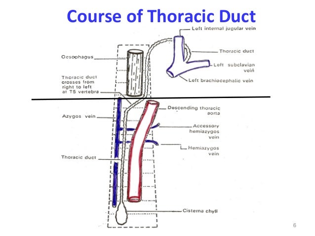
The thoracic duct is 38 to 45 centimeters long and 2 to 5 millimeters in diameter. It runs from the superior aspect of the cisterna chyli, a lymph sac at the L2 vertebral level, to the lower cervical spine. From the cisterna chyli, the duct continues superiorly, running between the aorta and the azygous vein and anterior to the vertebral column. The thoracic duct ascends through the aortic hiatus of the diaphragm entering the posterior mediastinum, still to the right of the vertebral column. It courses posterior to the esophagus at the T7 level and crosses over the midline to the left side of the thorax around the T5 vertebral level. As it continues upward, it runs behind the aorta and to the left of the esophagus ascending 2-3 cm above the clavicle. In the superior mediastinum, it passes behind the left common carotid artery, the vagus nerve, and the internal jugular vein. It then descends to empty into the junction of the left subclavian and internal jugular veins.
What is the superior mediastinum?
In the superior mediastinum, it passes behind the left common carotid artery, the vagus nerve, and the internal jugular vein. It then descends to empty into the junction of the left subclavian and internal jugular veins. The wall of the thoracic duct has three layers: the intima, the media, and the adventitia.
Which duct is responsible for lymph drainage?
The thoracic duct is the larger of the two and responsible for lymph drainage from the entire body except for the right sides of the head and neck, the right side of the thorax, and the right upper extremity which are primarily drained by the right lymphatic duct.[1][2]
Where does the thoracic duct start?
The thoracic duct typically starts at the second lumbar vertebra at the cisterna chyli, ending at the junction of the left subclavian and jugular veins. A total of 1.5 to 2 liters of lymph flows through the thoracic duct per day.
How to approach thoracic duct?
An alternative route to approach the thoracic duct is by retrograde transvenous approach or even direct ultrasound-guided puncture in the neck followed by embolization .[23] Thoracic duct embolization is usually performed by the percutaneous transabdominal method, and the supply route is blocked using N-butyl cyanoacrylate that is well mixed with lipiodol.[24] The success rate of thoracic duct embolization is almost 70%.[4] Postoperative complications of this procedure include chronic diarrhea, lower extremity edema, and abdominal ascites. [21]
How much chyle does the thoracic duct carry?
It carries almost 4 liters of chyle daily, most of which originates in the digestive tract. The flow rate can be up to 100 ml/kg/day, depending on the diet consumed. A combination of intrathoracic and intraabdominal pressures and arterial pulsations helps in maintaining lymph flow in the thoracic duct.
What is the largest lymphatic duct in the body?
The thoracic duct is the largest lymphatic duct in the body,[1] with a typical length of 45 cm and a diameter of 2 to 5 mm. It drains lymph from the whole body except the right hemithorax, the right side of the head and neck, and the right upper limb.
What are the effects of a thoracic duct leak?
Because of its unique contents, a thoracic duct leak can have multiple effects such as mechanical compression on the heart leading to tamponade, volume loss, pleural effusion, and pulmonary atelectasis, all of which can occur in an acute event. [10]
Which side of the thoracic duct is affected by pleural effusion?
The varied course of the thoracic duct and its site of leak dictates the side of pleural effusion. Hence, injury to the duct below the 5th thoracic vertebrae results in pleural effusion on the right side, and damage above this level occurs in a left-sided pleural effusion.[13] Diseases can also disrupt multiple tributaries to the thoracic duct that can produce pleural effusion unilaterally or bilaterally.
Why do ducts leak?
Thoracic duct leaks are most often due to traumatic injury and iatrogenic injury. Surgery of the esophagus and the heart are the most common source of injury. Untreated high volume leaks have a significant mortality rate.

Overview
In human anatomy, the thoracic duct is the larger of the two lymph ducts of the lymphatic system. It is also known as the left lymphatic duct, alimentary duct, chyliferous duct, and Van Hoorne's canal. The other duct is the right lymphatic duct. The thoracic duct carries chyle, a liquid containing both lymph and emulsified fats, rather than pure lymph. It also collects most of the lymph in the …
Structure
In adults, the thoracic duct is typically 38–45 cm in length and has an average diameter of about 5 mm. The vessel usually starts from the level of the twelfth thoracic vertebra (T12) and extends to the root of the neck. It drains into the systemic (blood) circulation at the angle of the left subclavian and internal jugular veins as a single trunk, at the commencement of the brachiocephalic vein.
The thoracic duct originates in the abdomen from the confluence of the right and left lumbar trunks and …
Function
The thoracic duct collects most of the lymph in the body other than from the right thorax, arm, head, and neck. These are drained by the right lymphatic duct.
The lymph transport, in the thoracic duct, is mainly caused by the action of breathing, aided by the duct's smooth muscle and by internal valves which prevent the lymph from flowing back down again. There are also two valves at the junc…
Clinical significance
The first sign of a malignancy, especially an intra-abdominal one, may be an enlarged Virchow's node, a lymph node in the left supraclavicular area, in the vicinity where the thoracic duct empties into the left brachiocephalic vein, right between where the left subclavian vein and left internal jugular join (i.e., the left Pirogoff angle). When the thoracic duct is blocked or damaged a large amount of lymph can quickly accumulate in the pleural cavity, this situation is called chylothorax.
Additional images
• Transverse section of thorax, showing relations of pulmonary artery.
• The arch of the aorta, and its branches.
• Deep lymph nodes and vessels of the thorax and abdomen (diagrammatic).
See also
• Lymph duct
• Lymphatic system
External links
• Anatomy figure: 21:05-02 at Human Anatomy Online, SUNY Downstate Medical Center — "The thoracic duct and azygos venous network"
• Anatomy image:8901 at the SUNY Downstate Medical Center
• figures/chapter_24/24-5.HTM: Basic Human Anatomy at Dartmouth Medical School