What is tympanometry?
Tympanogram: Interpreting impedance results Tympanometry is a test of middle ear functioning. It looks at the flexibility (compliance) of the eardrum to changing air pressures, indicating how effectively sound is transmitted into the middle ear.
Does tympanometry show middle ear pressure and static compliance?
Tympanometry showed no measurable middle ear pressure or static compliance, consistent with middle ear pathology. Tympanometry showed significant negative middle ear pressure in the presence of normal static compliance, consistent with Eustachian tube dysfunction or middle ear pathology.
How do you measure compliance on a tympanogram?
Compliance is shown in a vertical manner on the tympanogram and is measured in ml or mmho. In case the middle ear cavity pressure is the same as the external auditory canal pressure then the tympanogram shows maximum compliance of the middle ear system.
Why does my child show falsely increased compliance on a tympanometer?
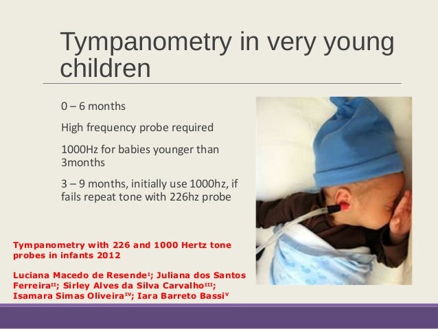
Children under six months of age may demonstrate falsely increased compliance, due to the fact that the ear canal skin and cartilage is quite lax. In this situation, the child may have a middle ear filled with fluid, but still register a compliance within the normal range. The tympanometer automatically calculates the canal volume.

What does compliance mean in tympanometry?
Tympanometry is a test of middle ear functioning. It looks at the flexibility (compliance) of the eardrum to changing air pressures, indicating how effectively sound is transmitted into the middle ear.
What is static compliance in tympanometry?
Static compliance (SC) “is the greatest amount of acoustic energy absorbed by the middle ear system (the vertical peak of the tympanic tracing)” (Onusko, 2004, p. 1716).
Which tympanogram shows normal middle ear pressure and compliance?
Immittance Audiometry There are three main types of tympanograms: A, B, and C. A Type A tympanogram indicates normal middle ear status. Reduced mobility of the tympanic membrane caused by a stiffened middle ear system can cause a shallow peak on the tympanogram, called a Type As tympanogram.
What causes highly compliant middle ear system?
Maximum compliance of the middle ear system occurs when the pressure in the middle ear cavity is equal to the pressure in the external auditory canal. The maximum compliance value occurs at the highest peak of the curve on the graph (figure 3).
What is normal middle ear compliance?
The compliance of the middle ear system is a measure of how well the system responds to sound. This is shown by the height of the “peak”. Middle ear compliance values from 0.3 to 1.5 cc are usually considered normal.
How do you read a tympanometry report?
How do you read a tympanogram report? A tympanogram will show the results of one eardrum at a time. An “L” on the tympanogram indicates the left eardrum; an “R” indicates the right eardrum. A clinician will mainly look at the peak of each graph.
What does Type C tympanogram mean?
Type C tympanograms indicate negative middle ear pressure and a retracted tympanic membrane.
What does a tympanometry test for?
Tympanometry. Tympanometry tests how well your eardrum moves.
How do I read my hearing test results?
The results of the hearing test are an indication for the degrees of hearing loss:Normal hearing: -10 to 20 dB.Mild hearing loss: 20 to 40 dB higher than normal.Moderate hearing loss: 40 to 70 dB higher than normal.Severe hearing loss: 70 to 90 dB higher than normal.Profound loss: 90 dB or more.
Is tympanometry a hearing test?
Often used to assess the function of the middle ear, tympanometry is one test that can determine whether your hearing loss can be helped by hearing aids or whether a medical treatment is available to treat your loss instead.
What type of tympanogram is conductive hearing loss?
Conductive hearing loss is often associated with Type B. Type C Tympanogram – This result tells us the person's Eustachian tube isn't working well. They could be just contracting or just recovering from otitis media.
What are the different types of Tympanograms?
Tympanogram tracings are classified as type A (normal), type B (flat, clearly abnormal), and type C (indicating a significantly negative pressure in the middle ear, possibly indicative of pathology).
What type of tympanogram is conductive hearing loss?
Conductive hearing loss is often associated with Type B. Type C Tympanogram – This result tells us the person's Eustachian tube isn't working well. They could be just contracting or just recovering from otitis media.
What does a Type C tympanogram mean?
Type C tympanograms indicate negative middle ear pressure and a retracted tympanic membrane.
What does a double peak tympanogram mean?
Double‑peak tympanometry with an intact tympanic membrane may indicate diseases causing severe erosion in the EAC.
What does tympanometry show?
The tympanometry result indicates; ear canal volume (cm3), the max pressure (daPa), and the peak compliance (ml). The test plays a crucial role in distinguishing between conductive hearing loss and sensorineural hearing loss.
Why is tympanometry important?
This test helps in determining various factors. Some of the factors are –. Presence of fluid in the ear. The tear in the eardrum or tympanic membrane perforation.
How Is Tympanometry Performed?
Tympanometry is performed by a hearing healthcare professional or a doctor. The test can be performed in any hearing clinic by any of the audiologist/professional.
What is the tymp test?
The graphics representation of tymp test data is the tympanogram. Tympanogram represents the relationship between the air pressure in the ear canal and the movement of the eardrum, or tympanic membrane, and the tiny bones in the air-filled middle ear space.
What is the role of tympanometry in the eardrum?
Tympanometry provides some extra information Eustachian Tube Functioning.
Why do you need a tymp test for otoscopy?
Otoscopy test with tympanometry enhances the reliability of the diagnosis as there are several ear canal and eardrum abnormalities that in some cases cause might cause different types of abnormalities which can easily be traced through tympanogram. The graphics representation of tymp test data is the tympanogram.
What is the middle ear test?
There are many tests that makeup through evaluation and act like pieces of a puzzle which when put together helps to determine the type and cause of hearing loss. One such middle ear test is tympanometry. Tympanometry is a type of test which is helpful in determining if the hearing loss can be helped by hearing aid or any medical treatment is ...
What is a tympanometry exam?
Tympanometry provides a way, along with a physical exam, for doctors to diagnose and monitor problems in the middle ear.
What does normal tympanometry test mean?
Normal tympanometry test results mean: There’s no fluid in the middle ear. The eardrum moves normally. There’s normal pressure in the middle ear. There’s normal movement of the ossicles (the small bones of the middle ear that conduct sound and aid in hearing) and the eardrum.
What is a tympanogram?
The results of tympanometry are recorded on a graph called a tympanogram. The test can help your doctor determine if you have: fluid in your middle ear. otitis media (a middle ear infection) a perforation (tear) in the tympanic membrane. a problem with the eustachian tube, which links the upper part of the throat and nose with the middle ear.
What is tympanic hearing test?
Tympanometry can help diagnose disorders that can lead to hearing loss, especially in children. The test measures the movement of your tympanic membrane in response to changes in pressure. The tympanic membrane is a thin tissue that separates the middle and outer segments of the ear.
How does a tympanogram work?
This test changes the air pressure in your ear to make the eardrum move back and forth. Measurements of the movement of your eardrum are recorded in a tympanogram.
What causes ear perforation?
perforation of the eardrum (tympanic membrane) scarring of the eardrum, which usually results from frequent ear infections. middle ear pressure beyond the normal range. growths in the middle ear. earwax blocking the eardrum. lack of mobility or other problems with the ossicles of the middle ear.
What happens if you test abnormally for tympanic membrane?
If your test results are persistently abnormal, or your doctor suspects that something other than fluid is behind the tympanic membrane, they may send you for additional testing and a follow-up appointment with a specialist. Last medically reviewed on June 1, 2018.
Why does a tympanometer measure canal volume?
Similarly, if there is a perforation of the tympanic membrane the tympanometer will measure an unusually large canal volume, because the space of the middle ear and mastoid air cells will be included in the volume calculation.
How does a tympanometer work?
The tympanometer measures the “admittance” or “compliance” of the tympanic membrane while different pressures are being applied to the external ear canal. The compliance of the TM is measured in cubic centimeters, and the pressure in the ear canal is measured in decapascals (daPa). The probe has different size “plugs” that provide a seal at the entrance to the external ear canal. The tip of the probe has a pressure transducer that changes the pressure in the external ear canal from negative, through atmospheric pressure, to positive pressure. While the pressure is changing, a sound transmitter sends a sound wave to the tympanic membrane. The wave that is reflected from the TM is then picked up by a microphone in the probe. The tympanometer measures the energy of the reflected sound.
What does a tympanometer do when a child has a middle ear filled with fluid?
The tympanometer automatically calculates the canal volume. If there is cerumen or other material occluding the ear canal, the volume will measure abnormally low.
What is the case of a one year old girl with bilateral pressure equalizing tubes?
You check a tympanogram to assure yourself that no fluid is present in the middle ear. Case 2. A one-year-old girl with bilateral pressure equalizing tubes. She has had recurrent infection in the left ear, and you suspect that the pressure equalizing tube may be obstructed.
What is a gradient of 150 dapa?
A gradient greater than 150 daPa is often associated with a collection of fluid in the middle ear space.
Is the tympanic membrane dull?
The tympanic membrane is dull and non-erythematous. The landmarks are easily visualized.
What are the problems with tympanometry?
One of the first and most obvious problems in discussing tympanometric data is the lack of standardization of acoustic-immittance instruments. As a consequence, the terminology associated with tympanometry has been inconsistent and, at times, confusing. The recently adopted American National Standard for acoustic-immittance instruments (ANSI S3.39, 1987) should help to rectify this long-standing problem. The International Electrotechnical Commission (IEC SC29C) also is nearing completion of an international standard for aural-impedance/admittance instruments. The two groups coordinated their efforts to ensure that the terminology and instrument specifications would be nearly identical in the two standards. To avoid further confusion, the terminology recommended in the ANSI standard will be used throughout this tutorial. The following list of terms pertains specifically to tympanometric measurements and instrumentation.
When was tympanometry first used?
Tympanometry has been performed routinely in clinical settings since the early 1970s. The first generation of acoustic-immittance instruments measured only the magnitude of acoustic immittance using a single low-frequency probe tone. The next generation of instruments was capable of making two-component, two-frequency measurements. Despite a significant body of literature demonstrating the advantage of using the more complex measurements for evaluating some types of middle-ear pathologies, many have persisted with the more familiar and simplified measurement techniques of the early 1970s. With the rapid integration of computers into the field, acoustic-immittance measurements are likely to become even more sophisticated, thus increasing the gap between advances in technology and instrumentation and an understanding of acoustic-immittance data. The primary objective of this tutorial is to enhance the understanding of two-component, multiple-frequency acoustic-immittance measurements in preparation for the next generation of computer-interfaced instruments and to demonstrate cases in which these measurements can be especially useful. It is recognized, however, that there are some instances in which only a screening procedure and a superficial analysis of the tympanometric data are necessary. Screening for middle-ear effusion has been the topic of several articles and will not be discussed further in the present paper.
How does a stiff ear affect the transmission system?
If the middle ear is stiffened because of a pathology such as otosclerosis, then the ear will remain stiffness-controlled over a wider frequency range than normal, and thus, the resonant frequency of the middle ear will be shifted toward a higher than normal frequency. In contrast, if the ear becomes mass controlled because of a pathology such as ossicular discontinuity, then the resonant frequency of the middle ear will decrease. The subsequent discussion will present examples of abnormal stiffness- and mass-controlled tympanograms plotted in a variety of forms that are being introduced with the new generation of microprocessor-based immittance instruments.
Why is immittance measured at ambient pressure?
Some authors have argued that immittance measures at ambient pressure are preferred because they represent the resting state of the ear under test or the normal mode of functioning. Immittance at ambient air pressure, however, can fluctuate in a given individual as the person swallows and breathes. Substantial differences in compensated ambient static immittance across time may occur that have no clinical relevance and are merely related to small shifts in middle-ear pressure. As discussed in a subsequent section, shifts in middle-ear pressure are not pathological unless the tympanogram is also reduced in amplitude.
How to interpret tympanograms?
Two approaches to the interpretation of tympanograms are in widespread use: (a) classification of tympanometric shape and (b) calculation of compensated static immittance. Calculation of static immittance can be of clinical value, particularly for low-frequency probe tones at which most tympanograms are single peaked, but an analysis of the morphology of the tympanograms may be more informative for high probe frequencies at which many of the tympanograms are notched in both normal and pathological ears. Several methods have been suggested for analyzing tympanometric shapes to aid in differentiating between normal and pathological middle ears. The typing of tympanometric patterns, popularized by Liden ( 1969 ); Liden, Peterson, and Bjorkman ( 1970 ); and Jerger ( 1970 ), probably is in the most widespread use. Feldman ( 1976a, 1976b, 1977) and Paradise, Smith, and Bluestone ( 1976) expanded these types to include a descriptive analysis of amplitude, peak pressure, and tympanometric width (gradient). With the exception of Feldman ( 1976a, 1976b, 1977 ), most classification procedures have been applied to single-component (admittance magnitude), single-frequency (226 Hz) tympanograms ( Margolis & Shanks, 1985, Figures 23.1, 23.2, & 23.4). For the most efficient clinical interpretation, however, low-frequency tympanograms may be best suited for static-immittance measurements. A descriptive analysis of tympanometric shape at 226 Hz is not particularly useful because normal low-frequency tympanograms are single peaked, except for some neonate ears. In general, pathological conditions of the middle ear do not alter the basic tympanometric shape for a low-frequency probe signal unless the impedance is so high as to obliterate the peak. Most abnormal 226-Hz tympanograms remain single peaked but have an abnormal peak amplitude. The best way to distinguish between normal and abnormal single-peaked tympanograms is by quantifying the peak amplitude (i.e., by calculating compensated static immittance).
What frequency is a mass pathology?
With currently available two-component instruments, changes in middle-ear resonance can be inferred by examining tympanometric shapes at two frequencies, 226 and 678 Hz. If the ear is abnormally mass controlled at 678 Hz, then a mass pathology is suspected. In Figure 7, examples of two-component, two-frequency admittance tympanograms reflecting a mass-controlled system because of ossicular discontinuity (left panel), tympanosclerosis (middle panel), and secretory otitis media (right panel) are shown. The increase in mass is obvious from an examination of tympanometric shapes at 660/678 Hz, but is not easily recognized from tympanometric shapes at 226 Hz. The 678 Hz tympanograms recorded in ossicular discontinuity and tympanosclerosis both can be classified as abnormally mass-controlled according to the Van Camp et al. ( 1986) criteria previously discussed. The tympanograms in the left panel of Figure 7 (ossicular discontinuity) are abnormal because the shapes are too complex (i.e., more than three extrema for the 678 Hz conductance tympanogram) and because the notches are too broad (i.e., the distance between the outermost maxima exceeds 75–100 daPa for both the 678 Hz susceptance and conductance tympanograms). The tympanograms in the right panel of Figure 7 (secretory otitis media) are abnormal because the notch width in the 678 Hz susceptance tympanogram exceeds 75 daPa. The tympanograms in the middle panel (tympanosclerosis) are abnormal because notching occurs at 226 Hz and because the shapes are too complex and broadly notched at 678 Hz. Pathologies of the tympanic membrane such as tympanosclerosis and monomeres often produce abnormal, mass-controlled tympanograms with little if any effect on hearing sensitivity. Low impedance tympanic-membrane pathologies present one of the most frustrating problems associated with tympanometric measures. A low impedance tympanic-membrane pathology will dominate completely the tympanometric pattern and will mask a more clinically important, high-impedance pathology such as otosclerosis. This case points out the importance of an otoscopic examination. The presence of a tympanic membrane pathology seriously restricts the use of tympanometry in the differential diagnosis of a conductive hearing loss because it may mask a coexisting, more medial pathology.
How does tympanometric pressure compare to middle ear pressure?
The error in estimating middle-ear pressure from tympanometric peak pressure is less than 30 daPa in normal ears and is largely because of movement of the tympanic membrane during tympanometry ( Eliachar & Northern, 1974; Flisberg, Ingelstedt, & Ortegren, 1963; Renvall & Holmquist, 1976 ). The difference between tympanometric peak pressure and middle-ear pressure, however, can be larger in abnormal ears. For example, Renvall and Holmquist ( 1976) found pressure differences in human temporal bones ranging from 0% to 70%. Flisberg et al. ( 1963) attributed the large discrepancy to a decrease in middle-ear volume due to inflammation of the middle-ear tissues that results in a large middle-ear pressure change with movement of the tympanic membrane during tympanometry. The disparity between tympanometric peak pressure and middle-ear pressure also is large in patients with monomeric tympanic membranes who have abnormally large eardrum displacement during tympanometry. Thus, the error in estimating middle-ear pressure from tympanometric peak pressure is largest in patients of greatest clinical interest (i.e., those with middle-ear disease).
The middle ear and its components
The tympanic membrane (TM) is a thin three-layer membrane, cone-like in shape, that separates the middle ear from the outer segments. During otoscopy, it is expected that a healthy, normal TM will reflect a cone of light, a reflection off of the membrane in the anterior inferior portion of the TM.
When is tympanometry conducted?
Tympanometry assesses the normal (or abnormal) functioning of the middle ear system. In other words, the efficiency of the middle ear. The test itself presents both positive and negative pressures accompanied by a constant probe tone. The test measures the amount of absorption or reflection of the probe tone from the middle ear space.
What does a tympanometer measure?
Tympanometry collects data to test four basic functions of the middle ear. The results of tympanometry are plotted on a graph called a tympanogram. A trained eye is needed to read and interpret a tympanogram, which can require some practice. A typical tympanometry result indicates the following:
Identifying the Data and Measurements on a Tympanogram
Tympanograms are classified by types – Type A, B, C, AS, and AD. Each classification indicates a range that falls between normal and abnormal.
How do you read a tympanogram report?
A tympanogram will show the results of one eardrum at a time. An “L” on the tympanogram indicates the left eardrum; an “R” indicates the right eardrum. A clinician will mainly look at the peak of each graph. The examples below use a 226 Hz probe tone. (Classifications can vary between audiologists, guidelines, countries, and clinics.)
A portable tympanometer that goes where you go
The KUDUwave Pro TMP is a portable audiometry system that integrates bilateral tympanometry for the very first time in the history of audiology. Dual tympanometers are integrated into each KUDUwave earcup enabling tympanometry of both ears without having to switch ears.
