What type of microscope should I use to view diatoms?
Brightfield and phase contrast microscopy can be used for observing diatoms. Here, phase contrast is particularly preferred when viewing specimens that are lightly silicified. For a dry specimen, 40X and 100X are commonly used. Take a look at our article in more depth about Pond Water Under the Microscope and Microorganisms.
What are the characteristics of a diatom?
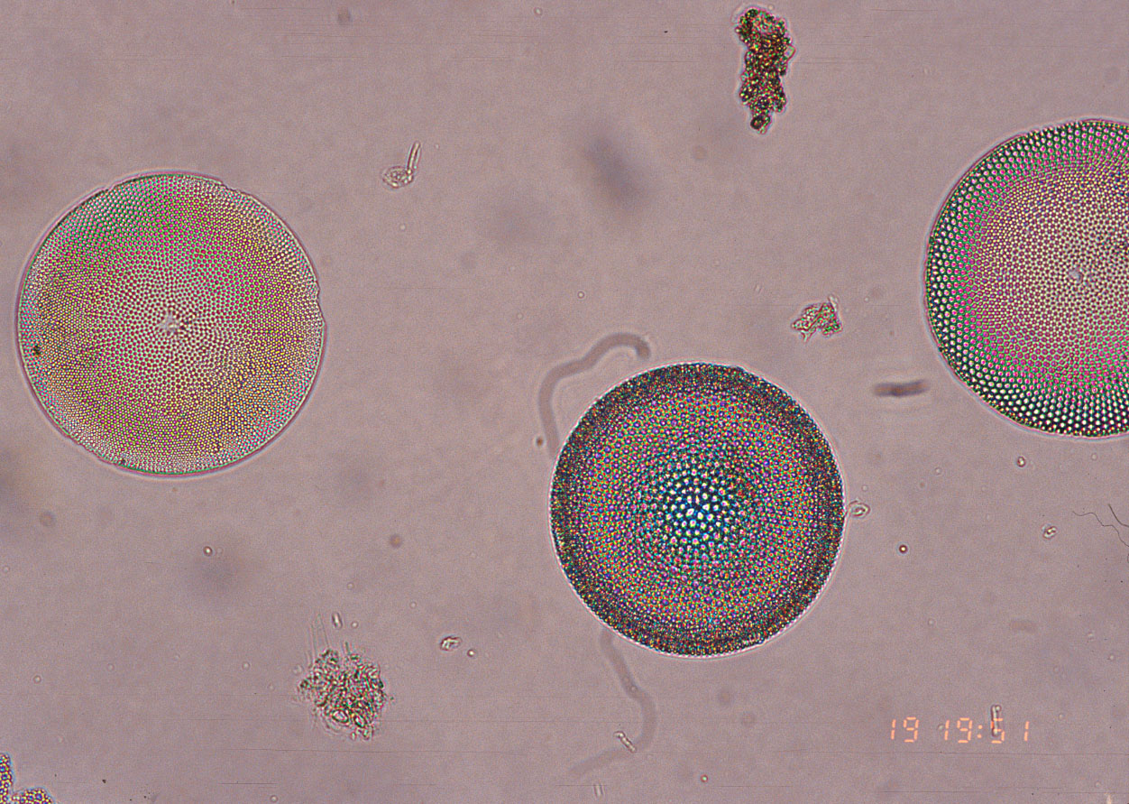
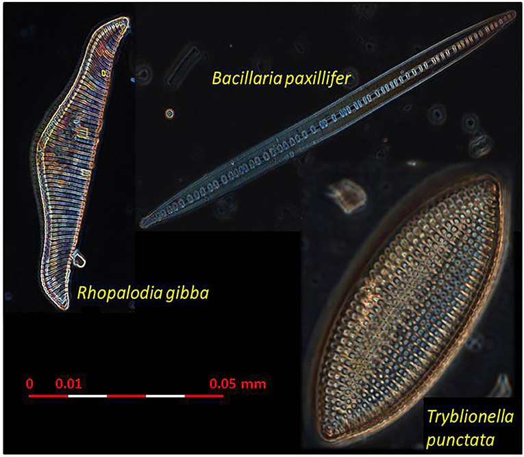
Diatoms are widely recognized by microbe enthusiasts and the general public for their wide range of attractive shapes and glassy appearance under a microscope. They occur in nature as circles, squares, ribbons, fans, zigzags, and stars, among other shapes. Diatoms are known as the “jewels of the sea.”
How much magnification do you need to see atoms?
No magnification will allow you to see atoms in the normal sense. Conventional microscopes focus light through a lens to magnify objects. However, this magnification is limited by the wavelength of light — you can’t see things that are only a tiny fraction of a wavelength in size.
How can i Zoom in with a microscope to 2NM?
Well you can use an atomic microscope which has the size of a building to zoom you till 2nm (nano meters) and if you are a science dude 2x10^-9 which is very small and see this: , First in biological Atomic resolution.
What are Diatoms?
What are the adaptations of diatoms?
What happens when diatoms die?
How do diatoms reproduce?
What are the two main orders of diatoms?
How do planktonic species stay afloat?
Is diatomaceous earth poisonous?
See 2 more
What microscope is used to see diatoms?
scanning electron microscopesScientists use light microscopes (LM) or scanning electron microscopes (SEM) to view diatoms. When diatoms are viewed with a light microscope, the silica cell walls appear transparent (because we are seeing through glass).
How do you view diatoms?
At this point you have to go to places where there is the presence of water, and observing the rocks and plants close to shore, look for those surrounded by the presence of a brown layer or one that is sometimes black. If you see such layers, you are most likely in the presence of diatoms.
How do you count diatoms under a microscope?
Once the slide is placed on the microscope stage, the starting coordinates are randomly chosen and recorded. For diatoms, 250 valves are counted and identified. Once 250 valves are reached, the ending coordinates are recorded and each count is multiplied by 2.
What type of microscope is used for plankton?
Observe the sample with a dissecting microscope. Since the plankton can move up and down in the drop, you will need to refocus your microscope to see plankton at different levels. 2.
How do you make diatom slides?
4:215:36Diatoms. Part 2: Preparation of permanent slide) - YouTubeYouTubeStart of suggested clipEnd of suggested clipThen take a cover slip from which the diluted diatoms solution has already been evaporated place itMoreThen take a cover slip from which the diluted diatoms solution has already been evaporated place it facedown on the plur acts place the slide on the ceramic tile and heat it gently.
How do you isolate diatoms?
The best and fast way to isolate diatoms from a natural sample for a clonal and axenic culture is using a flow cytometer equipped with cell sorting (FACS). Using this technique you can get a 96 wells plate with theoretically 96 isolated cells in only a few minutes. One of the ways is to use micropipette.
Can we see diatom with naked eye?
Individually, diatoms are invisible to the naked eye, but large concentrations can often be seen as brown or golden-brown discolouration on, for example, the surface of rocks, sand or mud. The structure of a diatom is similar to that of a pillbox or a petri dish.
What does diatomaceous earth look like under microscope?
7:128:37What is DIATOMACEOUS EARTH? | How to observe ancient fossils | 222YouTubeStart of suggested clipEnd of suggested clipAround here a little bit. Now i would like to now encourage you yourself to go out to collectMoreAround here a little bit. Now i would like to now encourage you yourself to go out to collect diatoms. Go out on a pond uh scratch the material off from a rock put it under the microscope.
How large is a diatom?
Diatoms are commonly between 20-200 microns in diameter or length, although sometimes they can be up to 2 millimeters long. The cell may be solitary or colonial (attached by mucous filaments or by bands into long chains).
What magnification do you need to see plankton?
40x magnificationFocus on the slide content using 10x magnification power. Some microscopes have a pointer that helps to identify the location of the plankton. They should observe the section now with 40x magnification power.
What magnification do you need to see zooplankton?
For viewing zooplankton you often don't need such a high magnification at all! I myself have a 5 x, 10 x, 20 x, 40 x and 100 x lens on my microscope and for zooplankton I use the 5 x and 10 x most. 40 x I only use them to look at details such as hairs on the legs of copepods.
Which type of microscope would you use to view the plankton and why?
Compound microscopes have very high magnification, which is essential to view these tiny phytoplankton.
Can we see diatom with naked eye?
Individually, diatoms are invisible to the naked eye, but large concentrations can often be seen as brown or golden-brown discolouration on, for example, the surface of rocks, sand or mud. The structure of a diatom is similar to that of a pillbox or a petri dish.
How do you collect diatom samples?
Collection of diatoms requires contact with stream water and stream sediment. Use precaution when reaching in streams to remove substrates to sample as glass or other sharp objects may be present and hard to see.
How are diatoms used in forensic science?
Detection of diatoms in various organs can contribute to the diagnosis of death by drowning, a process referred to as the 'diatom test'. The use of diatoms (the 'diatom test') to diagnose a cause of death by drowning is often one of a number of independent techniques utilized by the forensic pathologist.
What are the characteristics of diatom?
The diatom is unicellular, photosynthetic, free-moving, has a specialized frustule wall made of silica, has food reserves, has many different shapes, has few colors due to chlorophyll, a two-layered outer shell known as the thecae, and have both sexual and asexual reproductive cycles.
What are Diatoms?
Diatoms are photosynthetic organisms referred to as algae with a length/diameter of between 2 and 500 microns. They have a transparent cell wall (frustule) made of silicon dioxide, which is itself hydrated with a little amount of water. Therefore, diatoms are simply aquatic organisms, which can be found in such environments as fresh and marine (salty) waters and moist soils.
What are the adaptations of diatoms?
Different types of diatoms have different morphological adaptations that allow them to survive in their respective habitats . For instance, diatoms that live in such aquatic habitats as ponds, lakes and oceans possess morphological features that make it possible for them to remain suspended in water.
What happens when diatoms die?
When the aquatic diatoms die, they sink to the bottom of whatever habitat they are found in and collect to form what is known as diatomaceous earth. The shells (made of silica) cannot decay, and therefore collect together at the bottom of the lake. In some cases, they collect to form a soft, chalky light weight rock called diatomite.
How do diatoms reproduce?
Life Cycle. Typically, diatoms divide and reproduce by a process referred to as vegetative division, which involves the division of a single cell into two new cells. During the reproduction cycle, the new cell is formed inside the parent cell. The new cell is smaller in size given that it forms within the mother cell that has a rigid cell wall ...
What are the two main orders of diatoms?
Diatoms are also divided in to two main Orders, which include the Centrales and the Pennales.
How do planktonic species stay afloat?
By forming long chains that are linked to each other by silica spines, these planktonic species are able to remain suspended on water. Others will form zigzag/stellate colonies that keep them afloat. These species are often star-shaped. Other species grow and multiply on such surfaces as rocks and other aquatic plants.
Is diatomaceous earth poisonous?
Dust containing this substance can also be irritating to the eyes or cause skin irritation and dryness. However, it is not poisonous. See more information on Diatomaceous Earth here.
How to Observe Diatoms Under a Microscope?
As diatoms have been observed under a microscope since the early 18th century, solid protocols for diatom microscopy have been established. Diatoms are very common so don’t need to look hard to see them. Colonies, filaments, and flocks of diatoms are commonly viewed and photographed under 100X-400X magnification.
How Are Diatoms Classified?
Like other common microalgae and phytoplankton, diatoms belong to the Kingdom Protista. Diatom taxonomy, or the biological classification of diatoms in relation to other diatoms and the wider web of life, is complex and evolving. Diatom taxonomy is complicated by the sheer number of known and newly-discovered species of diatoms, which is estimated to be between 12,000 and 200,000 species.
What do Diatoms Eat?
To sustain themselves and produce energy, diatoms require carbon dioxide, water, and light. Like most microorganisms, they are responsive to their environment, and energy production can be influenced by water temperature, light availability, where they exist within the water column, and the speed at which they drift in water. Diatom energy is produced through photosynthesis, wherein energy from sunlight is converted into chemical energy to power a diatom’s cellular processes.
Where do Diatoms Live?
Since diatoms require only water, carbon dioxide, and light to produce energy, they live all over the world in oceans, bodies of freshwater, and damp soils. Free floating diatoms in oceans, called planktonic diatoms, exist in the upper mixed layer of the water column, where there is optimal exposure to nutrients and sunlight. Benthic diatoms, which exist closer to the seabed and are exposed to lower light levels, also exist but are less well-studied than their planktonic counterparts.
What happens to diatoms when they die?
In addition to increased sinking during the bloom to bust transition, when diatoms die, their frustule shells sink to the bottom of the waterbed and accumulate. Ocean and seabed are full of this diatom shell dust, which becomes white and chalky. On the seabed, this substance can accumulate up to 1,400 meters thick. Commercially, humans known this substance as diatomaceous earth, and use it for a variety of human uses.
What kingdom are diatoms in?
The study of diatoms falls within phycology or algology, the study of algae. Like other common microalgae and phytoplankton, diatoms belong to the Kingdom Protista. Diatom taxonomy, or the biological classification of diatoms in relation to other diatoms and the wider web of life, is complex and evolving.
How much oxygen does a diatom produce?
During photosynthesis, molecular oxygen is produced, and diatoms produce approximately 25% of the Earth’s oxygen. The ability of diatoms to undergo photosynthesis makes them similar to plants, but plants and diatoms evolved this ability from separate ancestral lineages.
What microscope can zoom in 2nm?
Well you can use an atomic microscope which has the size of a building to zoom you till 2nm (nano meters) and if you are a science dude 2x10^-9 which is very small and see this:
What microscope do you need to see DNA?
Now, if you want to visualise individual strands of double-stranded DNA (the most common form), you do need an advanced microscope. Either an atomic force microscope (AFM), which gives images like this:
How to see things at a nanometer?
To see things at a nanometer, which is a trillionth of a meter, you would need to increase magnification nearly 20,000,000 times. There are about 39 inches in a meter, meaning there are nearly 25,641,025,641 nanometers in an inch. This would give a field of view nearly 35x35 nanometer/inch, about 1282.1 nanometers total. At this range, atoms such as oxygen and hydrogen can appear, still kinda small, but hydrogen can look to be with oxygen ionizing in a chain, to be about 30x7 nanometer. Hydrogen 2/5 of the chain, and oxygen 3/5. This would appear about as wide as a pencil held at arms length.
How to trap atoms far away from each other?
It turns out that you can create systems to trap atoms far away from each other in a regular lattice and build a microscope to image them. This is done using so called optical lattices, standing waves of light formed by lasers, where the regions of minimum (or maximum depending on the frequency) intensity can form “wells” in which you can drop atoms. This is most easily done fo
Can you see atoms under a microscope?
Yes it is possible to see an atom under a microscope but not the particles of atoms. In fact the particles are used to probe the location of a single atom or the group of atom which is to be imaged. A group of scientists at UCLA [1] were able to image the arrangement of Platinum atoms.
Is seeing distorted?
Anyway, Seeing, that concept can be long and distorted depending on the definition. To see, some electromagnetic stimulation has to happen, at that level, visible ranges of light frequencies are wider than the sample, so coordination to the light refracting, is important. There is a way to accomplish it, but it's not easy at all, with the naked eye or in a sample, which allude to how I make it work, but as of yet, I haven't divulged precisely all my data. Though, there has been ample provided throughout Quora.
Can you see atoms with light?
Most of the answers here suggest that you can’t see atoms with light, but that’s not completely true. It is true for atoms in a lattice where the spacing is too small, such as a crystal and you hit the diffraction limit.
What objective to use for diatoms?
If you are working with cleared diatom material, an oil immersion x63 or x100 objective, will help enormously.
Can you use LM for coccolithophorids?
If you have access to SEM, this is really the best for coccolithophorids, but you can resolve some of the larger species with good LM. If you are using LM for diatoms, DIC or phase contrast is essential. Fluorescence will be very important for resolving the plate tabulation of dinoflagellates, but you may not need this level of discrimination?
Do Goryaev chambers have holes?
Neubauer, Nageotte, Goryaev chambers have holes on the chamber side. When I investigate the sample after less than one hour more than half of sampled water evaporates. So they don’t help me.
Is a 1000x binocular microscope good?
Usb binocular digital microscope yj-2005dn. Work best and is also not expensive. 1000x is more than enough. Because more than 1000x not increase any resolution and only enlarge image.
What are the two types of diatoms?
Diatoms can be ecologically separated in two broad categories: planktonic species that live in the water column and benthic species that live in the sediments or attached to rock, plant, or other substrates. A ratio called P:B or the planktonic:benthic ratio provides a simple metric of possible changes in the paleoenviroment based on lake level change or nutrients. The ratio is often expressed as a percentage, i.e. a P:B ratio of 1.0 would equal 50% percent planktonic species. Depending on lake morphometry lake level changes can result in dramatic shifts in the proportion of open water and littoral zone habitat (Wolin and Stone 2010). Sediments preserve a temporal and spatial integration of a lake's diatom communities so periods with greater littoral zone extent will generally have great benthic diatom numbers. The other major factor that drives changes in P:B is lake eutrophication. Nutrient addition to lakes often results in proportionally greater planktonic diatom growth and higher P:B ratios (Edlund et al. 2009).
What are the conditions of diatoms?
Several environmental conditions enhance the dissolution of diatoms: breakage, salinity, alkalinity, and silica-poor environments. Diatom valves are composed of biogenic opaline silica and each valve has areas where silica is deposited more thickly (e.g., central areas, raphe ribs) as well as areas with patterns of ornamentation comprising holes (areolae, pore fields) and lines (striae). The dissolution of diatoms generally follows a pattern where highly ornamented or thin areas are lost first, followed by sequential loss depending on silica thickness, until only the most heavily silicified areas of the diatom remain. Diatom species are also differentially susceptible to dissolution. A simple metric (% pristine) based on the condition of diatom microfossils in smear slides can be used to summarize the level of dissolution in your samples.
What are diatoms made of?
Diatoms are one of the most abundant biological microfossils in sediment cores and on smear slides. Diatoms are one group of microscopic algae and characterized by their golden-brown pigmentation and having a cell wall made of biologically produced glass or biogenic opaline silica. Because they are made of opaline silica, diatom cell walls are fairly resistant to breakage and dissolution and can form up to 60% of the dry weight in some lake cores (Edlund et al. 2000). The cell wall in a living diatom is put together like a Petri dish; the two halves are called valves with one valve slightly larger and overlapping the other valve. There are a few other parts in the cell wall, primarily hoop or band-like structures called girdle bands that form the overlap between the two valves. Collectively, the siliceous parts surrounding a single cell are termed the frustule. Size, shape, and ornamentation on the valves distinguish the more than 40,000 diatom species in the world. For detailed diatom analysis, sediments are specially prepared using peroxide or strong acids to digest the cell contents; the "cleaned" cell walls are mounted on microscope slides using specialized mountants that optimize resolution of valve ornamentation for species-level identification. More detailed information on diatom preparation and species identification can be found here.
What are Diatoms?
Diatoms are photosynthetic organisms referred to as algae with a length/diameter of between 2 and 500 microns. They have a transparent cell wall (frustule) made of silicon dioxide, which is itself hydrated with a little amount of water. Therefore, diatoms are simply aquatic organisms, which can be found in such environments as fresh and marine (salty) waters and moist soils.
What are the adaptations of diatoms?
Different types of diatoms have different morphological adaptations that allow them to survive in their respective habitats . For instance, diatoms that live in such aquatic habitats as ponds, lakes and oceans possess morphological features that make it possible for them to remain suspended in water.
What happens when diatoms die?
When the aquatic diatoms die, they sink to the bottom of whatever habitat they are found in and collect to form what is known as diatomaceous earth. The shells (made of silica) cannot decay, and therefore collect together at the bottom of the lake. In some cases, they collect to form a soft, chalky light weight rock called diatomite.
How do diatoms reproduce?
Life Cycle. Typically, diatoms divide and reproduce by a process referred to as vegetative division, which involves the division of a single cell into two new cells. During the reproduction cycle, the new cell is formed inside the parent cell. The new cell is smaller in size given that it forms within the mother cell that has a rigid cell wall ...
What are the two main orders of diatoms?
Diatoms are also divided in to two main Orders, which include the Centrales and the Pennales.
How do planktonic species stay afloat?
By forming long chains that are linked to each other by silica spines, these planktonic species are able to remain suspended on water. Others will form zigzag/stellate colonies that keep them afloat. These species are often star-shaped. Other species grow and multiply on such surfaces as rocks and other aquatic plants.
Is diatomaceous earth poisonous?
Dust containing this substance can also be irritating to the eyes or cause skin irritation and dryness. However, it is not poisonous. See more information on Diatomaceous Earth here.
