The stylopharyngeus muscle of the pharynx is innervated by the glossopharyngeal nerve. This muscle acts to shorten and widen the pharynx, and elevate the larynx during swallowing. Fig 1.2 – Path of the parasympathetic fibres to the parotid gland. The glossopharyngeal nerve provides parasympathetic innervation to the parotid gland.
What are the 4 functions of the glossopharyngeal nerve?
Function 1 Sensory Function. The glossopharyngeal nerve plays a sensory role in numerous important structures. ... 2 Specialized Sensory Function. The lingual branch performs the specialized task of transmitting taste information to your brain. ... 3 Motor Function. ... 4 Parasympathetic Function. ...
What nerve innervates the stylopharyngeus?
Stylopharyngeus Muscle Nerve. This is the motor branch that provides the motor innervation for the stylopharyngeus muscle. It reaches the muscle from its lateral surface, in the part of the glossopharyngeal nerve course between the stylopharyngeus and the styloglossus muscle.
Where does the glossopharyngeal nerve travel?
Meanwhile, the main glossopharyngeal nerve travels downward between the internal carotid artery and the internal jugular vein and then curves forward to form an arch on the side of your neck on top of the stylopharyngeus muscle and the middle pharyngeal constrictor muscles high in the throat.
What is the difference between sensory and motor division of glossopharyngeal nerve?
The motor division of the glossopharyngeal nerve is derived from the basal plate of the embryonic medulla oblongata, while the sensory division originates from the cranial neural crest .

What areas are innervated by the glossopharyngeal nerve IX?
These nerves are sensory and they innervate the mucosa of the posterior one-third of the tongue, starting from the terminal sulcus of the tongue, and up to the epiglottis.
What does glossopharyngeal nerve supply?
The glossopharyngeal nerve (CN IX) supplies organs, muscles and other structures in your mouth and throat. It helps you taste food and sense pain in your throat.
What is the function of the glossopharyngeal nerve quizlet?
The glossopharyngeal nerve is cranial nerve IX. Its major motor function is to help in swallowing.
What is the function of glossopharyngeal and vagus nerve?
The glossopharyngeal and vagus cranial nerves provide the brainstem with sensory inputs from different receptors in the heart, lung, and vasculature. This afferent information is critical for the short-term regulation of arterial blood pressure and the buffering of emotional and physical stressors.
What happens when the glossopharyngeal nerve is damaged?
Glossopharyngeal nerve lesions produce difficulty swallowing; impairment of taste over the posterior one-third of the tongue and palate; impaired sensation over the posterior one-third of the tongue, palate, and pharynx; an absent gag reflex; and dysfunction of the parotid gland.
Which nerve is responsible for gag reflex?
As mentioned above, Following intraoral stimulation, afferent fibers from the trigeminal, glossopharyngeal, and vagus nerves pass to the medulla oblongata. From here, efferent impulses give rise to spasmodic and uncoordinated muscle movements characteristic of gagging.
What does the 11th cranial nerve control?
This nerve supplies the sternocleidomastoid and trapezius muscles, which have the following functions: Rotation of head away from the side of the contracting sternocleidomastoid muscle. Tilting of the head toward the contracting sternocleidomastoid muscle. Flexion of the neck by both sternocleidomastoid muscles.
What does a Glossopharyngeal block anesthetize?
The glossopharyngeal nerve block (GPNB) is used primarily in pain management in cases of neuralgia as well as to abolish the gag reflex for anesthetic, endoscopic, or dental procedures. Traditionally, an extraoral and an intraoral techniques have been described citing soft-tissue landmarks.
Overview
Nerves are bundles of thread-like fibers that make up part of your nervous system. Your brain’s chemical and electrical messengers (neurons) travel along these fibers. Nerves help your brain communicate with different parts of your body.
Function
There are many glossopharyngeal nerve functions. The glossopharyngeal nerve affects muscles, organs and body processes near your throat, such as the:
Anatomy
The glossopharyngeal nerve starts in the lower part of your brainstem (medulla oblongata). It passes through many structures in your neck before reaching your pharynx (throat).
Conditions and Disorders
Many conditions can affect CN IX, some of which can impact quality of life. They include:
Care
It might not be possible to prevent some causes of CN IX disease. Conditions such as glossopharyngeal neuralgia can occur for no known reason.
Where does the glossopharyngeal nerve go?
Meanwhile, the main glossopharyngeal nerve travels downward between the internal carotid artery and the internal jugular vein and then curves forward to form an arch on the side of your neck on top of the stylopharyngeus muscle and the middle pharyngeal constrictor muscles high in the throat. At that point, the glossopharyngeal nerve sends off the carotid sinus nerve, which then runs downward in the neck to the carotid artery.
Where do the vagus and glossopharyngeal nerves travel?
Some research has shown that a small percentage of people have abnormal connections between the glossopharyngeal and vagus nerves where they travel close together inside the skull. That's especially important during surgery in that area to keep the nerve fibers from being cut. 2
What causes the glossopharyngeal nerve to be damaged?
Damage to the nerve can be caused by injury or surgery to the head and neck, as well as by strokes, diseases that affect nerve function, or tumors that grow on or compress the nerve.
What nerve is responsible for swallowing?
The glossopharyngeal nerve provides motor function to the stylopharyngeus muscle. Located in the pharynx, which is the portion of your throat behind the nose and mouth, this muscle is involved in swallowing. It shortens and widens the pharynx and lifts the larynx (commonly called the voice box) when you swallow. 3 .
What are the symptoms of glossopharyngeal neuralgia?
Some people with glossopharyngeal neuralgia may also have vagus nerve involvement, which leads to symptoms including: 1 Abnormal heart rhythms 2 Low blood pressure 3 Fainting 4 Seizures 5 Cardiac arrest 5
Which nerve is responsible for sensory function in the middle ear?
The glossopharyngeal nerve plays a sensory role in numerous important structures. In the middle ear, via its tympanic branch, it becomes part of the tympanic plexus. That's a network of nerves that provides sensory function to the middle ear, the eustachian tube, and the internal surface of the tympanic membrane (your eardrum).
Which branch of the cranial nerve joins with the vagus nerve?
Pharyngeal branch: Joins with fibers of the vagus nerve (the tenth cranial nerve) to form the pharyngeal plexus.
What nerve is the glossopharyngeal?
The glossopharyngeal nerve as noted above is a mixed nerve consisting of both sensory and motor nerve fibers. The sensory fibers' origin include the pharynx, middle ear, posterior one-third of the tongue (including taste buds); and the carotid body and sinus. These fibers terminate at the medulla oblongata.
Where does the motor division of the glossopharyngeal nerve originate?
The motor division of the glossopharyngeal nerve is derived from the basal plate of the embryonic medulla oblongata, while the sensory division originates from the cranial neural crest .
What nerve enters the jugular foramen?
The visceral motor fibers pass through both ganglia without synapsing and exit the inferior ganglion with CN IX general sensory fibers as the tympanic nerve. Before exiting the jugular foramen, the tympanic nerve enters the petrous portion of the temporal bone and ascends via the inferior tympanic canaliculus to the tympanic cavity. Within the tympanic cavity the tympanic nerve forms a plexus on the surface of the promontory of the middle ear to provide general sensation. The visceral motor fibers pass through this plexus and merge to become the lesser petrosal nerve. The lesser petrosal nerve re-enters and travels through the temporal bone to emerge in the middle cranial fossa just lateral to the greater petrosal nerve. It then proceeds anteriorly to exit the skull via the foramen ovale along with the mandibular nerve component of CN V (V3).
How to evaluate glossopharyngeal nerve?
The integrity of the glossopharyngeal nerve may be evaluated by testing the patient's general sensation and that of taste on the posterior third of the tongue. The gag reflex can also be used to evaluate the glossphyaryngeal nerve.
How to tell if glossopharyngeal nerve is damaged?
The clinical tests used to determine if the glossopharyngeal nerve has been damaged include testing the gag reflex of the mouth, asking the patient to swallow or cough, and evaluating for speech impediments. The clinician may also test the posterior one-third of the tongue with bitter and sour substances to evaluate for impairment of taste.
Where does the glossopharyngeal fiber travel?
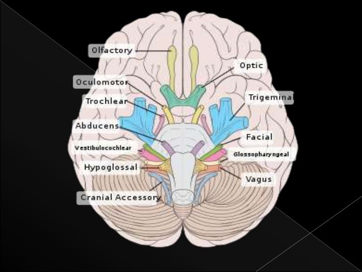
The glossopharyngeal fibers travel just anterior to the cranial nerves X and XI, which also exit the skull via the jugular foramen .
Which nerve is responsible for the formation of the pharyngeal plexus?
A communicating branch to the vagus nerve. Note: The glossopharyngeal nerve contributes in the formation of the pharyngeal plexus along with the vagus nerve. The glossopharyngeal nerve has five distinct general functions: Branchial motor ( special visceral efferent ) – supplies the stylopharyngeus muscle.
Which muscle innervates the stylopharyngeus muscle of the pharynx?
Motor: Innervates the stylopharyngeus muscle of the pharynx.
How does the glossopharyngeal nerve terminate?
The glossopharyngeal nerve terminates by splitting into several sensory branches:
What is the ninth paired cranial nerve?
The glossopharyngeal nerve, CN IX, is the ninth paired cranial nerve. In this article, we shall look at the anatomical course of the nerve, and the motor, sensory and parasympathetic functions of its terminal branches.
What muscle is in the neck?
Fig 1 – Lateral view of the neck, showing the innervation of the stylopharyngeus muscle.
Which nerve terminates by splitting into several sensory branches?
The glossopharyngeal nerve terminates by splitting into several sensory branches: Pharyngeal branch - combines with fibres of the vagus nerve to form the pharyngeal plexus. It innervates the mucosa of the oropharynx. Lingual branch - provides the posterior 1/3 of the tongue with general and taste sensation.
Which nerve is responsible for sensory innervation of the head and neck?
The glossopharyngeal nerve provides sensory innervation a variety of structures in the head and neck. The tympanic nerve arises as the nerve traverses the jugular foramen. It penetrates the temporal bone and enters the cavity of the middle ear.
Which nerve is associated with the derivatives of the third pharyngeal arch?
Embryologically, the glossopharyngeal nerve is associated with the derivatives of the third pharyngeal arch. Sensory: Innervates the oropharynx, carotid body and sinus, posterior 1/3 of the tongue, middle ear cavity and Eustachian tube. Special sensory : Provides taste sensation to the posterior 1/3 of the tongue.
Where does the glossopharyngeal nerve originate?
The glossopharyngeal nerve originates in the medulla oblongata of the brain.
How to determine the integrity of the glossopharyngeal nerve?
The integrity of the glossopharyngeal nerve may be evaluated by testing the patient’s general sensation and that of taste on the posterior third of the tongue.
What nerve can cause loss of taste sensation?
Damage to the glossopharyngeal nerve can result in loss of taste sensation to the posterior one third of the tongue, and impaired swallowing.
Which nerve is located outside the jugular foramen?
Immediately outside the jugular foramen are placed two sensory ganglia that belong to the glossopharyngeal nerve, the superior and the inferior ganglia.
Which nerve provides taste sensation to the posterior 1/3 of the tongue?
The glossopharyngeal nerve provides taste sensation to the posterior 1/3 of the tongue, via its lingual branch (Note: not to be confused with the lingual nerve).
Where does the glossopharyngeal nerve enter the tympanic canal?
It separates from the glossopharyngeal nerve directly under the jugular foramen, and then it courses forward and laterally across the inferior side of the temporal pyramid, where it enters the tympanic canal.
Which nerve contains both motor and sensory fibers?
The glossopharyngeal nerve is a mixed nerve that contains both motor and sensory fibers.
Where does the glossopharyngeal nerve originate?
Nerves. The glossopharyngeal nerve has its origin in the medulla oblongata and exits the skull via the jugular foramen, which is where the tympanic nerve branches off to give parasympathetic innervation to the parotid gland.
Which cranial nerve is the glossopharyngeal?
Introduction. The glossopharyngeal nerve is the 9th cranial nerve (CN IX). It is one of the four cranial nerves that has sensory, motor, and parasympathetic functions. It originates from the medulla oblongata and terminates in the pharynx.
What nerves are affected by carotid endarterectomy?
Glossopharyngeal Nerve Dysfunction Following Carotid Endarterectomy. During a carotid endarterectomy, injury can occur to different cranial nerves, including the glossopharyngeal nerve (although this is less common than injury to others, such as the hypoglossal and vagus nerves).[8] .
What causes glossopharyngeal neuralgia?
Idiopathic glossopharyngeal neuralgia is caused by compression of cranial nerve IX by a vessel or dysfunction of the central pons , whereas secondary glossopharyngeal neuralgia can result from trauma, neoplasm, infection of the throat, surgery, or malformations. [5] .
What is the 9th cranial nerve?
Neuroanatomy, Cranial Nerve 9 (Glossopharyngeal) - StatPearls - NCBI Bookshelf. The glossopharyngeal nerve is the 9th cranial nerve (CN IX). It is one of the four cranial nerves that has sensory, motor, and parasympathetic functions. It originates from the medulla oblongata and terminates in the pharynx. This nerve is most clinically relevant in ...
Which branch of the glossopharyngeal nerve carries parasympathetic fibers?
The branches of the glossopharyngeal nerve are: Tympanic nerve(AKA nerve of Jacobson) – carries parasympathetic fibers and eventually becomes the lesser petrosal nerve, exiting the skull via the foramen ovale and synapses in the otic ganglion. Stylopharyngeal nerve– provides motor innervation to the stylopharyngeus muscle.
Which nerve carries sensory, efferent motor, and parasympathetic fibers?
The glossopharyngeal nerve carries sensory, efferent motor, and parasympathetic fibers. Its branches consist of tympanic, tonsillar, stylopharyngeal, carotid sinus nerve, branches to the tongue, lingual branches, and a communicating branch to cranial nerve X (vagus nerve).
Which nerve innervates the stylopharyngeus?
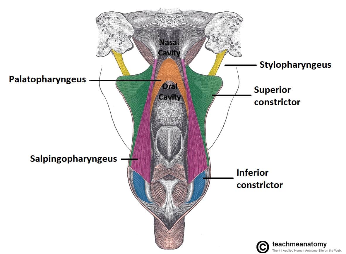
They are all innervated by the pharyngeal plexus and pharyngeal branch of the vagus nerve, except the stylopharyngeus which is innervated by the glossopharyngeal nerve. Functions. They all act on the pharynx, either constricting or elevating it.
Which muscles make up the outer layer of the pharyngeal wall?
The muscles that make up the pharyngeal walls run both circularly on the outside and longitudinally on the inside. The three pharyngeal constrictor muscles make up the outer layer of the wall while the inner layer is comprised of paired muscles. The superior, middle and inferior pharyngeal constrictor muscles form a muscular sleeve ...
What muscles are used to elevate the larynx?
These muscles are known as the stylopharyngeus, the palatopharyngeus and the salpingopharyngeus. There are several gaps that exist between the folds of the pharyngeal constrictor muscles which ...
Which muscle attaches to the stylohyoid ligament?
Middle pharyngeal constrictor muscle. The middle pharyngeal constrictor muscle proximally attaches to the stylohyoid ligament and the greater and lesser cornu of the hyoid bone. It distally attaches to the median pharyngeal raphe, as does the inferior pharyngeal constrictor muscle.
Which muscle passes through the pharyngeal constrictor?
Between the superior and middle pharyngeal constrictor muscles, the stylopharyngeus muscle, the glossopharyngeal nerve and the stylohyoid muscle pass through. Between the middle and inferior pharyngeal constrictor muscles, the internal laryngeal nerve and the superior laryngeal artery and vein pass through. Lastly, below the inferior pharyngeal ...
Where does the inferior pharyngeal constrictor muscle come from?
The inferior pharyngeal constrictor muscle arises from the oblique line of the thyroid cartilage of the larynx and the lateral aspect of the cricoid cartilage of the larynx. It acts by constricting the lower portion of the pharynx.
Where does the stylopharyngeus muscle come from?
Stylopharyngeus muscle. Lastly, the stylopharyngeus muscle comes from the medial aspect of the base of the styloid process and functions by elevating the pharynx and expanding it laterally. Key facts about the stylopharyngeus muscle. Origins.
Overview

Structure
Functions
Clinical significance
Additional images
External links
From the anterior portion of the medulla oblongata, the glossopharyngeal nerve passes laterally across or below the flocculus, and leaves the skull through the central part of the jugular foramen. From the superior and inferior ganglia in jugular foramen, it has its own sheath of dura mater. The inferior ganglion on the inferior surface of petrous part of temporal is related with a triangular depression into which the aqueduct of cochlea opens. On the inferior side, the glossopharyngea…