What makes up the perineal body?
The perineal body is the point of junction of the superficial and deep transverse perineal muscles, perineal membrane, external anal sphincter, posterior vaginal muscularis, and fibers from the puborectalis and pubococcygeus muscles.
Is ischiocavernosus attached to perineal body?
The ischiocavernosus muscle (erectores penis or erector clitoridis in older texts) is a muscle just below the surface of the perineum, present in both men and women....Ischiocavernosus muscleInsertionCrus of penis (male) or Crus of clitoris (female)ArteryPerineal arteryNervePudendal nerve11 more rows
Where are the perineum muscles?
9:2510:30Anatomy of the Perineum (3D tutorial) - YouTubeYouTubeStart of suggested clipEnd of suggested clipAnd the clitoris or penis. The internal pedental artery passes deep to the membrane. And givesMoreAnd the clitoris or penis. The internal pedental artery passes deep to the membrane. And gives branches to the erectile tissue the urethra. And clitoris or penis. So what about the superficial pouch
Where does the ischiocavernosus attach?
Perineal striated muscles 3.5). The paired fusiform ischiocavernosus muscles attach to the ischial tuberosities and ischiopubic rami of the pubic bone and partially cover the penile crurae.
What are the superficial perineal muscles?
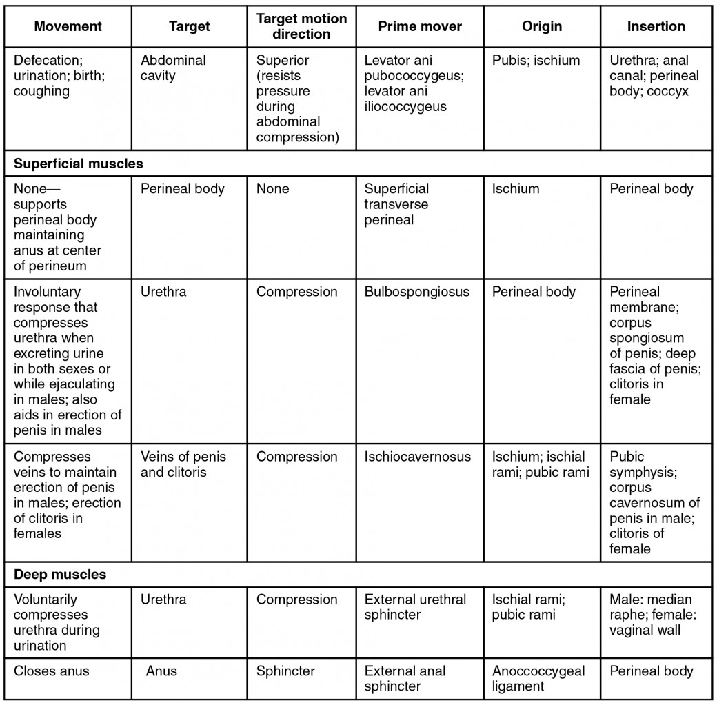
The superficial transverse perineal muscle is a transverse strip of muscle that runs across the superficial perineal space anterior to the anus. It originates from the anterior and medial aspect of the ischial tuberosity and inserts at the perineal body. It is also innervated by the deep branch of the perineal nerve.
What muscle separates pelvis from perineum?
The pelvic diaphragmThe pelvic diaphragm separates pelvis from perineum. Structures leave the pelvis and enter the perineum via the greater and lesser ischiadic foramina, respectivley.
What organs are in the perineum?
OrgansUreters.Urinary Bladder.Urethra.
What is behind the perineum?
The anorectal triangle is the posterior part of the perineal region and is generally identical in both males and females. The triangle is limited anteriorly by the posterior boundary of the urogenital triangle (the interischial line and perineal body) and its apex (located posteriorly) is at the tip of the coccyx.
What is the ischiocavernosus muscle?
Ischiocavernosus is a bilateral perineal muscle in both males and females. In males, it arises from the medial aspect of the ischial tuberosity and more anteriorly, from the ischial ramus. It inserts into the aponeurosis over the medial and lateral sides of the crus penis.
What are the actions of the bulbospongiosus and ischiocavernosus muscles?
In males, the function of bulbospongiosus is to aid in emptying the penile urethra during urination and ejaculation. Together with the ischiocavernosus muscle, it compresses the bulb of penis and the deep dorsal vein of penis.
What is in the deep perineal pouch?
Contents of the deep perineal pouch in males: The muscular components sphincter urethrae and deep transverse perinei with perineal membrane together are known as the urogenital diaphragm. a detailed description of these structures is given in the muscles section below.
What is the perineal nerve?
The perineal nerve is the terminal branch of the pudendal nerve. It typically originates in the last portion of the pudendal canal (within the Alcock's canal) or just as the pudendal nerve exits the canal. It runs anteriorly through the perineum, accompanied by the perineal artery.
What is the perineal body?
The perineal body is the space in which the connective tissue of muscles and septa join. It is the meeting point of the superficial and deep layers of the pelvis contributing the balance of biomechanical forces. It integrates the excretory functions of the urogenital and anorectal organs absorbing the posterior visceral movements. Continence is maintained by the integrity of this system related to the respiratory function of the diaphragm and the postural and locomotor functions of the trunk and the inferior limbs.[5] In the female, hormones during pregnancy influence tissue density and regulate the elongation properties of the perineal body and the pelvic soft tissues, stretching during childbirth. [6]
Why is the perineal body important?
The perineal body is critical for maintaining the integrity of the pelvic floor, especially in females.
What is the lateral smooth muscle?
The perineal lateral smooth muscles (PSMs): Covered by the skin, these are the lateral aspect of the perineal body. They receive the LAM bundles that continue anteriorly to the vestibular area of the superficial perineal fascia. [11]
Which nerve is innervated by the perineal artery?
The perineal body is innervated mainly by the perineal nerve, the broader and inferior terminal branch of the pudendal nerve, directed forward under the internal pudendal artery. The perineal artery and nerve accompany each other, and the nerve divides into the posterior scrotal (or labial) branches and muscular branches:
Which vessels provide blood to the perineum?
The blood supply depends upon the perineal branches of the pudendal vessels that nourish the superficial and muscular tissue of the perineum and the tissues placed inferiorly to the plane of the levator anis muscle.
Which muscle runs from the LAM laterally to the bulbourethral glands?
The recto-urethralis muscle (perin eal smooth muscle) runs from the LAM laterally to the bulbourethral glands adheres anterolaterally to the puborectalis muscle.
Which muscle group arises during the concomitant formation of the first anlage of the pelvic organs?
During the concomitant formation of the first anlage of the pelvic organs, two muscle groups arise the pubis-caudal group and the cloacal group, or Gegenbauer's muscle.
Which nerve is the deep branch of the perineal nerve?
deep branch of perineal nerve from pudendal nerve
Which two organs combine to form the pelvic diaphragm?
coccygeus and levator ani combined form the pelvic diaphragm
How does the piriformis leave the pelvis?
piriformis leaves the pelvis by passing through the greater sciatic foramen
Which nerve is inferior rectal?
inferior rectal nerves (from the pudendal nerve)
Where is the greater trochanter on its medial surface?
greater trochanter on its medial surface above the trochanteric fossa
What is the perineal body?
Anatomically, the perineal body lies just deep to the skin. It acts as a point of attachment for muscle fibres from the pelvic floor and the perineum itself: Levator ani (part of the pelvic floor).
Where is the perineum located?
The perineum is an anatomical region in the pelvis. It is located between the thighs, and represents the most inferior part of the pelvic outlet. The perineum is separated from the pelvic cavity superiorly by the pelvic floor. This region contains structures that support the urogenital and gastrointestinal systems – and it therefore plays an ...
What is the perineum subdivided by?
Roof – pelvic floor. Base – skin and fascia. The perineum can be subdivided by a theoretical line drawn transversely between the ischial tuberosities.
What is the urogenital triangle?
The urogenital triangle is the anterior half of the perineum. It is bounded by the pubic symphysis, ischiopubic rami, and a theorectical line between the two ischial tuberosities. The triangle is associated with the structures of the urogenital system – the external genitalia and urethra.
What is the deep perineal pouch?
Deep perineal pouch - a potential space between the deep fascia of the pelvic floor (superiorly) and the perineal membrane (inferiorly). It contains part of the urethra, external urethral sphincter, and the vagina in the female.
Where are the Bartholin glands located?
The Bartholin’s glands are located within the superficial perineal pouch of the urogenital triangle. Their role is to make a small amount of mucus-like fluid.
What are the boundaries of the perineum?
There are two main ways in which the boundaries of the perineum can be described. The anatomical borders refer to its exact bony margins, whilst the surface borders describe the surface anatomy of the perineum.
Where is the transverse perineal muscle?
The Transversus perinæi superficialis ( Transversus perinæi; Superficial transverse perineal muscle) in the female is a narrow muscular slip, which arises by a small tendon from the inner and forepart of the tuberosity of the ischium, and is inserted into the central tendinous point of the perineum, joining in this situation with the muscle of the opposite side, the Sphincter ani externus behind, and the Bulbocavernosus in front.
Which muscle is smaller, the erector clitoridis or the ischiocavernos?
The Ischiocavernosus ( Erector clitoridis) is smaller than the corresponding muscle in the male. It covers the unattached surface of the crus clitoridis. It is an elongated muscle, broader at the middle than at either end, and situated on the side of the lateral boundary of the perineum.
Where is the sphincter urethra located?
The Sphincter urethræ membranaceæ surrounds the whole length of the membranous portion of the urethra, and is enclosed in the fasciæ of the urogenital diaphragm. Its external fibers arise from the junction of the inferior rami of the pubis and ischium to the extent of 1.25 to 2 cm., and from the neighboring fasciæ. They arch across the front of the urethra and bulbourethral glands, pass around the urethra, and behind it unite with the muscle of the opposite side, by means of a tendinous raphé. Its innermost fibers form a continuous circular investment for the membranous urethra.
Where is the fibers of the Sphincter ani externus decussate?
In some cases, the fibers of the deeper layer of the Sphincter ani externus decussate in front of the anus and are continued into this muscle. Occasionally it gives off fibers, which join with the Bulbocavernosus of the same side. Variations are numerous.
What is the superior fascia of the urogenital diaphragm?
The superior fascia of the urogenital diaphragm is continuous with the obturator fascia and stretches across the pubic arch. If the obturator fascia be traced medially after leaving the Obturator internus muscle, it will be found attached by some of its deeper or anterior fibers to the inner margin of the pubic arch, while its superficial or posterior fibers pass over this attachment to become continuous with the superior fascia of the urogenital diaphragm. Behind, this layer of the fascia is continuous with the inferior fascia and with the fascia of Colles; in front it is continuous with the fascial sheath of the prostate, and is fused with the inferior fascia to form the transverse ligament of the pelvis.
How deep is the inferior fascia?
The superficial of these two layers, the inferior fascia of the urogenital diaphragm, is triangular in shape, and about 4 cm. in depth. Its apex is directed forward, and is separated from the arcuate pubic ligament by an oval opening for the transmission of the deep dorsal vein of the penis.
Which fascia is connected to the middle line?
In the middle line, it is connected with the superficial fascia and with the median septum of the Bulbocavernosus. This fascia not only covers the muscles in this region, but at its back part sends upward a vertical septum from its deep surface, which separates the posterior portion of the subjacent space into two.
