The myenteric plexus is the major nerve supply to the gastrointestinal tract and controls GI tract motility. According to preclinical studies, 30% of myenteric plexus' neurons are enteric sensory neurons, thus Auerbach's plexus has also a sensory component.
What is the innervation of the GI tract?
Innervation of the GI tract • The autonomic nervous system (ANS) of the GI tract comprises both extrinsic and intrinsic nervous systems. 1. Extrinsic innervation (parasympathetic and sympathetic nervous systems) • Efferent fibers carry information from the brain stem and spinal cord to the GI tract.
How is the digestive system innervated by the brain?
The digestive system is innervated through its connections with the central nervous system (CNS) and by the enteric nervous system (ENS) within the wall of the gastrointestinal tract. The ENS works in concert with CNS reflex and command centers and with neural pathways that pass through sympathetic ganglia to control digestive function.
Which nerve innervates the lower large intestine?
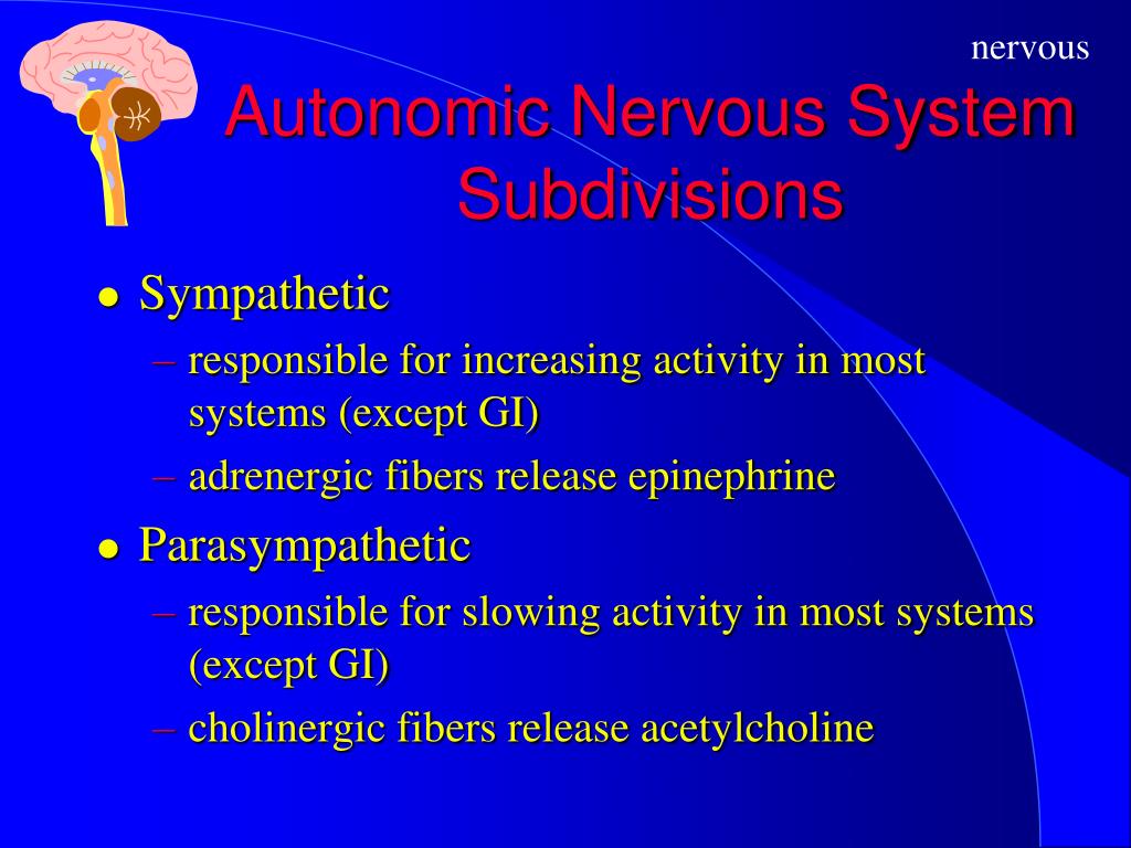
(2) The pelvic nerve innervates the lower large intestine, rectum, and anus. b. Sympathetic nervous system • is usually inhibitory on the functions of the GI tract (the) • Fibers originate in the spinal cord between T-8 and L-2. • Pre-ganglionic sympathetic cholinergic fibers synapse in the pre-vertebral ganglia.
What are the intrinsic enteric and extrinsic nerves of the GI tract?
See other articles in PMC that cite the published article. The gastrointestinal (GI) tract is innervated by intrinsic enteric neurons and by extrinsic projections, including sympathetic and parasympathetic efferents as well as visceral afferents, all of which are compromised by age to different degrees.

Which nerves supply the GI tract?
The GI tract is innervated by intrinsic neurons of the enteric nervous system (ENS) and by the axons of extrinsic sympathetic, parasympathetic, and visceral afferent neurons.
What Innervates the lower GI tract?
pelvic nervesIn the case of the GI tract, the parasympathetic tract is typically excitatory. The parasympathetic system exerts its effects primarily via the vagus (innervates the esophagus, stomach, pancreas, upper large intestine) and pelvic nerves (innervates the lower large intestine, rectum, and anus.)
What cranial nerve controls the GI tract?
The vagus nerveThe vagus nerve (VN), the longest cranial nerve in the body, not only regulates gut physiology, but is also involved in controlling the cardiovascular, respiratory, immune and endocrine systems.
What is the most important nerve of the digestive tract?
The vagus nerve, also known as the vagal nerves, are the main nerves of your parasympathetic nervous system. This system controls specific body functions such as your digestion, heart rate and immune system.
What nerve innervates the large intestine?
(A) The colon and rectum are innervated by two distinct spinal pathways, the lumbar splanchnic and sacral pelvic nerves.
Can the vagus nerve affect your bowels?
The vagus nerve is a communication super highway that runs from your brain to your bowel, and the messages it sends up and down between the two have a significant impact on your IBS symptoms.
What are symptoms of vagus nerve damage?
Potential symptoms of damage to the vagus nerve include:difficulty speaking.loss or change of voice.difficulty swallowing.loss of the gag reflex.low blood pressure.slow or fast heart rate.changes in the digestive process.nausea or vomiting.More items...
What happens when the vagus nerve is overstimulated?
When the vagus nerve is overstimulated, the body's blood vessels dilate, especially those in the lower extremities, and the heart temporarily slows down. The brain is deprived of oxygen, causing the patient to lose consciousness.
What organs are in the lower GI tract?
The lower gastrointestinal tract, commonly referred to as the large intestine, begins at the cecum and also includes the appendix (humans only) colon, rectum, and anus. The primary function of the large intestine in all three species is to dehydrate and store fecal material.
What separates the lower and upper GI tract?
From the point of view of GI bleeding, however, the demarcation between the upper and lower GI tract is the duodenojejunal (DJ) junction (ligament of Treitz); bleeding above the DJ junction is called upper GI bleeding, and that below the DJ junction is called lower GI bleeding.
What is the difference between upper GI and lower GI?
An “upper GI test” examines your esophagus, stomach and the first part of your small intestine (duodenum). A “lower GI test” examines the lower part of your small intestine (ileum) and your large intestine, including your colon and rectum.
Which is an organ of the lower GI tract quizlet?
What organs are considered part of the lower GI tract? The small and large intestines comprise the organs of the lower gastrointestinal tract.
What is the digestive system?
The digestive system is innervated through its connections with the central nervous system (CNS) and by the enteric nervous system (ENS) within the wall of the gastrointestinal tract. The ENS works in concert with CNS reflex and command centers and with neural pathways that pass through sympathetic ganglia to control digestive function.
Where is the myenteric plexus?
The myenteric plexus forms a continuous network that extends from the upper esophagus to the internal anal sphincter. Submucosal ganglia and connecting fiber bundles form plexuses in the small and large intestines, but not in the stomach and esophagus. The connections between the ENS and CNS are carried by the vagus and pelvic nerves ...
How is the striated muscle esophagus determined?
Movements of the striated muscle esophagus are determined by neural pattern generators in the CNS. Likewise the CNS has a major role in monitoring the state of the stomach and, in turn, controlling its contractile activity and acid secretion, through vago-vagal reflexes.
Which part of the body contains reflexes?
In contrast, the ENS in the small intestine and colon contains full reflex circuits, including sensory neurons, interneurons and several classes of motor neuron, through which muscle activity, transmucosal fluid fluxes, local blood flow and other functions are controlled.
What is the innervation of the GI tract?
The GI tract is innervated by intrinsic neurons of the enteric nervous system (ENS) and by the axons of extrinsic sympathetic, parasympathetic, and visceral afferent neurons. Both the intrinsic and the extrinsic innervation, to different degrees, are affected by age. Earlier experimental reports describing the changes in the innervation of the GI tract during healthy aging have been extensively reviewed, summarized, interpreted, and discussed elsewhere (e.g., Santer and Baker, 1993; Hall, 2002; Wade, 2002; Wiley, 2002; Saffrey et al., 2004; Wade and Cowen, 2004; Wade and Hornby, 2005); thus, these observations are not simply repeated in the present survey. Rather, the goals of the present survey are threefold: First, we summarize recent observations, including those from our laboratory as well as those reported by others, and place these findings on age-related changes in the innervation of the GI tract in context with the earlier material covered by the previous reviews. Key structural changes in the aging autonomic innervation of the gut are illustrated. Second, we suggest a provisional list of general patterns of aging of the GI innervation. Third, and lastly, we consider methodological issues that need to be addressed in order to accurately and fully characterize the neurobiology of the aging gut.
Where are the extrinsic innervation of the gut located?
The gut’s extrinsic innervation is supplied by neurons located in prevertebral ganglia, the brainstem, and peripheral afferent ganglia. Though some observations on the effects of aging on these different sites are available (Baker and Santer, 1988b; Schmidt et al., 1990; Sturrock, 1990; Gai et al., 1992; Soltanpour et al., 1996; Soltanpour and Santer, 1997; Bergman and Ulfhake, 1998; Santer et al., 2002; Schmidt, 2002a), these same ganglia and brainstem nuclei also contain neurons that project to non-GI viscera. The absence of practical techniques to readily distinguish those neurons within a nucleus that innervate the gut from those that innervate non-gut tissues, makes it difficult to survey target- or organ-specific neuronal subpopulations. Thus, studies of the aging of the extrinsic innervation of the GI tract have not examined neuronal somata and have instead been largely limited to examinations of the extrinsic axons as they course through gastrointestinal tract tissues. As outlined below, dilated and dystrophic profiles occur in both the efferent and afferent extrinsic projections to the gut.
What is the MP in the intestine?
The myenteric plexus (MP), the outer of the two major ENS plexuses, is the network of neurons situated between the muscle layers of the GI tract, and is primarily involved in the initiation and control of smooth muscle motor patterns such as peristalsis. Loss of neurons in the aged MP was first described by Santer and Baker (1988)in the rat, and verified as a general phenomenon of aging in the intestines of numerous species including guinea pig (e.g., Gabella, 1989), humans (e.g., De Souza et al., 1993), and mice (El-Salhy et al., 1999). Most relevant to the current review, cell death in the MP of the small and large intestines has been demonstrated in several strains of rats raised under a wide range of housing conditions (Fischer 344: Phillips and Powley, 2001; Phillips et al., 2003, 2004b, 2006a,b; Sprague-Dawley: Cowen et al., 2000; Thrasivoulou et al., 2006; Wistar: Santer and Baker, 1988); Figure 1. Because species, strain, staining method, and sampling strategy all potentially influence the patterns of results observed and complicate comparisons among experiments, we report, in the course of this survey, some of the more critical variables for the individual experiments. We also consider the effects of these variables more explicitly in Section 7.
What are the two proteins that regulate cholergic neurons?
Cholinergic neurons can be further subdivided based on their expression of one of the calcium -binding proteins, calretinin and calbindin. Calcium-binding proteins are thought to be primarily involved in the sensing and regulation of calcium pools critical for synaptic plasticity (Schwaller et al., 2002). Neurons positive for calretinin form a much larger proportion of myenteric neurons compared to calbindin-positive neurons (Thrasivoulou et al. 2006); Figure 4D,E. Both of the subpopulations distinguished by the expression of calcium binding proteins seem either to die with age or to have down-regulated expression of the peptides. In the rat ileum, approximately 30% of the neurons positive for calretinin are lost by 17 months of age (Thrasivoulou et al., 2006) with similar findings reported for the guinea-pig ileum (Abalo et al., 2005). Calbindin-positive neurons also die with a loss of over 50% of the neurons in the ileum of 17-month-old rats (Thrasivoulou et al., 2006). While we have not quantitatively evaluated changes with age in the calbindin neurons in the MP of F344 rats, we have observed the presence of markedly swollen calbindin-positive axons in the MP ganglia and connectives of middle-aged rats; Figure 4F. No comparable observations describing age-related changes in the different chemical phenotypes of the SMP presently exist.
Where are enteric glia located?
Enteric glia are prominently located within the ganglia of both the MP and SMP where they interdigitate with neurons; Figure 1A. While located primarily within the ganglia, glia are also present in the interconnecting nerve strands of both plexuses, and within the villi of the mucosa where they make close contacts with the epithelial cell layer (Rühl, 2005). Classically, enteric glia were thought to act only as structural support for neurons, but recently the list of their candidate functions has expanded to include metabolic, trophic, and protective support of neurons (Gershon and Rothman, 1991; Cabarrocas et al., 2003; Rühl et al., 2004; Nasser et al., 2006). Additionally, it is clear that glia play an important role in the health of the enteric innervation of the GI tract (Bush et al., 1998; Cabarrocas et al., 2003; Steinkamp et al., 2003; Rühl et al., 2004; Nasser et al., 2006).
Where are sympathetic axons found?
A distinct axonopathy represented by markedly swollen sympathetic axons was commonly found in the innervation of the submucosal plexus. All of the ganglia (blue neuronal counterstain, Cuprolinc Blue) within the submucosal plexus were innervated by sympathetic (brown, TH) axons (A-F). Cases of well-labeled sympathetic axons that were healthy in appearance (i.e., identical to the innervation seen at younger ages; A) were routinely found in aged tissue (B). In contrast, however, swollen sympathetic axons and terminals also became both more common and more pronounced with age (C-F). Ganglia were often observed to be innervated by sympathetic axons that were both markedly swollen and healthy looking (C,D). Scale bar = 25 μm in F (applies to A-F).
What are the four major functions of the GI tract?
These processes all work together to achieve four major actions required for a proper functioning GI tract: motility, secretion, digestion, and absorption . This activity will primarily focus on neural control, specifically the physiologic function of the enteric nervous system and autonomic nervous system, and their associated pathology.
How does the small intestine move?
The small intestine utilizes two different mechanisms regarding motility. First is the pacemaker activity which propagates slow waves. Second is the migrating motility complex (MMC). This process is dependent on the enteric nervous system and has three phases. The first is the quiet phase in which there is minimal propulsion, which lasts approximately 70 minutes. The second phase includes intermittent motor activity, in which there are one to five contractions with each slow wave. This entire phase lasts between 10 and 20 minutes. Last, there is the regular, propagating contractile activity phase in which there are regular contractions, and the bulk of the food gets moved through the small intestine in a peristaltic pattern, which lasts a total of five minutes. This peristaltic pattern is under the mediated of the “law of the intestine” in which distension of one area is sensed by mechanoreceptors, leading to contraction above the area of distension, and relaxation below the area. This phase is mediated predominately by the autonomic and enteric nervous systems, and repeat every 90 to 120 minutes[8].
How does swallowing affect the esophagus?
Initially, swallowing induces a stimulus that begins the sequence of peristalsis within the esophagus. This stimulus activates the lower motor neurons in the nucleus ambiguous in the brainstem. When the peripheral end of these neurons is stimulated via the vagus nerve, different segments of the esophagus contract. Initially, the caudal end of the dorsal nucleus of the vagus (DMN) is activated via an inhibitory pathway. This inhibition is exerted on all the parts of the esophagus. However, the inhibition lingers for a longer time in the distal areas of the esophagus. Once the inhibition ceases, there is excitatory input leading to sequential activation of the neurons in the rostral zone of the DMN leading a contraction wave that is considered peristaltic. This action allows the area proximal to the food bolus to contract while the area distal remains relaxed, propelling the food down the esophagus. The nerves that allow for this peristaltic motion within the esophagus consists of the myenteric plexus and its association with the circular and longitudinal muscular layers. To continue from the esophagus to the stomach, the food bolus must propel through the lower esophageal sphincter. While this sphincter typically contracted via the effects of acetylcholine on its intrinsic muscle activity, the neurological sequelae of swallowing inhibit this normally remains contracted sphincter, allowing it to relax before the peristaltic wave reaches down the esophagus. [6]
How does the stomach work?
The stomach then acts as a sieve, mixing food particles with gastric fluids, and breaking those particles down into smaller parts.
Which system utilizes cholecystokinin, gastrin, and secretin?
Hormonal control: Utilizes various hormones including cholecystokinin, gastrin, and secretin, among multiple others for a myriad of functions. Neural control: including the GI's intrinsic enteric nervous system and the autonomic nervous system. [2]
Which organ system is responsible for digestion, absorption, and excretion of matter vital for energy expenditure and compatibility?
The gastrointestinal (GI) tract is the body’s organ system responsible for digestion, absorption, and excretion of matter vital for energy expenditure and compatibility with life.
Why does my esophageal spasm?
Diffuse esophageal spasm which can present as dysphagia, heartburn, and regurgitation, is primarily caused by an aberrant response to esophageal distension. Instead of relaxing in response to swallowing and consequent esophageal distension, the esophagus is unable to do so and remains in the contracted state, preventing proper peristaltic movement.
Which nerves are responsible for GI pain?
Behavioural, neurophysiological and clinical evidence shows that most forms of GI pain are mediated by activity in visceral afferent fibres running in sympathetic nerves and that the afferent innervation of the gut mediated by parasympathetic nerves is not primarily concerned with the signalling and transmission of GI pain.
What is the pain of the GI tract?
The only non-general sensation that can be evoked from the gastrointestinal (GI) tract is that of pain ranging from mild discomfort to intense pain.
What is the only non-general sensation that can be evoked from the gastrointestinal tract?
The only non-general sensation that can be evoked from the gastrointestinal (GI) tract is that of pain ranging from mild discomfort to intense pain. However, in certain regions of the gut, such as the rectum and gastro-oesophagus, the feeling of pain can be preceded by non-painful sensations of dist ….
What is the effect of divergent input on motor and autonomic systems?
Such a divergent input can activate many different systems, motor and autonomic as well as sensory, and thus trigger the general reactions that are characteristic of visceral nociception: a diffuse and ill-localized pain sometimes referred to somatic areas, and autonomic and somatic reflexes that result in prolonged motor activity.
Where is GI pain projected?
In some cases, GI pain is projected to areas of the body away from the originating viscus ('re ferred' pain). These properties indicate that the representation of internal organs within the central nervous system is very imprecise.
Is GI pain dull?
However, in certain regions of the gut, such as the rectum and gastro-oesophagus, the feeling of pain can be preceded by non-painful sensations of distension at lower stimulus intensities. GI pain is often dull, aching, ill-defined and badly localized. In some cases, GI pain is projected to areas of the body away from the originating viscus ...
How many sets of plexi are there in the GI tract?
2 primary sets of plexi in the GI tract (Make up enteric i.e. intrinsic nervous system)
What causes red cells to release neuroendocrine signaling factors into the wall of the gut?
1. Stimuli (distension, low pH) causes red cells to release neuroendocrine signaling factors into the wall of the gut
Where are neurotransmitters found?
Almost every known neurotransmitter can be found in the ENS
Where are efferent and afferent limbs located?
Reflex in which both afferent and efferent limbs are contained in the vagus nerve (eg in stomach when food bolus sends afferent signal via vagus nerve to CNS, and efferent signaling relaxes stomach wall)
Which nerve is activated to modify only digestive activity?
The autonomic nerves, especially the vagus nerve, can be discretely activated to modify only digestive activity. In GIT the neurotransmitter are either major neurotransmitters (like (1) acetylcholine, (2) norepinephrine, (3) adenosine triphosphate, (4) serotonin, (5) dopamine, (6) cholecystokinin, (7) sub-
How do neuronal innervation of gastrointestinal smooth muscle occur?
Two mechanisms for neuronal innervation of gastrointestinal smooth muscle exist. Most innervation occurs through interstitial cells of Cajal. Neurons can also directly innervate intestinal smooth muscle cells.
What are the intrinsic plexuses?
The intrinsic plexuses influence all facets of digestive tract activity. Various types of neurons are present in the intrinsic plexuses. Some are sensory neurons; other local neurons innervate the smooth muscle cells and exocrine and endocrine cells of the digestive tract to directly affect digestive tract motility, secretion of digestive juices, and secretion of gastrointestinal hormones.
Which system is primarily responsible for the coordination of local activity within the digestive tract?
As with the central nervous system, these input and output neurons of the enteric nervous system are linked by inter-neurons. Some of the output neurons are excitatory, and some are inhibitory. These intrinsic nerve networks primarily coordinate local activity within the digestive tract. 3. Interstitial cells of Cajal (Gastrointestinal action potential and Mechanical contraction): •The interstitial cells of cajal are specialized pacemaker cells located between the smooth muscle in the wall of the stomach, small intestine, and large intestine. These cells are connected to the smooth muscle and the myentric plexus via gap junctions.
Where is gastrin produced?
Gastrin is produced by cells called G cells in the lateral walls of glands in the antral portion of the gastric mucosa. Gastrin found in other organs in addition to stomach, including pituitary gland, , hypothalamus, medulla oblongata, vagus and sciatic nerve. Gastrin contains 17 amino acids ("little gastrin"). Little gastrin is the form secreted in response to a meal. "Big gastrin" contains 34 amino acids, although it is not a dimer of little gastrin. All of the biologic activity of gastrin resides in the four C-terminal amino acids. The different forms of gastrin suggest that different forms are tailored for different action. G
How to tell the difference between gastrointestinal muscle and nerve fibers?
Important difference between the action potentials of the gastrointestinal smooth muscle and those of nerve fibers 1. The higher the amplitude of slow wave (i.e. the higher the slow wave rise), the higher the frequency of spike potential usually ranging between 1 and 10 spikes per second. The spike potentials last 10 to 40 times as long in gastrointestinal muscle as the action potentials in large nerve fibers, with each gastrointestinal spike lasting as long as 10 to 20 milliseconds. 2. Ionic bases of slow wave Slow wave without spike potential: Depolarization: opening Fast Na channels, repolarization: opening of K channels Slow wave with spike potential: Depolarization: opening Ca-Na channels, repolarization: opening of K channels
What are the layers of the GI tract?
Structure of the gastrointestinal (GI) tract, Layers of the Gastrointestinal Tract 1. Mucosa: including A lining epithelium, including glandular tissue, an underlying layer of loose connective tissue called the lamina propria, which provides vascular support for the epithelium, and often contains mucosal glands. Products of digestion pass into these capillaries. Lymphoid follicles and plasma cells are also often found here. Finally, a thin double layer of smooth muscle is often present; the muscularis mucosa for local movement of the mucosa . 2. Sub-mucosa: A loose connective tissue layer, with larger blood vessels, lymphatic vessels, nerves, and can contain mucous secreting glands. 3. Muscularis propria (externa): There are usually two smooth muscle layer; the inner layer is circular (contraction causes decrease diameter), and the outer layer is longitudinal (contraction causes shorting). 4. Adventia layer (or serosa): Outermost layer of loose connective tissue; covered by the visceral peritoneum. Contains blood vessels, lymphatics and nerves