What nerve supplies the maxillary incisors?
The anterior superior alveolar nerve branches off the infraorbital nerve just before it exits through the infraorbital foramen. It travels in the anterior wall of the maxillary sinus and supplies the mucous membrane. It gives off branches to the superior dental plexus which supplies the upper incisor and canine teeth.
What nerve innervates the maxillary premolar teeth?
Posterior superior alveolar nerveA sketch of the Posterior Super Nasal NerveDetailsInnervatesmaxillary sinus, molars, dental alveolusIdentifiers6 more rows
Which nerves supply teeth?
The inferior alveolar nerve will be responsible for sensory innervation to the cheek, lips, chin, teeth, and gingivae.
What are the nerves of the maxilla?
The maxillary nerve is the second division of the trigeminal nerve and is also known as the V2 division. This nerve is the middle division of the trigeminal nerve and is attached to the distal convex border of the trigeminal ganglion. The maxillary nerve exits from the cranial cavity through the foramen rotundum.
How do you anesthetize maxillary teeth?
The techniques most commonly used in maxillary anesthesia include supraperiosteal (local) infiltration, periodontal ligament (intraligamentary) injection, PSA nerve block, MSA nerve block, anterior superior alveolar nerve block, greater palatine nerve block, nasopalatine nerve block, local infiltration of the palate, ...
What does the maxillary nerve control?
Maxillary: This nerve branch is responsible for sensations in the middle part of your face. Maxillary refers to the upper jaw. The maxillary nerves extend to your cheeks, nose, lower eyelids and upper lip and gums.
Which nerve supply lower teeth?
inferior alveolar nerveThe inferior alveolar nerve supplies feeling to your lower teeth. It's a branch of the mandibular nerve, which itself branches off from the trigeminal nerve. It's sometimes called the inferior dental nerve.
How many maxillary nerves are there?
In neuroanatomy, the maxillary nerve (V2) is one of the three branches or divisions of the trigeminal nerve, the fifth (CN V) cranial nerve....Maxillary nerveFromTrigeminal nerveToInfraorbital nerve, zygomatic nerve, palatine nerve, nasopalatine nerve, sphenopalatine ganglionIdentifiers9 more rows
Where does the maxillary nerve come from?
The maxillary nerve arises from the anterior edge of the trigeminal ganglion. It courses forward through the lateral dural wall of the cavernous sinus, inferiorly and laterally to the ophthalmic nerve.
What does nasopalatine nerve supply?
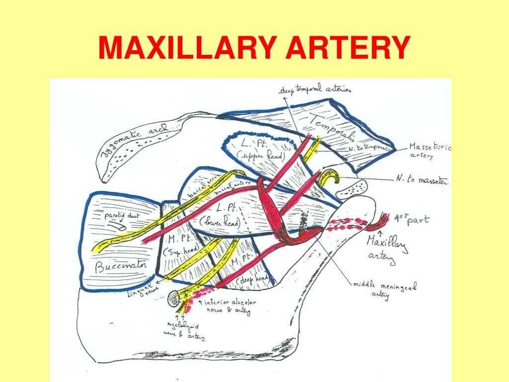
The nasopalatine (or sphenopalatine) artery is a branch of the internal maxillary artery that enters the nasal cavity via the sphenopalatine foramen to supply the frontal, maxillary, ethmoid, and sphenoid sinuses.
What artery supplies the maxillary molars and premolars and the gingiva?
The posterior superior alveolar artery supplies the maxillary molar and premolars and the adjacent gingiva. The infraorbital artery supplies the maxillary canines and incisors and the skin of the infraorbital region of the face.
What does the nasopalatine nerve innervate?
The nasopalatine nerve innervates the anterior part of the hard palate and the mucosa of the nasal septum. A nasopalatine nerve block may be used as local anesthesia for some dental procedures, though it is often painful for the patient.
Which of the following nerves Innervates the upper molars?
Innervation of the mandibular teeth The mandibular teeth are primarily supplied by the inferior alveolar nerve which is a branch of the mandibular nerve (third division of the trigeminal nerve).
What happens if the maxillary nerve is damaged?
As a branch of the trigeminal nerve, the maxillary nerve is often implicated in trigeminal neuralgia, a rare condition characterized by severe pain in the face and jaw. 1 In addition, lesions of this nerve can cause intense hot and cold sensations in the teeth.
Which division of the mandibular nerve supplies sensation to the mandible and the teeth?
c. Mandibular Nerve or Third Division. This division supplies sensation to the mandible and the teeth. It contains somatic motor fibers as well as sensory nerve fibers. (The first two divisions are primarily sensory.) This nerve passes downward to enter the mandible through the mandibular foramen.
Which nerve gives off the anterior superior alveolar branch to the maxillary incisors and cus?
Then, the maxillary nerve gives off an anterior superior alveolar branch to the maxillary incisors and cuspid. (2) The palatal area. The maxillary nerve on each side gives off a palatine nerve, which has an anterior, middle, and posterior branch.
Why is the fifth cranial nerve important?
It is of particular importance in dentistry since it provides the nerve supply to the jaws and the teeth. The fifth cranial nerve contains both motor and sensory fibers. Thus, it has a motor root supplying motor impulses to the muscles of mastication and a sensory root supplying sensory impulses from the structures of the head and face. ...
What are the twelve pairs of cranial nerves?
Twelve pairs of cranial nerves arise in the brain and give off branches to the structures of the head and face . These nerves leave the cranial cavity through foramina in the base of the cranium. The fifth cranial nerve (the trigeminal nerve) is the largest of the twelve pairs. See figure 2-13. It is of particular importance in dentistry since it provides the nerve supply to the jaws and the teeth. The fifth cranial nerve contains both motor and sensory fibers. Thus, it has a motor root supplying motor impulses to the muscles of mastication and a sensory root supplying sensory impulses from the structures of the head and face. Before leaving the cranial cavity, the sensory root divides into three branches or divisions.
Which nerve is located on the roof of the mouth?
Another branch of the maxillary nerve gives rise to the nasopalatine nerve. This nerve descends to the roof of the mouth through the incisive canal and communicates with the corresponding nerve of the opposite side and with the anterior palatine nerve. c. Mandibular Nerve or Third Division.
Where does the anterior palatine nerve end?
The anterior palatine nerve emerges upon the hard palate through the greater palatine foramen, and passes forward nearly to the incisor teeth where it ends with fibers of the nasopalatine nerve. It supplies the gingiva (gum tissue), the mucous membrane, and the glands of the hard palate and part of the soft palate.
Which nerve has no somatic motor fibers?
It contains no somatic motor fibers. (1) The maxillary teeth. The maxillary nerve on each side passes forward in the floor of the orbit of the eye. It first gives off the posterior superior alveolar branch to the three maxillary molars.
What is the maxillary nerve?
The maxillary nerve is one of the branches of the trigeminal nerve, otherwise known as the fifth cranial nerve (CN V).
Which branch of the maxillary nerve carries sensory impulses?
While coursing through the middle cranial fossa, the maxillary nerve extends to the meningeal branch that carries the sensory impulses from the dura mater of the middle cranial fossa.
What is trigeminal nerve?
The trigeminal nerve (cranial nerve V) is a mixed nerve, meaning that it is made of both afferent and efferent neuronal fibers.
Where does the zygomatic nerve enter the orbit?
Soon after that, the zygomatic nerve passes through the inferior orbit al fissure and enters the orbit. While inside the orbit, the nerve courses along its lateral wall and then enters the canal present in the zygomatic bone .
Which nerve innervates the adjacent parts of the skin?
These will innervate the adjacent parts of the skin. On the lateral wall of the orbit, the zygomatic nerve makes anastomosis with the lacrimal nerve through their common connective branch. Thanks to this anastomosis, parasympathetic fibers from the pterygopalatine ganglion reach the lacrimal gland .
Where does the maxillary nerve enter the infratemporal fossa?
In the infratemporal fossa, the nerve is located adjacent to the maxillary tuberosity. From that position, the nerve turns medially and enters the orbit through the inferior orbital fissure, where it is recognized by the name infraorbital nerve. This nerve represents the terminal branch of the maxillary nerve.
Which nerve is responsible for feeling the pain of a fly?
The maxillary nerve is one of the branches of the trigeminal nerve, otherwise known as the fifth cranial nerve (CN V). Supplying sensory innervation to certain parts of the face, the mucosa of the nose, together with the teeth, this nerve allows you to feel that annoying fly landing underneath your eye or that annoying pain caused by your dentist.
What is the maxillary nerve?
The maxillary nerve is the second division of the trigeminal nerve and is also known as the V2 division. This nerve is the middle division of the trigeminal nerve and is attached to the distal convex border of the trigeminal ganglion. The maxillary nerve exits from the cranial cavity through the foramen rotundum. From this point, the nerve traverses the superior part of the pterygopalatine fossa and swings laterally to traverse the inferior orbital fissure toward the maxillary sinus. As the nerve runs along the roof of the maxillary sinus, it supplies the maxillary sinus itself and the anterior teeth of the upper jaw via the anterior and middle superior alveolar nerves. The nerve then exits through the infraorbital foramen to innervate the skin of the face and the underlying mucosa extending from the lower eyelid to the upper lip. While the nerve is at the pterygopalatine fossa, it is connected to the pterygopalatine ganglion, through which it gives off branches to the nasal cavity, pharynx, and palate. In addition, the nerve gives off the zygomatic nerve and the posterior superior alveolar nerve. The zygomatic nerve supplies the lateral portion of the face and the posterior superior alveolar nerve, which supplies the upper molar region.2
Where do the posterior dental branches come from?
The posterior dental branches arise from the maxillary nerve just in front of the infra-orbital groove (Fig. 16.3 ). These branches supply the molar teeth, before issuing branches to the upper gum and the adjoining part of the cheek. There are nerve fibers for: the maxilla bone.
What nerve is blocked in the infraorbital canal?
The infraorbital nerve (the rostral progression of the maxillary nerve) is blocked as it exits the infraorbital foramen, which is identified as a depression 1 to 3 centimeters (cm) (depending on the size of the camelid) directly above the premolars (Figure 48-2 ). The needle should be inserted into the canal, and 1 to 2 milliliters (mL) (depending on the size of the camelid) of local anesthetic should be injected. The tissues that are desensitized in small animals includes the ipsilateral maxilla and intraoral soft tissues; dentition; and extraoral soft tissues, including nose, upper lip, and skin from the site of the infraorbital foramen cranial to midline. 4 It is assumed that the same structures would be desensitized in camelids.
What is the zygomaticofacial nerve?
The zygomaticofacial nerve is a terminal branch of the zygomatic nerve, which in turn is a branch of the maxillary nerve. The zygomaticofacial nerve emerges on the face through the zygomaticofacial foramen, where it pierces the orbicularis oculi muscle and supplies the skin on the prominence of the cheek.
How long does it take for a maxillary nerve to be desensitized?
Structures innervated by the maxillary nerve are desensitized within 15 minutes.
What is the major palatine foramen?
Major palatine foramen approach to the maxillary nerve. This approach is sometimes used to anesthetize the maxillary nerve. Because of the thickness of the palatine soft tissue it is difficult to palpate this foramen. It lies at the midpoint between the mesial border of the maxillary first molar tooth and midline.
Which nerve is the terminal branch of the zygomatic nerve?
The zygomaticotemporal nerve is another terminal branch of the zygomatic nerve. It traverses the zygomaticofacial canal, emerges into the anterior part of the temporal fossa, and ascends between the bone and the substance of the temporalis muscle, piercing the temporal fascia approximately 2 cm above the zygomatic arch and supplying the skin of the temple. 36
What is the maxillary nerve?
The maxillary nerve is the second branch of the trigeminal nerve, which originates embryologically from the first pharyngeal arch. Its primary function is sensory supply to the mid-third of the face. In this article, we shall look at the anatomy of the maxillary nerve – its anatomical course, sensory and parasympathetic functions.
What is the function of the maxillary division of the trigeminal nerve?
Its primary function is sensory supply to the mid-third of the face.
What are the three sensory nuclei?
Three sensory nuclei: Mesencephalic nucleus. Principle sensory nucleus. Spinal nucleus. Motor nucleus of the trigeminal nerve. At the level of the pons, the sensory nuclei merge to form a sensory root. The motor nucleus continues to form a separate motor root.
What causes a swollen trigeminal nerve to swell?
Its cause is unknown but it is thought to be the result of damage to trigeminal nerve due to conditions such as multiple sclerosis, stroke or trauma.
Which nerve originates from the first pharyngeal arch?
The maxillary nerv e is the second branch of the trigeminal nerve, which originates embryologically from the first pharyngeal arch.
Where is the trigeminal ganglion located?
The trigeminal ganglion is located lateral to the cavernous sinus, in a depression of the temporal bone known as the trigeminal cave or Meckel’s cave. The motor root passes inferiorly to the sensory root, along the floor of the trigeminal cave. Motor fibres are only distributed to the mandibular division (V3).
Which division of the trigeminal nerve is responsible for motor fibres?
Motor fibres are only distributed to the mandibular division ( V3). From the trigeminal ganglion, the three terminal divisions of the trigeminal nerve arise; the ophthalmic (V1), maxillary (V2) and mandibular (V3) nerves.
What are the branches of the maxillary nerve?
The other branches of the maxillary nerve are the ganglionic branches, the posterior superior alveolar nerve and the zygomatic nerve. The infraorbital nerve forms a plexus with the posterior superior alveolar nerve, which is known as the superior dental plexus.
Which nerve supplies the mandibular nerve?
Inferior alveolar nerve (branch of mandibular nerve V3) Blood supply of mandibular teeth. Inferior alveo lar artery (branch of maxillary artery) This article will highlight the main neurovasculature that supplies both the maxilla and the mandible, including the venous drainage of the teeth and will finish by explaining the pathological implications ...
What nerve innervates the mesiobuccal root of the first molar?
The middle superior alveolar nerve varies upon its path and as it descends to form the middle portion of the superior dental plexus it innervates the medial and lateral aspects of the maxillary sinus and the premolars. It may, in some cases, also innervate the mesiobuccal root of the first molar, if it is not covered by the posterior superior alveolar nerve.
Which nerve innervates the anterior aspect of the maxillary sinus?
Lastly, the anterior superior alveolar nerve descends to form the anterior portion of the superior dental plexus. It innervates the anterior aspect of the maxillary sinus as well as the incisors and the canines. Before we continue, a few additional words must be said about the infraorbital nerve.
Which part of the dental plexus is the posterior superior alveolar?
The posterior superior alveolar turns laterally into the pterygomaxillary fissure and into the infratemporal fossa. It descends via the infratemporal surface of the maxilla to form the posterior portion of the superior dental plexus and innervates the posterior aspect of the maxillary sinus as well as the maxillary molars.
Which nerve carries sensory fibers to the maxillary arch?
The maxillary nerve, which is the second division of the trigeminal nerve (CN V/II) carries sensory fibers teeth of the maxillary dental arch. It runs laterally to the cavernous sinus and exits the skull via the foramen rotundum in the middle cranial fossa, leading into the pterygopalatine fossa.
Which bone is the mirror of the maxillary and mandibular nerves?
While it is true that within the alveolar bone the maxillary and mandibular nerves and vessels mirror one another, there are anatomical differences with extra branches and adjacent structures, such as the mental foramen of the mandible or the greater palatine foramen of the hard palate.
What nerve supplies the upper teeth?
The branches of the maxillary nerve supply the upper teeth, the nasal cavity and palate, and the upper part of the cheek.
What does the maxillary nerve supply?
In sum, the maxillary nerve innervates the skin of the lower eyelid, the prominence of the cheek, the alar part of the nose, part of the temple, and the upper lip (Figures 2.2 and 2.3).
What nerves supply teeth?
The inferior alveolar nerve will be responsible for sensory innervation to the cheek, lips, chin, teeth, and gingivae.
Which cranial nerve supplies the teeth and jaws?
The trigeminal nerve (the fifth cranial nerve, or simply CN V) is a nerve responsible for sensation in the face and motor functions such as biting and chewing; it is the most complex of the cranial nerves.
What causes inflammation of the trigeminal nerve?
There are inflammatory causes of trigeminal neuralgia because of systemic diseases including multiple sclerosis, sarcoidosis, and Lyme disease. There also is an association with collagen vascular diseases including scleroderma and systemic lupus erythematosus.
How can I increase blood flow to my teeth?
While brushing and flossing also help to bring oxygen to the gums, rubber tipping brings it a step further by stimulating blood flow. Rubber tipping helps to prevent periodontal disease by keeping your gums clean – free of plaque and food debris – and by stimulating blood flow to the area.
What is the function of the maxillary nerve?
It comprises the principal functions of sensation from the maxilla, nasal cavity, sinuses, the palate and subsequently that of the mid-face, and is intermediate, both in position and size, between the ophthalmic nerve and the mandibular nerve.
