Two solid organs, the liver and the pancreas (both of which are embryologically derived from the digestive tract), produce digestive juices that reach the intestine through small tubes known as ducts. In addition, parts of other organ systems (for instance, nerves and blood) play a major role in the digestive system.
Full Answer
What are the organs of the digestive system?
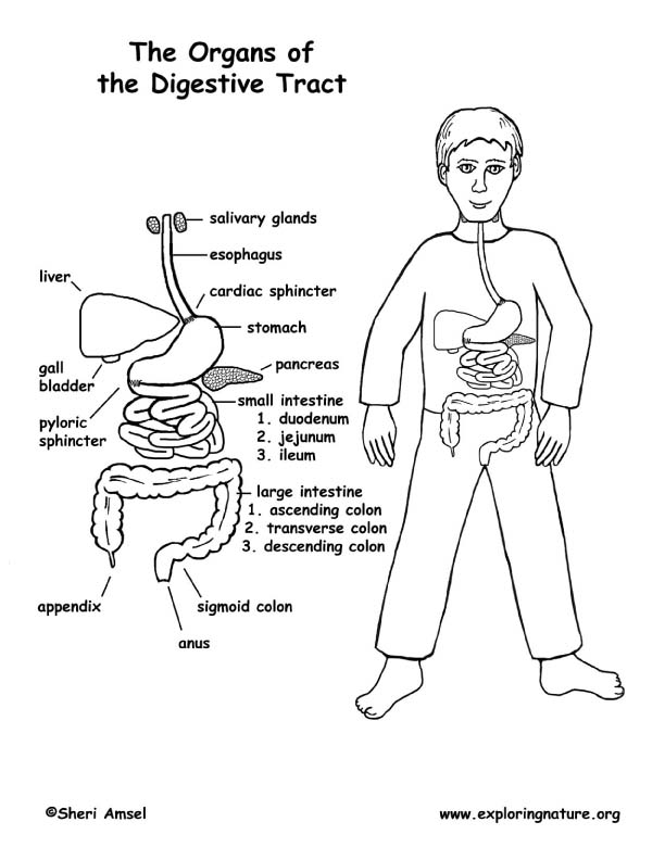
The main organs that make up the digestive system (in order of their function) are the mouth, esophagus, stomach, small intestine, large intestine, rectum and anus. Helping them along the way are the pancreas, gall bladder and liver. Here’s how these organs work together in your digestive system.
How is the digestive tract formed in embryos?
The Digestive Tract in Human Embryos Between Carnegie Stages 11 and 13 [3] "The digestive tract was initially formed by a narrowing of the yolk sac, and then several derived primordia such as the pharynx, lung, stomach, liver, and dorsal pancreas primordia differentiated during 12 (21-29 somites) and CS13 (≥ 30 somites).
Where is the lining of the digestive tract derived from?
Embryologically, almost all the lining of the digestive tract is derived from endoderm. The only exceptions to this rule are the very first part of the digestive system and the very last part: the anterior portion of the mouth develops from an ectodermal depression called the stomodeum.
What is the development of the digestive system?
The development of the digestive system begins as a simple blind-ended gut tube. The accessory digestive organs form as outpouchings from the primitive gut tube, whereas formation of the intestines require them to first protrude out into the umbilical cord (physiological herniation) before retracting back into the abdominal cavity.
What organs are derived from the foregut?
The foregut gives rise to the esophagus, stomach, liver, gallbladder, pancreas and the caudal portion of the duodenum.
Where is the digestive system derived from?
The development of the digestive system in the human embryo concerns the epithelium of the digestive system and the parenchyma of its derivatives, which originate from the endoderm. Connective tissue, muscular components, and peritoneal components originate in the mesoderm.
What is recanalization in embryology?
During the solid stage of development the endoderm of the gut tube proliferates until the gut is a solid tube. A process of recanalization restores the lumen.
What structures are derived from the midgut?
Midgut derived structures include the duodenum distal to the ampulla of Vater, the jejunum, ileum, cecum, ascending colon, and foremost two-thirds of the transverse colon.
What are the 5 parts of the digestive tract?
The hollow organs that make up the GI tract are the mouth, esophagus, stomach, small intestine, large intestine, and anus.
What are the 4 main components of the digestive system?
Regions of the Digestive SystemMouth.Pharynx & Esophagus.Stomach.Small and Large Intestine.
What is a recanalization?
Recanalization is the reestablishment of blood flow into a formerly occluded region (Hall et al., 1989). This phenomenon destabilizes the occluded region and may lead to significant rebleeding at the treatment site.
Is spleen derived from foregut?
The foregut organs are the stomach, the first half of the duodenum, and the liver, gallbladder, pancreas, and spleen.
Is pancreas derived from midgut?
the pancreas remains retroperitoneal throughout its development. the liver is derived from the midgut. the hindgut is supplied by the celiac artery....FOREGUTMIDGUTHINDGUTGallbladder & bile ductsAscending colonUrogenital sinusPancreas (dorsal & ventral)Proximal 2/3 of transverse colonUpper duodenum*8 more rows
What are the derivatives of the hindgut?
The hindgut gives rise to the distal third of the transverse colon, the descending colon, the sigmoid colon, the rectum, and the upper portion of the anal canal. The hindgut endoderm also lines the bladder and the urethra.
What is midgut and hindgut?
The midgut is from the mid-duodenum to the initial two-thirds of the transverse colon. The hindgut is from the later one-third transverse colon to the upper portion of the anus.
What organs are in the hindgut?
The hindgut is composed of the cecum, large colon, small colon and the rectum.
What is digestive system in anatomy?
What is the digestive system? Your digestive system is made up of the gastrointestinal (GI) tract and your liver, pancreas and gallbladder. The GI tract is a series of hollow organs that are connected to each other from your mouth to your anus.
Which organ is responsible for digestion?
The digestive tract includes the mouth, esophagus, stomach, intestines, and anus. So-called "accessory" organs include the liver, pancreas, and gallbladder; food doesn't move through these organs, but they secrete hormones and chemicals that are essential to digestion.
What is the science behind digestion?
Digestion of food involves both mechanical and chemical processes. Through digestion, large food particles are converted into smaller components that can be readily absorbed into the bloodstream. We look at mechanical digestion and the chemical digestion (enzymatic hydrolysis) of carbohydrates, proteins and fats.
What is part of the digestive system?
The digestive system includes the mouth, pharynx (throat), esophagus, stomach, small intestine, large intestine, rectum, and anus. It also includes the salivary glands, liver, gallbladder, and pancreas, which make digestive juices and enzymes that help the body digest food and liquids.
When does the digestive system develop?
The first important step in development of the digestive system occurs during folding of the embryonic disc as neurulation takes place (weeks 4 and 5) - the thin endodermal layer is ‘gathered up' inside the embryonic body to form a tubular structure.
How many portions are there in the embryonic gut tube?
It is usual and helpful to subdivide the embryonic gut tube into three portions:
Which septum separates the hindgut from the urogenital system?
explain separation of the hindgut from the urogenital system by the urorectal septum
Where does the liver develop?
The liver begins its development as a small endodermal outgrowth - the hepatic diverticulum - which arises from the caudal part of the foregut. This tubular outgrowth extends through the ventral mesentery into the septum transversum, branching as it grows. One branch is formed close to the septum transversum, and this subsequently develops into the gall bladder and cystic duct. The remaining branches are formed within the septum, and the mesodermal cells which surround this branching tubular system assist in the formation of the liver tissue. Thus, the endodermal hepatic diverticulum forms the duct system and storage organ of the biliary system, while the mesodermal cells contributed by the septum transversum form the liver cells.
What are the structures that support the large intestine?
Postnatally, the stomach, the jejunum and ileum, and some parts of the large intestine are supported from the posterior wall of the abdomen by membranous structures called mesenteries. It is through these mesenteries that blood vessels, lymph vessels, and nerves are distributed to and from the gut wall. The mesenteries allow mobility to the gut - an important aid to digestive processes. In the adult, the mesenteries have a complex arrangement, but this can be understood by working through the embryological processes which produce them.
What is the role of neural crest cells in the digestive system?
Migrating into the splanchnic mesoderm of the developing digestive system will be many neural crest cells - these will play an important part in establishing the autonomic nerve supply to the gut. Can you think of an abnormality of the digestive system where this process of innervation has failed - usually in the distal portion of the large intestine? (clue: look up Hirschsprung's disease.)
Which organs are formed from splanchnic mesoderm?
The other layers of the digestive tract - for example: the muscular wall - are developed from splanchnic mesoderm. Similarly, organs such as the liver and pancreas arise as endodermal outgrowths from the gut tube, but mesoderm contributes to their development. (Recall that when the lateral mesoderm was split by the intra-embryonic coelom, one layer remained in association with the ectoderm - the somatic mesoderm, and one layer remained in contact with the endoderm - the splanchnic mesoderm.)
Which abdominal contents fail to return to the abdomen and are sealed by peritoneum?
Omphalocele: abdominal contents fail to return to the abdomen and are sealed by peritoneum
Why are there no neurons in the colon?
Lack of neural crest-derived- ganglion neurons in a part of the colon due to abnormal migration, differentiation, or proliferation of neural crest cells during embryogenesis. [7]
What is the ectoderm?
Ectoderm further separates into the surface ectoderm, neural tube, and neural crest. The surface ectoderm is the precursor to the epidermis, lens of eyes, nails, hair. The neural tube differentiates into the brain and spinal cord. The neural crest is the source of the peripheral nervous system, including the neurons of the GI tract (also called the enteric nervous system). [1]
How long does it take for epithelial cells to form?
Different epithelial cell types are present by 12 weeks and are resembling adult cells of the intestine by 22 weeks.
Which connective tissue gives rise to the connective tissue?
Mesoderm gives rise to the connective tissue, including the wall of the gut tube and the smooth muscle.
Can GI tract defects cause fetal demise?
Many different pathophysiological processes can occur during embryogenesis of the GI tract. Many of these can are correctable by surgery and do not cause fetal demise in utero. A few of these embryological defects have associations with other diseases, such as cystic fibrosis and Down syndrome. Therefore, babies with meconium ileus and duodenal atresia require close monitoring and further evaluation for cystic fibrosis and Down syndrome, respectively.
Which organ is the first embryonic division of the gastrointestinal tract?
First embryonic division of gastrointestinal tract, extending from the oral (buccopharyngeal) membrane and contributing oesophagus, stomach, duodenum (to bile duct opening), liver, biliary apparatus (hepatic ducts, gallbladder, and bile duct), and pancreas. The forgut blood supply is the celiac artery (trunk) excluding the pharynx, lower respiratory tract, and most of the oesophagus.
Where does the gastrointestinal tract originate?
The tract and associated organs later have contributions from all the germ cell layers.
How is the midgut generated?
The large mid-gut is generated by lateral embryonic folding which "pinches off" a pocket of the yolk sac, the 2 compartments continue to communicate through the vitelline duct.
What stage is the digestive tract formed?
The Digestive Tract in Human Embryos Between Carnegie Stages 11 and 13 "The digestive tract was initially formed by a narrowing of the yolk sac, and then several derived primordia such as the pharynx, lung, stomach, liver, and dorsal pancreas primordia differentiated during 12 (21-29 somites) and CS13 (≥ 30 somites).
How many pairs of pharyngeal pouches were formed in all 13 embryos?
The differentiation of four pairs of pharyngeal pouches was complete in all 13 embryos. The respiratory primordium was recognized in ≥ 26-somite embryos and it flattened and then branched at 13. The trachea formed and then elongated in ≥ 35-somite embryos.
What are the three regions of the GIT?
During the 4th week three distinct regions (fore-, mid- and hind-gut) extend the length of the embryo and will contribute different components of the GIT. The large mid-gut is generated by lateral embryonic folding which "pinches off" a pocket of the yolk sac, the 2 compartments continue to communicate through the vitelline duct.
How is the stomach curvature generated?
stomach curvature is generated by left-right asymmetric gut morphogenesis "Left-right (LR) asymmetry is a fundamental feature of internal anatomy, yet the emergence of morphological asymmetry remains one of the least understood phases of organogenesis. Asymmetric rotation of the intestine is directed by forces outside the gut, but the morphogenetic events that generate anatomical asymmetry in other regions of the digestive tract remain unknown. Here, we show in mouse and Xenopus that the mechanisms that drive the curvature of the stomach are intrinsic to the gut tube itself. The left wall of the primitive stomach expands more than the right wall, as the left epithelium becomes more polarized and undergoes radial rearrangement. These asymmetries exist across several species, and are dependent on LR patterning genes, including Foxj1, Nodal and Pitx2 Our findings have implications for how LR patterning manifests distinct types of morphological asymmetries in different contexts."
What is the digestive system?
See additional information. Digestive system: The system of organs responsible for getting food into and out of the body and for making use of food to keep the body healthy. The digestive system includes the salivary glands, mouth, esophagus, stomach, liver, ...
Which organs produce juices?
In the mouth, stomach, and small intestine, the mucosa contains tiny glands that produce juices to help digest food. Two solid organs, the liver and the pancreas (both of which are embryologically derived from the digestive tract), produce digestive juices that reach the intestine through small tubes known as ducts.