What are the problems with the diaphragm?
Nov 15, 2021 · They sit to the left and right of the heart, within a space called the thoracic cavity. The cavity is protected by the rib cage. A sheet of muscle called the diaphragm serves other parts of the respiratory system, such as the trachea, or windpipe, and bronchi, conduct air to the lungs. How does the diaphragm work with the heart?
What does the diaphragm do in the digestive system?
The diaphragm is a thin dome-shaped muscle that separates the thoracic cavity (lungs and heart) from the abdominal cavity (intestines, stomach, liver, etc.). Participate in the breath, pulling your chest down when you inhale and up when you exhale.
What causes pressure on diaphragm and swollen abdomen?
What organs does the diaphragm work with? The diaphragm separates the thoracic cavity, containing the heart and lungs, from the abdominal cavity and performs an important function in respiration: as the diaphragm contracts, the volume of the thoracic cavity increases, creating a negative pressure there, which draws air into the lungs.
What does the diaphragm do in the body?
The diaphragm is a thin skeletal muscle that sits at the base of the chest and separates the abdomen from the chest. It contracts and flattens when you inhale. This creates a …
What does the diaphragm connected to?
The diaphragm has a dome-like structure with the peripheral segment attached to the chest wall and abdominal cavity. The muscle fibers from these attachments converge in a central tendon, which forms the crest of the dome.Aug 15, 2018
Does the heart move with the diaphragm?
As you can see, the heart, which is attached to the diaphragm via its pericardium (a membranous sac that envelops the heart), moves up and down with the diaphragm.
Where does the diaphragm attach?
Origin and insertion. The diaphragm is a musculotendinous structure with a peripheral attachment to a number of bony structures. It is attached anteriorly to the xiphoid process and costal margin, laterally to the 11th and 12th ribs, and posteriorly to the lumbar vertebrae.
Is the diaphragm an organ?
Abstract. The diaphragm is the only organ which only and all mammals have and without which no mammals can live. The human is the only mammal which keeps the diaphragm parallel to the ground even during locomotion.
Where is the diaphragm in relation to the liver?
The liver is located under the ribs on the right hand side of the body. It lies just below the lungs, under the top of the diaphragm to which it is attached. The diaphragm is the muscle beneath the lungs which regulates our breathing. The liver is partly protected by the rib cage.
Which muscles attach to the diaphragm?
The fascia involving the diaphragm posteriorly, ie, at the retroperitoneal level, is separated in four parts. It joins the aortic system, inferior vena cava, liver, psoas muscles, quadratus lumborum, cardiac area, phrenic-esophageal ligaments and, finally, the kidneys.Jul 25, 2013
Where does the aorta pass through the diaphragm?
The aortic hiatus is one of the three major apertures through the diaphragm and lies at the level of T12.Jan 5, 2019
What are the 3 openings in the diaphragm?
There are a number of openings in the diaphragm through which structures pass between the thorax and abdomen. There are three large openings — one for the aorta, one for the esophagus, and one for the inferior vena cava (the caval opening), plus a series of smaller ones.
What is the function of the diaphragm?
Function. The diaphragm plays an integral role in respiration (breathing). Most of the time, the diaphragm moves involuntarily. Your thoracic diaphragm also plays a role in helping the movement of muscles during childbirth, having a bowel movement, urinating, and lifting heavy objects.
What is the diaphragm?
Anatomy. The diaphragm is a parachute-shaped fibrous muscle that runs between the chest and abdomen, separating these two large cavities. It is asymmetric, as the right dome is larger than the left dome. The diaphragm has openings that allow certain structures to span the chest and abdominal cavities.
Where is the diaphragm located?
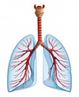
Location. The diaphragm spans across the body from the front to the back. It is the floor of the thoracic cavity and the ceiling of the abdominal cavity. 2 . Your heart, lungs, and the upper part of your esophagus (food pipe) are in the thoracic cavity above the diaphragm.
What is the central tendon?
The central tendon is a large part of the diaphragm that anchors the diaphragm to the ribs. There are three large openings (holes) through the diaphragm: The esophageal opening (esophageal hiatus), through which the esophagus, right and left vagus nerves, and left gastric artery and vein pass. The aortic opening (aortic hiatus), through which ...
What happens when the diaphragm is activated?
When the diaphragm is activated by a nerve, it contracts and flattens. This action decreases pressure and increases the space in the thoracic cavity, allowing your lungs to expand as you inhale. When the diaphragm relaxes, your chest cavity becomes smaller and your lungs release air. 2
What causes the diaphragm to move?
There are several medical conditions that involve the thoracic diaphragm. Traumatic injuries or anatomical defects can interfere with the muscle's function, and the movement of the diaphragm can also be impaired by issues like nerve disease or cancer.
What is a hernia in the chest?
Diaphragmatic hernias are structural defects that allow abdominal organs to enter the chest cavity. They may be present from birth, or, less commonly, can result from trauma. Congenital: The diaphragm doesn't develop as it should in roughly 1 in 2,000 births.
What is the function of the diaphragm?
It is not directly a part of the digestive system but serves the important purpose of keeping the abdominal cavity, and all the organs of the digestive system separated from the respiratory system, so both can function properly. It also allows the esophagus to run through ...
Where is the diaphragm located?
The diaphragm is located between the thoracic and abdominal cavities [3], with important organs like the lungs and heart located superior to it, and the liver (proximal position), kidney and stomach being inferior to it. The curved muscle is inserted into the lower part of the rib cage. Diaphragm Muscle Location Picture.
What is the thoracic diaphragm?
The thoracic diaphragm is a large, flat muscle that plays a vital role in the respiratory system, and is located just beneath the two lungs, dividing the chest cavity from the abdominal cavity [1]. With its characteristic dome shape, it is the primary respiratory muscle, also supporting the lungs and heart [2]. Diaphragm.
How long do diaphragm spasms last?
Diaphragm Spasms: Sometimes, the diaphragm spasms causing harmless hiccups may last for days or weeks, indicating some underlying health condition. Sometimes, this abnormally contracted muscle may make it difficult to breathe deeply, leading to other problems [24].
Which nerve innervates the diaphragm?
What Nerves Innervate the Diaphragm. The phrenic nerve, originating at the C3-C5 vertebral level, provides motor supply to the diaphragm [2], and controls its movement [15]. The medullary inspiratory neuron (responsible for regulating breathing) sends information to the diaphragm via this nerve, enabling us to breathe in. ...
What is the central tendon?
An aponeurosis, (interlacing white, fibrous tissues with a wide area of attachment, taking the place of tendons in flat muscles), the central tendon forms the central upper surface of the diaphragm [9]. It fuses with the pericardium (the outer membrane of the heart) located just above it and helps the heart to stay in place [10].
What happens to the diaphragm during exhalation?
The diaphragm then relaxes and comes back to its natural dome shape, reducing the space within the chest cavity, thus putting pressure on the lungs so the air can be pushed out [3].
