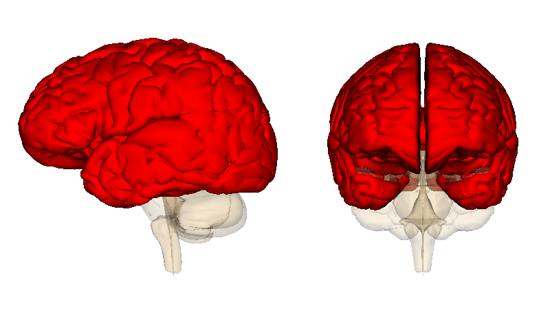
What part of the brain is responsible for sensory perception?
At the lowest level, sensory information is mapped separately in the visual and auditory cortexes. Following this, this information is automatically integrated in the parietal lobe, which is located in the upper area of the brain.
What part of the brain is responsible for visual information?
Visual cortex The visual cortex commonly known as cortex visualis in Latin is part of the sensory cortex found in the occipital lobe (2). Furthermore, the occipital lobe is one of the four primary lobes of the human brain and it acts as the visual processing center. Therefore, the visual cortex is responsible for processing visual information.
What is the function of the sensory cortex?
The sensory cortex is defined as all cortical areas linked with sensory functions (1). In another definition, the sensory cortex is a section of the cerebral cortex which is responsible for receiving and interpreting sensory information from different parts of the body. neuron to a specific section of the brain (3).
What part of the brain is responsible for sense of touch?
The middle part of the brain, the parietal lobe helps a person to identify objects and understand spatial relationships (where one's body is compared to objects around the person). The parietal lobe is also involved in interpreting pain and touch in the body. Occipital lobe.

What is the brain's function?
Together with the spinal cord, brain structure and function helps control the central nervous system— the main part of two that make up the human nervous system. (The other part, the peripheral nervous system, is made up of nerves and neurons that connect the central nervous system to the body's limbs and organs.)
What Are the Regions of the Brain and How Do They Fit Into the Brain Structure?
The three main parts of the brain are split amongst three regions developed during the embryonic period: the forebrain, midbrain and hindbrain. Together, these regions act as a useful map to understanding the various parts of the brain's structure and functions.
What Is the Brain and Why Does It Matter?
The brain is a three-pound organ that serves as headquarters for our bodies. Without it, we wouldn't be able to process information, move our limbs, or even breathe. Together with the spinal cord, brain structure and function helps control the central nervous system—the main part of two that make up the human nervous system. (The other part, the peripheral nervous system, is made up of nerves and neurons that connect the central nervous system to the body's limbs and organs.) The human nervous system is responsible for helping us think, breathe, move, react and feel.
What Are the 4 Lobes of the Brain?
The cerebrum's left and right hemispheres are each divided into four lobes: the frontal, parietal, occipital and temporal lobes . The lobes generally handle different functions, but much like the hemispheres, the lobes don't function alone. The lobes are separated from each other by depressions in the cortex known as sulcus (or sulci) and are protected by the skull with bones named after their corresponding lobes.
What Are the Main Parts of the Brain Stem?
The brain stem is made up of three parts: the midbrain, the pons and the medulla.
What Is the Cerebellum?
The cerebellum stands for "little brain" in Latin. It looks like a separate mini-brain behind and underneath the cerebrum (beneath the temporal and occipital lobes) and above the brain stem. The cerebellum (along with the brain stem) is considered evolutionarily to be the oldest part of the brain.
What are the parts of the brain?
There are three main parts of the brain: the cerebrum, cerebellum and the brain stem.
Which part of the brain controls movement?
The largest part of the brain, the cerebrum initiates and coordinates movement and regulates temperature. Other areas of the cerebrum enable speech, judgment, thinking and reasoning, problem-solving, emotions and learning. Other functions relate to vision, hearing, touch and other senses.
How does the brain work?
The brain sends and receives chemical and electrical signals throughout the body. Different signals control different processes, and your brain interprets each. Some make you feel tired, for example, while others make you feel pain.
What is the brain made of?
Weighing about 3 pounds in the average adult, the brain is about 60% fat. The remaining 40% is a combination of water, protein, carbohydrates and salts. The brain itself is a not a muscle. It contains blood vessels and nerves, including neurons and glial cells.
What organ controls memory, emotion, touch, motor skills, vision, breathing, temperature, hunger, and every other process?
The brain is a complex organ that controls thought, memory, emotion, touch, motor skills, vision, breathing, temperature, hunger and every process that regulates our body. Together, the brain and spinal cord that extends from it make up the central nervous system, or CNS.
Where is the spinal cord located?
The spinal cord extends from the bottom of the medulla and through a large opening in the bottom of the skull. Supported by the vertebrae, the spinal cord carries messages to and from the brain and the rest of the body.
How many halves are there in the cerebral cortex?
The cerebral cortex is divided into two halves, or hemispheres. It is covered with ridges (gyri) and folds (sulci). The two halves join at a large, deep sulcus (the interhemispheric fissure, AKA the medial longitudinal fissure) that runs from the front of the head to the back.
Why are the two different shades of gray on a neuron scan?
Gray matter is primarily responsible for processing and interpreting information, while white matter transmits that information to other parts of the nervous system.
How does the brain integrate sensory input?
How the brain integrates sensory input. Hearing, sight, touch - our brain captures a wide range of distinct sensory stimuli and links them together. The brain has a kind of built-in filter function for this: sensory impressions are only integrated if it is necessary and useful for the task at hand. Researchers from Bielefeld University, Oxford ...
Where is sensory information mapped?
At the lowest level, sensory information is mapped separately in the visual and auditory cortexes. Following this, this information is automatically integrated in the parietal lobe, which is located in the upper area of the brain. Only at a higher level of processing does the brain parse out the information from the previous stages and, ...
What happens when you see an image and hear a sound?
For example, if you always hear a sound and see an image at the same time, the brain integrates this information. However, if the sound and image appear together, they will not be integrated -- even though they were previously separate from each other. "In the "causal inference" model, the brain infers that the source of ...
Which part of the brain is responsible for abstract thinking?
This flexibility in perception is located in special areas of the frontal lobe that are responsible for abstract thinking.
Is auditory information always useful?
It is not always useful, however, for auditory and visual information to be automatically integrated in the brain: one example of this would be watching a foreign-language film that is dubbed and the movements of the actors' lips do not match the spoken sounds.
Is sensory input automatically integrated?
Sensory stimuli are therefore not automatically integrated -- this is only the case if they do originate from the same source," says Kayser. To test these three models, study participants were exposed to visual and auditory stimuli.
Which lobe of the brain is responsible for spatial information?
The parietal lobe processes sensory information for cognitive purposes and helps coordinate spatial relations so we can make sense of the world around us. The parietal lobe resides in the middle section of the brain behind the central sulcus, above the occipital lobe. The temporal lobe is located on the bottom of the brain below the lateral fissure.
Which lobe of the brain is responsible for forming our personality and influencing out decisions?
The frontal lobe is the emotional control center of the brain responsible for forming our personality and influencing out decisions. The frontal lobe is located at the front of the central sulcus where it receives information signals from other lobes of the brain. The parietal lobe processes sensory information for cognitive purposes ...
What is the smallest lobe of the brain?
The occipital lobe, the smallest of the four lobes of the brain, is located near the posterior region ...
What are the four lobes of the brain?
Brain Lobes and their Functions. The brain is divided into four sections, known as lobes (as shown in the image). The frontal lobe, occipital lobe, parietal lobe, and temporal lobe have different locations and functions that support the responses and actions of the human body.
Why is it more common to injure the frontal lobe than the other lobes of the?
It is more common to injure the frontal lobe than the other lobes of the brain because the lobe is located at the front of the skull. The effects of damage to the frontal lobe often result in personality changes, difficulty controlling sexual urges, and other impulsive and risk-taking behaviors. The parietal lobe has several functions ...
What are the functions of the temporal lobe?
The temporal lobe. There are two temporal lobes located on both sides of the brain that are in close proximity to the ears. The primary function of the temporal lobes is to processing auditory sounds. Other functions of the temporal lobe include: 1 Since the hippocampus, or part of the brain responsible for transferring short-term memories into long-term memories, is located in the temporal lobe, the temporal lobe helps to form long-term memories and process new information. 2 The formation of visual and verbal memories. 3 The interpretation of smells and sounds.
What is the parietal lobe?
This lobe is responsible for processing sensory information from various parts of the body. Here are some of the functions of the parietal lobe: Sensing pain, pressure, and touch. Regulating and processing the body's five senses.
Which lobe of the brain processes auditory information?
This is located in the temporal lobe. This processes auditory information.
What part of the brain is responsible for voluntary movements?
This is the "little brain" attached to the rear of the brainstem that coordinates voluntary movements and balance. The cerebellum helps you learn how to ride a bike.
What is the role of the horn in the brain?
This is a nerve network in the brainstem that plays an important role in controlling arousal. An example of this is if you live near a train and in the middle of the night the horn blows your reticular formation system will allow you to keep sleeping and disregard the sound.
Which lobe of the brain controls social interaction?
This is one of four lobes located at the forehead. The frontal lobe is in charge of higher cognitive functioning. An example of this is it controls social interaction like when and how to speak to someone.
Where is the sensory switchboard located?
This is the brain's sensory switchboard, located on the top of the brainstem. It directs messages to the sensory areas in the cortex and transmits replies to the cerebellum and medulla. An example of this at work is when visual information is obtaining from the retina and specialized and then sent on to the primary visual cortex.
Which lobe receives information from the body?
located in the front of the parietal lobe. This receives information from skin and organs. For example, when you touch something it tells your brain that you are feeling something and activates sensors.
Which lobe controls voluntary movements?
This is located in the back of the frontal lobe. This controls voluntary movements. An example of this is moving a leg or arm it tells the sensors and muscles when and how to move.
Which part of the brain is responsible for creating memories?
They concluded that the hippocampus is involved in creating memories, specifically normal recognition memory as well as spatial memory (when the memory tasks are like recall tests). The hippocampus also projects information to cortical regions that give memories meaning and connect them to other bits of information.
What part of the brain is involved in memory?
They have argued that memory is located in specific parts of the brain, and specific neurons can be recognized for their involvement in forming memories. The main parts of the brain involved with memory are the amygdala, the hippocampus, the cerebellum, and the prefrontal cortex.
What is the role of the amygdala in memory?
The main job of the amygdala is to regulate emotions, such as fear and aggression. The amygdala plays a part in how memories are stored as information storage is influenced by emotions and stress. Jocelyn (2010) paired a neutral tone with a foot shock to a group of rats to evaluate the rats fear related to the conditioning with the tone. This produced a fear memory in the rats. After being conditioned, each time the rats heard the tone, they would freeze (a defense response in rats), indicating a memory for the impending shock. Then the researchers induced cell death in neurons in the lateral amygdala, which is the specific area of the brain responsible for fear memories in rats. They found the fear memory became extinct (the fear memory faded). Because of its role in processing emotional information, the amygdala is also involved in memory consolidation: the process of transferring new learning into long-term memory. The amygdala seems to facilitate encoding memories at a deeper level when the event is emotionally arousing. For instance, in terms of the Craik and Lockhart’s (1972) depth of processing model, recent research has demonstrated memories encoded of images that elicit an emotional reaction tend to be remembered more accurately and easier compared to neutral images (Xu et al., 2014). Additionally, fMRI research has demonstrated stronger coupled activation of the amygdala and hippocampus while encoding predicts stronger and more accurate recall memory ability (Phelps, 2004). Greater activation of the amygdala predicting higher probabilities of accurate recall provides evidence illustrating how association with an emotional response can create a deeper level of processing during encoding, resulting in a stronger memory trace for later recall.
Which part of the brain is involved in fear and fear memories?
The amygdala is involved in fear and fear memories. The hippocampus is associated with declarative and episodic memory as well as recognition memory. The cerebellum plays a role in processing procedural memories, such as how to play the piano. The prefrontal cortex appears to be involved in remembering semantic tasks.
How many neurons are there in the brain?
Recent estimates of counts of neurons in various brain regions suggests there are about 21 to 26 billion neurons in the human cerebral cortex (Pelvig et al., 2008), and 101 billion neurons in the cerebellum (Andersen, Korbo & Pakkenberg, 1992), yet the cerebellum makes up roughly only 10% of the brain (Siegelbaum et al., 2013). The cerebellum is composed of a variety of different regions that receive projections from different parts of the brain and spinal cord, and project mainly to motor related brain systems in the frontal and parietal lobes.
What are episodic memories?
Within the category of explicit memories, episodic memories represent times, places, associated emotions and other contextual information that make up autobiographical events. These types of memories are sequences of experiences and past memories that allows the individual to figuratively travel back in time to relive or recall the event that took place at a particular time and place. Episodic memories have been demonstrated to rely heavily on neural structures that were activated during a procedure when the event was being experienced. Gottfried and colleagues (2004) used fMRI scanners to observe brain activity when participants were trying to remember images they had first viewed in the presence of a specific scent. When recalling the images participants had viewed with the accompanying smell, areas of the primary olfactory cortex (the prirform cortex) were more active compared to no scent pairing conditions (Gottfried, Smith, Rugg & Doland, 2004), suggesting memories are retrieved by reactivating the sensors areas that were active while experiencing the original event. This indicates sensory input is extremely important for episodic memories which we use to try to recreate the experience of what had occurred.
Which theory explains how strong emotions trigger the formation of strong memories and weaker emotional experiences form weaker memories?
arousal theory: strong emotions trigger the formation of strong memories and weaker emotional experiences form weaker memories. engram: physical trace of memory. equipotentiality hypothesis: some parts of the brain can take over for damaged parts in forming and storing memories.
