Why is intraocular pressure important for vision?
Why Eye Pressure Matters. Normal intraocular pressure helps support the shape of the eye, which in turn supports the 2 million parts of the eye that help you see. High pressure. When the fluid in the front of your eye doesn’t drain as well as it should, pressure can get too high.
What does eye pressure mean?
Eye pressure—also called intraocular pressure or IOP—is a measurement of the fluid pressure inside the eye. Measuring it is like measuring blood pressure. The eye has a jelly-like substance called vitreous humorfilling most of the back part of the eye. A more-watery liquid called aqueous humoralso is present.
What part of the eye produces clear fluid Quizlet?
Clear fluid that is produced in the ciliary processes and fills the space from the posterior cornea to the anterior vitreous; maintains the intraocular pressure; nourishes the cornea, iris, and lens. Nice work! You just studied 37 terms! The area inside the eye, behind the cornea, and in front of the iris.
What is intraocular pressure (IOP)?
Intraocular pressure (IOP) is the fluid pressure of the eye. As pressure is a measure of force per area, IOP is a measurement involving the magnitude of the force exerted by the aqueous humor on the internal surface area of the anterior eye.

How is intraocular pressure regulated?
IOP is controlled by the balance between aqueous humor secretion from the ciliary body (CB) and its drainage through the trabecular meshwork (TM).
What produces the intraocular pressure?
High pressure inside the eye is caused by an imbalance in the production and drainage of fluid in the eye (aqueous humor). The channels that normally drain the fluid from inside the eye do not function properly.
What is the main function of the rods in the eye?
rod, one of two types of photoreceptive cells in the retina of the eye in vertebrate animals. Rod cells function as specialized neurons that convert visual stimuli in the form of photons (particles of light) into chemical and electrical stimuli that can be processed by the central nervous system.
Where is intraocular fluid produced?
the ciliary bodyIntraocular fluid is produced in outcrops of the ciliary body. It circulates in the anterior segment of the eye. It fills the anterior and posterior chamber of the eye and is drained into the scleral blood vessels.
What is intraocular pressure?
Intraocular pressure (IOP) is the fluid pressure of the eye. As pressure is a measure of force per area, IOP is a measurement involving the magnitude of the force exerted by the aqueous humor on the internal surface area of the anterior eye.
Why does intraocular pressure matter?
High pressure. When the fluid in the front of your eye doesn’t drain as well as it should, or your eye is producing too much fluid, pressure can get too high.
What is the normal range for intraocular pressure?
It shows how firm your eyeball is with the same measurement units used to check blood pressure. The normal range for intraocular pressure is about 10-20 mm HG.
What does an eye doctor check?
At your regular eye exam, one thing your eye doctor always checks is your intraocular pressure. It gives an important picture of your eye health and can find signs of optic nerve damage that might affect your eyesight. Your eyes are filled with fluid that helps keep them inflated like a ball.
What does it mean when your pressure is below 5 mm?
When the pressure is below 5 mm HG, doctors call it ocular hypotony. It can make you more likely to get several eye problems, including:
Why do my eyes have fluid?
Your eyes are filled with fluid that helps keep them inflated like a ball. The “normal” pressure in the eyes can change during the day and differ from person to person. In healthy eyes, the fluids drain freely to keep the eye pressure steady.
What is the damage to the macula?
Damage to the macula, the light-sensing part of the retina that allows you to see. Discomfort . Unlike with blood pressure, the danger zones for eye pressure can be tricky to pin down. People can have different ranges for what’s normal.
Can steroids cause high eye pressure?
You might get ocular hypertension after an eye injury or disease. Some medications, such as steroids, also can raise your eye pressure. It might also happen after certain medical procedures, such as when you get a tube put into your throat.
What is intraocular pressure?
Intraocular pressure is carefully regulated, and disturbances are often implicated in the development of pathologies such as glaucoma, uveitis, and retinal detachment. IOP exists as a fine-tuned equilibrium between the production and drainage of aqueous humor. The balance between IOP increases with increased systemic blood pressure. Sudden increases in IOP can cause mechanical stress and ischemic effects on the retinal nerve fiber layer, while sudden decreases in IOP can cause micro-bubbles to form from dissolved gases in microvasculature with resultant gas emboli and ischemic tissue damage.[1] Chronic elevation of IOP has been infamously implicated in the pathogenesis of primary open-angle glaucoma (POAG) and other vision-damaging problems.
How to determine intraocular pressure?
The IOP can be theoretically determined by the Goldmann equation , which is IOP = (F/C) + P, where F represents aqueous flow rate, C represents aqueous outflow, and P is the episcleral venous pressure. A change or fluctuation in any of these variables will inevitably alter the IOP.
What causes elevated IOP?
Anterior uveitis, an inflammation of the anterior uveal tract, frequently leads to derangement of intraocular pressure homeostasis. In some cases, inflammation of the pars plicata of the ciliary body can result in the ciliary body to shut down, resulting in IOP that is below the normal range. In other cases, inflammation of the anterior uveal tract disrupts aqueous outflow, resulting in elevated IOP. In this case, prolonged elevation in IOP will result in glaucomatous optic neuropathy (inflammatory glaucoma). [11]
What is OHT in medical terms?
The definition of ocular hypertension (OHT) is intraocular pressure that is two standard deviations above the mean IOP (16 mmHg) with normal visual fields and no detectable glaucomatous damage.[7] When IOP is high above the normal range, there is an increased incidence of development of primary open-angle glaucoma.
How does homeostatic regulation of intraocular pressure work?
Homeostatic regulation of IOP, however, relies primarily on the regulation of aqueous outflow through the trabecular meshwork. This regulation occurs through modulation of the resistance of the TM outflow tract in the juxtacanalicular region (region bordering SC), likely at the level of the inner wall basement membrane.[2] IOP forces produce mechanical stress of the cells of this layer, which initiates a signal cascade leading to increased activity of matrix metalloproteinases (specifically MMP14 and MMP2) with a resultant increase in cell turnover at the level of the TM, allowing increased aqueous humor outflow. [3]
What are the risk factors for glaucoma?
Other risk factors for glaucoma include older age, central corneal thickness, cup/disc ratio, and pattern standard deviation. In the clinical setting, a critical relevance of intraocular pressure is its utility for diagnosis and treatment of ocular hypertension before the development of glaucoma. The landmark Ocular Hypertension Treatment Study showed that elevated IOP in the setting of decreased corneal thickness carries a significant risk for the development of glaucoma. It has also been shown that treatment of ocular hypertension in this setting with topical ocular hypotensive medication is effective for delaying or preventing the development of primary open-angle glaucoma. [13]
Where does humor drain?
The vast majority of aqueous humor drains through the trabecular meshwork at the angle of the anterior chamber and into the Schlemm canal where it enters episcleral veins.
Which part of the eye is responsible for central vision?
A tiny but very specialized area of the retina called the macula is responsible for giving us our detailed, central vision.
What is the muscle that controls the movement of the eyeball?
This is a strong layer of tissue that covers nearly the entire surface of the eyeball. This illustration shows eye muscles , which control eye movement.
What is the role of the cornea and lens in the eye?
By helping to focus light as it enters the eye, the cornea and the lens both play important roles in giving us clear vision. In fact, 70% of the eye's focusing power comes from the cornea and 30% from the lens.
What part of the retina is responsible for the transmission of light?
The retina has special cells called photoreceptors. These cells change light into energy that is transmitted to the brain. There are two types of photoreceptors : rods and cones.
What part of the eye is the orbit?
Eye Anatomy: Parts of the Eye Outside the Eyeball. The eye sits in a protective bony socket called the orbit. Six extraocular muscles in the orbit are attached to the eye. These muscles move the eye up and down, side to side, and rotate the eye. The extraocular muscles are attached to the white part of the eye called the sclera.
What is the function of the pupil?
Directly behind the pupil sits the lens. The lens focuses light toward the back of the eye. The lens changes shape to help the eye focus on objects up close.
What is the drainage angle of the eye?
The eye is always producing aqueous humor. To maintain a constant eye pressure, aqueous humor also drains from the eye in an area called the drainage angle. Behind the anterior chamber is the eye’s iris (the colored part of the eye) and the dark hole in the middle called the pupil. Muscles in the iris dilate (widen) or constrict (narrow) ...
What is Intraocular Pressure?
This aqueous needs to drain from the eye in a controlled manner. The fluid is made in the ciliary body, just behind the iris and drains from the eye through the trabecular meshwork which is in front to he iris. That way a flow of fluid is maintained that nourishes the eye structures. This also has the affect of keeping the eye inflated. If the eye did not have its own pressure, it would not properly maintain its shape and vision would decline. So, at each visit to the eye doctor, your intraocular pressure is measured to make sure the eye is healthy.
Why did my Intraocular Pressure Increase?
As retina specialists, we perform many procedures and prescribe some medications that can cause the intraocular pressure to rise. Retinal surgery, intravitreal injections, and use of any type of steroid medications in the eye can cause a temporary elevation of intraocular pressure. If the optic nerve is healthy and the pressure rise moderate, it does not always have to be treated. But, if the optic nerve is not healthy (like in a person who already has glaucoma) or the intraocular pressure rise is significant (like over 30 mmHg), then topical pressure lowering or oral pressure lowering medications are usually prescribed. In addition, rarely, the intraocular pressure rise can be so dangerously high and not responsive to medical treatment that urgent pressure lowering surgery is recommended. There is some evidence that chronic intravitreal injections, like those given for age-related macular degeneration, causes intraocular pressure to increase in 10 percent of patients.
Why does glaucoma happen?
The reason this happens is because the little space where the optic nerve enters the eye is sensitive to high intraocular pressures.
What is the name of the blue light device used to measure intraocular pressure?
Most offices use either a blue light device which is called applanation tonometry or a handheld device which is called a tonopen. Since the intraocular pressure fluctuates throughout the day, the time of measurement is usually recorded. Most people's intraocular pressure fluctuates 4 to 6 mmHg during the day.
How long does it take for an intraocular pressure to be damaged?
When the intraocular pressure is very high the nerve can be damaged quickly, sometimes in a few hours or days. When the intraocular pressure is a little high, the nerve can be damaged slowly. Optic nerve damage is not reversible.
What happens if your eye pressure is not properly maintained?
This also has the affect of keeping the eye inflated. If the eye did not have its own pressure, it would not properly maintain its shape and vision would decline. So, at each visit to the eye doctor, your intraocular pressure is measured to make sure the eye is healthy.
Where does aqueous fluid drain from the eye?
The fluid is made in the ciliary body, just behind the iris and drains from the eye through the trabecular meshwork which is in front to he iris. That way a flow of fluid is maintained that nourishes the eye structures.
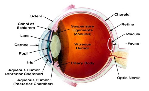
Overview
The eye is one of the sense organs of the body. It is responsible for sight. This page contains a description of the structure of the eye and its components
The inner part of the eye
The inner part of the eye is the area surrounded by the 3 layered wall. It contains the lens and spaces (chambers) filled with aqueous and vitreous humour.
Accessory structures
These are structures external to the eyeball that work with other components of the eye to ensure good vision. They include conjunctiva, eyelid, lacrimal system and extraocular muscles.
Which structure in the eye focuses light by changes of curvature of its front surface?
The resilient, transparent structure in the eye that focuses light by changes of curvature of its front surface.
What is the fluid in the eye called?
The area inside the eye, behind the cornea, and in front of the iris. This area is filled with clear, watery fluid called "aqueous humor."
What muscles control the size of the pupil?
The colored part of the eye. It consists of two circular muscles with a hole in the middle, called the "pupil." The iris sphincter and dilator muscles control the size of the pupil for maximum visual performance.
What is the white part of the eye made of?
The white portion of the eye made up of a tough, fibrous tissue that gives structure to the eyeball.
What is the primary action of the ophthalmologist?
It's primary action is to move the eye upward (elevation). It also abducts the eye. Additionally, it can rotate the top of the eye toward the nose and the bottom of the eye toward the temple (intorsion).
Where is the most powerful refractive tissue in the eye?
The clear, transparent tissue that is located on the very front (anterior) portion of the eye. It is the most powerful refractive media of the eye. It provides most of the eye's ability to focus light.
What is the primary action of the adductor?
It's primary action is to turn the eye downward (depression). It also adducts the eye. Additionally, it can rotate the top of the eye toward the temple and the bottom of the eye toward the nose (extorsion).
How Is Intraocular Pressure Measured?
Intraocular pressure is measured with a tonometer as part of a thorough eye assessment.
What is the average intraocular pressure?
The average worth of intraocular pressure is 15.5 mmHg with changes of about 2.75 mmHg.
What is the definition of hypotension?
Ocular hypotension, Hypotony, or ocular hypotony, is typically defined as intraocular pressure equivalent to or less than 5 mmHg. Such low intraocular pressure might suggest fluid leak and deflation of the eyeball.
What is IOP in glaucoma?
IOP is an important aspect in the examination of patients at risk from glaucoma. Most tonometers are adjusted to measure pressure in millimeters of mercury (mmHg).
Why is intraocular pressure so high?
Intraocular pressure might become raised due to anatomical issues, swelling of the eye, hereditary factors, or as a side-effect from medication. Intraocular pressure laws follow fundamentally from physics. Any kinds of intraocular surgery should be done by considering the intraocular pressure fluctuation. All of a sudden increase of intraocular pressure leads to intraocular micro barotrauma and causing anemia impact and mechanical stress to retinal nerve fiber layer. Rapid intraocular pressure drop leads to intraocular decompression that produces micro bubble that potentially triggering numerous micro emboli and resulting in hypoxia, anemia and retinal micro structure damage.
Can refractive surgery cause intraocular pressure to be abnormal?
As an outcome, some kinds of refractive surgery (such as photorefractive keratectomy) can cause standard intraocular pressure measurements to appear normal when in fact the pressure might be unusually high.
Is intraocular pressure a secondary result?
Intraocular pressure has been measured as a secondary result in a systematic review comparing the result of neuroprotective agents in slowing the progression of open angle glaucoma.