
Where does the thoracic outlet pass through the neck?
The lower four cervical nerves and the first thoracic nerve emerge from the spinal cord, pass in between the scalene muscles in the neck, through the thoracic outlet, under the pectoralis minor, into the armpit, and down the arm. If the brachial plexus is compressed at any point along its path, you may feel the symptoms of thoracic outlet syndrome.
What is the function of the thoracic outlet?
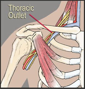
The thoracic outlet is a small space between the clavicle (collarbone) and the first rib (see image below). A bundle of nerves and blood vessels passes through the thoracic outlet and provides sensation, motor control, and circulation of blood to the chest, shoulder, arm, and hand. The network...
What network of nerves passes through the thoracic outlet?
The network of nerves that passes through the thoracic outlet is called the brachial plexus. The lower four cervical nerves and the first thoracic nerve emerge from the spinal cord, pass in between the scalene muscles in the neck, through the thoracic outlet, under the pectoralis minor, into the armpit,...
What is thoracic outlet syndrome (TOS)?
This helps them comprehend how the compression of nerves, arteries, veins, and blood vessels in this area affects an individual’s health and well-being and can lead to TOS. The thoracic outlet region is located within the lower part of the neck, beginning just above and behind the clavicle and extending to the upper part of the arm.
What travels through thoracic outlet?
The thoracic outlet is defined as the space in the lower neck between the thorax and axilla through which the subclavian vein, subclavian artery, and brachial plexus travel from their central origins to their peripheral termini.
What passes through the inferior thoracic outlet?
The thoracic outlet or inferior thoracic aperture is the space between the first rib and the collarbone. The thoracic outlet is the space where the nerves and blood vessels enter the arms.
What structures are transmitted through the thoracic inlet?
Structures that pass through the thoracic inlet include:trachea.oesophagus.thoracic duct.apices of the lungs.nerves. phrenic nerve. vagus nerve. recurrent laryngeal nerves. sympathetic trunks.vessels. arteries. left and right common carotid arteries. left subclavian arteries. veins. ... lymph nodes and lymphatic vessels.
What muscles are involved in TOS?
The thoracic outlet is the ring formed by the top ribs, just below the collarbone. Thoracic outlet syndrome (TOS) occurs when nerves or blood vessels are compressed by the rib, collarbone or neck muscles at the top of the outlet.
What nerves are affected by TOS?
Thoracic outlet syndrome (TOS) is a group of disorders that occur when blood vessels or nerves in the space between your collarbone and your first rib (thoracic outlet) are compressed. This can cause shoulder and neck pain and numbness in your fingers.
What passes through the scalene triangle?
The scalene triangle is bound by the anterior and middle scalene muscles, with the first rib at the base. The brachial plexus nerves (yellow) and the subclavian artery (red) pass through the scalene triangle, while the subclavian vein (blue) passes in front.
Which of the following structures does not pass through the superior thoracic aperture?
All of the following structures pass through the superior aperture of thorax except? The right recurrent laryngeal nerve loops around the right subclavian artery and hence does not pass through the superior aperture of thorax.
How do you test for thoracic outlet syndrome?
To confirm the diagnosis of thoracic outlet syndrome, your doctor may order one or more of the following tests:Ultrasound. An ultrasound uses sound waves to create images of your body. ... X-ray. ... Computerized tomography (CT) scan. ... Magnetic resonance imaging (MRI). ... Arteriography and venography. ... Electromyography (EMG).
Does the thoracic duct pass through the thoracic inlet?
It crosses to the left of midline at the thoracic plane (range T4-T6 vertebral body) and continues superiorly through the thoracic inlet, posterior to the left subclavian artery and continues in the neck anterior to the anterior scalene muscle and phrenic nerve.
What are 3 of the special tests for thoracic outlet syndrome?
Adson's, Eden's and Wright's tests are special orthopedic assessment test for Thoracic Outlet Syndrome (Fig. 10).
What muscles are affected by thoracic outlet syndrome?
The muscles in this group include the supraspinatus, infraspinatus, teres minor, and subscapularis.
What structures are compressed in thoracic outlet syndrome?
The thoracic outlet comprises the space from the supraclavicular fossa to the axilla. The symptoms of TOS arise from compression of the brachial plexus nerves, subclavian artery and vein, and axillary artery and vein.
What passes through the superior thoracic aperture?
The right and left subclavian arteries and veins pass through the superior thoracic aperture. These vessels may be compromised by pathologic conditions of the lower cervical or upper thoracic viscera.
What are the boundaries of the inferior thoracic aperture?
Boundariesposteriorly: 12th thoracic vertebral body and transverse processes.posterolaterally: 11th and 12th ribs.anterolaterally: the costal margins formed by the conjoint costal cartilages of 7th to 10th ribs.anteriorly: xiphisternal joint.
What forms the boundary of the inferior thoracic aperture?
The diaphragm occupies and closes the inferior thoracic aperture, thereby separating the thoracic and abdominal cavities.
What are 3 of the special tests for thoracic outlet syndrome?
Adson's, Eden's and Wright's tests are special orthopedic assessment test for Thoracic Outlet Syndrome (Fig. 10).
Where does the thoracic outlet pass?
The lower four cervical nerves and the first thoracic nerve emerge from the spinal cord, pass in between the scalene muscles in the neck, through the thoracic outlet, under the pectoralis minor, into the armpit, and down the arm. If the brachial plexus is compressed at any point along its path, you may feel the symptoms of thoracic outlet syndrome.
What is the bundle of nerves that passes through the thoracic outlet?
The network of nerves that passes through the thoracic outlet is called the brachial plexus. The lower four cervical nerves and the first thoracic nerve emerge from ...
Why do we develop the chronic muscle tightness that causes thoracic outlet syndrome?
The muscles of the neck, shoulders, and chest that we discussed in the last section become tight as a result of the way we use our body every day. If we engage in activities or adopt habitual postures that involve flexing the head and neck forward, rounding the shoulders and upper back, and rotating the shoulders inward and bringing our arms in toward our body, we can easily develop the patterns of muscular tightness that lead to thoracic outlet syndrome.
What causes thoracic outlet syndrome?
Thoracic outlet syndrome can be caused by a structural issue, such as an extra rib, the growth of a tumor, or an injury to the area. But more often, thoracic outlet syndrome is a functional issue, caused by poor posture or chronic muscle tension in the neck, shoulder, and chest. The nerves that form the brachial plexus emerge from ...
Which muscle is most commonly implicated in thoracic outlet syndrome?
The pectoralis minor is the muscle most commonly implicated in thoracic outlet syndrome. However, before the brachial plexus reaches the pectoralis minor, it passes through the thoracic outlet. The clavicular fibers of the pectoralis major attach to the clavicle, as does the deltoid muscle. Chronic tightness in these two muscles creates ...
Can thoracic outlet syndrome be prevented?
Thoracic Outlet Syndrome: Anatomy, Causes, and Treatment with Clinical Somatics. Thoracic outlet syndrome might sound a little scary, but most cases are functional—meaning they can be prevented and often eliminated completely. When tight muscles or poor posture compress the thoracic outlet, you may experience symptoms such as pain ...
Where is the thoracic outlet?
The thoracic outlet is defined as the space in the lower neck between the thorax and axilla through which the subclavian vein, subclavian artery, and brachial plexus travel from their central origins to their peripheral termini.
Where does the subclavian artery exit the thoracic outlet?
Each subclavian artery ascends superiorly into the neck before arching laterally and traveling posterior to the anterior scalene muscle through the scalene triangle and exiting the thoracic outlet via the costoclavicular space (above the first rib, below the clavicle) to become the axillary artery.
What is the subclavian artery?
The subclavian arteries are the primary blood supply to the upper extremities. The left subclavian artery branches directly from the aorta, whereas the right subclavian artery arises from the brachiocephalic artery. Each subclavian artery ascends superiorly into the neck before arching laterally and traveling posterior to the anterior scalene muscle through the scalene triangle and exiting the thoracic outlet via the costoclavicular space (above the first rib, below the clavicle) to become the axillary artery. Because of the close proximity of the clavicle, first rib, and anterior and middle scalene muscles, the costoclavicular space is the most frequent site of arterial compression.
What muscle is used to compress the brachial plexus?
Compression with movement of the scalene muscle. As the anterior scalene muscle goes from rest (A) to contracted (B), it pulls the first thoracic rib cranially and compresses the brachial plexus and subclavian within the costoclavicular space. Presence of a supernumerary cervical rib, as demonstrated in this figure, may constrict this space further. (FromAdson AW. Surgical treatment for symptoms produced by cervical ribs and the scalenus anticus muscle. Surg Gynecol Obstet. 1947;85:687. Reprintedwith permission from the Journal of the American College of Surgeons, formerly Surgery Gynecology & Obstetrics.)
Where is the subclavian vein located?
The subclavian vein develops in the fourth gestational week and is formed by the fusion of venous tributaries from the upper limb bud.3The vein can be found in an anomalous location posterior to the anterior scalene muscle, immediately adjacent to the subclavian artery. More rarely, the subclavian vein splits to form a “clavicular loop” or travels between the clavicle and the subclavius muscle.
Which artery is the left subclavian artery?
The left subclavian artery develops from the left seventh intersegmental artery, whereas the right subclavian artery arises from the fourth aortic arch, right dorsal aorta, and right seventh intersegmental artery.3Anomalies of the distal subclavian artery are rare, although anomalies of the aortic arch frequently encompass the origin of the either artery. In the thoracic outlet, the subclavian artery occasionally penetrates the anterior scalene muscle and rarely may pass entirely anterior to it.
Where does the vein go in the neck?
On either side, the vein ascends superiorly with the subclavian artery into the neck. It then travels anterior to the anterior scalene muscle (outside of the technical scalene triangle) and continues in parallel to the artery with the anterior scalene muscle separating the two structures.
What is the thoracic outlet?
Thoracic outlet syndrome (TOS) refers to a condition involving the compression of nerves and blood vessels as they pass through a part of the body called the thoracic outlet . This area is bordered by the collar bone, side of the neck, shoulder blade and top of the first rib. 1 TOS can lead to symptoms located in a variety of areas ranging from your neck to finger tips. These symptoms include aching pain mostly along the side of the neck to the shoulder. Other symptoms can include numbness, tingling sensations, headaches, muscle weakness, changes in skin color and changes in skin temperature.
Who gets Thoracic Outlet Syndrome?
Most people who develop TOS are in their 20s and 30s. Women develop this condition slightly more often than men; those who are overweight are also at greater risk. Prolonged poor sitting and poor posture combined with repetitive hand activities such as typing on a computer are prime contributors to the gradual development of TOS. Trauma to the neck such as whiplash during an auto accident or the shoulder can lead to TOS developing more suddenly. About half of diagnosed TOS cases are caused by trauma. 2
How to diagnose TOS?
TOS is clinically diagnosed through a physical examination that may include assessing strength, sensation, and placing your neck, arms, and hands in positions that may momentarily provoke symptoms. 4 Other methods involved in diagnosing TOS focus on ruling out other conditions such as cervical disc pathologies with the use of medical imaging. Nerve conduction tests may be useful in assessing patients with neurogenic TOS. Vascular tests such as a brachial artery angiography, venography, doppler ultrasonography can help diagnosis arterial or venous TOS. 1,2 TOS can be misdiagnosed as carpal tunnel syndrome, cervical disc pathologies, or other conditions that may cause pain and tingling in the upper extremity. 5 The frequency of the misdiagnosis of TOS has been difficult to pinpoint but researchers believe that anywhere from 3 to 80 out of every 1,000 have this condition. 6
How to treat TOS?
Conservative treatments such as physical therapy are most often successful in treating TOS. Physical therapy treatment for TOS will vary from person to person but may include correction of posture with an emphasis on strengthening muscles necessary to maintain posture throughout the day. 4 Physical therapists often have expertise in assessing postural and functional abnormalities that may contribute to TOS.
Where is the anterior scalene located?
Anterior Scalene Syndrome: The compression of nerves or blood vessels occurs between 2 of the scalene muscles, which are located on the sides of the neck.
Where is the thoracic outlet?
Specifically, the thoracic outlet is the hollow space above your collarbone, between your shoulder and your neck. Slide your hand down either side of your neck to find the thoracic outlet. Three vital structures pass from the neck and chest through each thoracic outlet to enter each arm.
When does thoracic outlet syndrome occur?
Thoracic outlet syndrome occurs in women more than men, usually between the ages of 20 to 40. We currently recognize an increased risk of developing thoracic outlet syndrome in the following situations: Neck injuries resulting from motor vehicle accidents. Work environments with repetitive stress injures.
What causes TOS?
The nerves, artery and vein must be free to pass through the thoracic outlet. However, anatomic variants, acquired abnormalities, and abnormal arm motion may cause compression of these vital structures. In that case, patients may experience the symptoms of thoracic outlet syndrome. People with neck injuries, occupational injuries or overuse, and overhead athletes are all at risk for developing thoracic outlet syndrome.
What are the symptoms of TOS?
Symptoms of TOS are different in each type of TOS. Patients with venous TOS suffer swelling of the arm from decreased blood flow returning to the heart. Patients with arterial TOS suffer loss of blood flow to all or part of the arm. Patients with neurogenic TOS suffer a broad range of sensory and motor disturbances of the affected arm and hand
What is TOS in surgery?
TOS can range from an emergent surgical condition to a progressive, chronic pain syndrome that alters the lives of TOS patients. Three vital structures pass from the neck and chest to reach each upper extremity. When compression of these vital structures causes symptoms, TOS is present.
What is a TOS patient?
Doctors use the abbreviation ‘TOS’ instead of the full name of the disease, ‘Thoracic Outlet Syndrome.’ TOS patients suffer pain, tingling, numbness, weakness, or abnormal blood flow in one or both upper extremities. TOS can range from an emergent surgical condition to a progressive, chronic pain syndrome that alters the lives of TOS patients.
What is a TOS?
What is TOS? TOS stands for Thoracic Outlet Syndrome. The thoracic outlet is the hollow space between your neck and shoulder on each side, above the collar bone. Three vital structures pass through the thoracic outlet on each side: an artery, a vein, and a nerve plexus.
Where is the thoracic outlet?
The term “thoracic outlet” is the area between the neck and shoulder, over the top of the thorax, and under the clavicle. Knowledge of thoracic outlet anatomy and the four major areas of compression is cardinal for the surgeon to perform thoracic outlet syndrome decompression without injuring major structures, having a minimal number of complications, and avoiding malpractice litigation.
Why is thoracic outlet anatomy important?
Knowledge of the thoracic outlet anatomy and its many variations is essential for surgical decompression to avoid injuries to vital structures, minimizing complications and lawsuits.
What are the branches of the subclavian artery?
Branches of the subclavian artery include the internal mammary artery proceeding inferiorly under the sternum. Just before passing over the first rib, the subclavian gives off the vertebral artery, which is directed cephalad toward the brain. The thyrocervical trunk branches distally. The subclavian artery is joined by the nerves of the brachial plexus before passing beneath the clavicle over the top of the first rib. It also carries branches of the sympathetic nerves peripherally. Pressure on this artery from the scalenus anticus or any other area of compression in the four major areas may produce poststenotic dilatation if it is a minor compression, or a subclavian artery stenosis, aneurysm, or occlusion if it is major compression. Emboli may occur distally in the axillary-subclavian artery from stenosis, occlusion, or aneurysm. After removing the compressed area, poststenotic dilatations generally regress gradually over a period of time and do not usually require further therapy. All of the other anomalies of compression usually need medical or surgical treatment ( Box 2 ).
What is the clavicle attached to?
The clavicle attaches to the sternum and to the first rib anteriorly through the costoclavicular and clavicular-sternal ligaments medially (see Fig. 1 ). It proceeds laterally to articulate with the acromial process of the scapula. The medial part of the clavicle moves very little; however, the lateral part is more mobile because of the scapula. Removal of the clavicle provides decompression of the thoracic outlet and was used early in the therapeutic history of TOS; however, removal of the first rib achieves the same end without as much cosmetic morbidity. There are marked variations in the ligaments, tendons, and muscles attaching to the bony structures in the thoracic outlet. Roos [3] has listed 10 types of anomalous congenital bands and ligaments within the thoracic outlet, most of which are not frequently seen clinically.
What are cervical ribs?
Cervical ribs usually elevate the neurovascular structures, decreasing the thoracic outlet space, and are either extensions of the C-7 transverse process (>2.5 cm) or articulated like any other rib to the C7 vertebra ( Fig. 2 ). The distal end of the cervical rib may be free, fuse directly with the first rib, or connect to it through fibrous ligaments ( Box 1 ). The incidence of cervical ribs in the population is roughly 0.5% to 1%, whereas those associated with TOS occur in approximately 5% to 10% of patients. There is a slightly higher incidence in women and a greater occurrence on the left side.
Why is the transaxillary approach preferred?
For primarily nerve (most frequent) and vein decompression, the transaxillary approach is favored because the neurovascular structures are away from the first rib and do not require retraction for rib removal. Also, it is easier to remove the first rib completely.
Where does the subclavian vein pass?
Fig. 3. The subclavian vein and artery pass over the first rib and under the clavicle. The brachial plexus traverses the top of the bony circle to join the artery. The apex of the pleura (cupula) is depicted on the left side.
What is the thoracic inlet?
The thoracic inlet, also known as the superior thoracic aperture, refers to the opening at the top of the thoracic cavity. It is also clinically referred to as the thoracic outlet, in the case of thoracic outlet syndrome; this refers to the superior thoracic aperture, and not to the lower, larger opening, the inferior thoracic aperture .
What is the structure of the thoracic inlet?
Structure. The thoracic inlet is essentially a hole surrounded by a bony ring, through which several vital structures pass. The thoracic inlet is bounded by: the first thoracic vertebra (T1) posteriorly; the first pair of ribs laterally, forming lateral C-shaped curves posterior to anterior; and the costal cartilage of the first rib and ...
Which muscle is superior to the thoracic inlet?
Superior to the thoracic inlet is the root of the neck, and the superior mediastinum is inferiorly related. The brachial plexus is a superolateral relation of the thoracic inlet. The brachial plexus emerges between the anterior and middle scalene muscles, superior to the first rib, and passes obliquely and inferiorly, underneath the clavicle, into the shoulder and then the arm. Impingement of the plexus in the region of the scalenes, ribs, and clavicles is responsible for thoracic outlet syndrome .
Where is the brachial plexus?
The brachial plexus emerges between the anterior and middle scalene muscles, superior to the first rib, and passes obliquely and inferiorly, underneath the clavicle, into the shoulder and then the arm.
Where is the oesophagus located?
The oesophagus lies against the body of the T1 vertebra, separated from it by the prevertebral fascia, and the trachea lies in front of the oesophagus, in the midline, and may touch the manubrium. The apices of the lungs lie to either side of the oesophagus and trachea, and is separated from them by the other vessels and nerves listed above. Furthermore, they extend slightly superior past the level of the inlet (e.g. the horizontal plane of the first rib).
Can a thyroid gland extend inferiorly?
There are several other minor, but important, vessels and nerves passing through, and an abnormally large thyroid gland may extend inferiorly through the thoracic inlet into the superior mediastinum.
What structures pass through the thoracic outlet?
Structures passing through the thoracic outlet between the thorax and abdomen include the inferior vena cava and esophagus, both of which pass through the diaphragm, and the abdominal aorta and thoracic duct which pass through the diaphragm, through the aortic hiatus .
Which is larger, the thoracic outlet or inferior thoracic aperture?
It is closed by the diaphragm, which separates the thoracic cavity from the abdominal cavity. The thoracic outlet or inferior thoracic aperture is much larger than the thoracic inlet (superior thoracic aperture). The thoracic outlet is bounded by: the 12th thoracic vertebra posteriorly, 11th and 12th pairs of ribs laterally,
What is the posterior aspect of the thorax?
The posterior aspect of the thorax. (Inferior thoracic aperture visible at bottom.) The thora cic outlet is the lower opening of the thoracic cavity whose edges are the lowest ribs. It is closed by the diaphragm, which separates the thoracic cavity from the abdominal cavity.
Which joint is the articulation between the xiphoid process and the inferior border of the stern?
11th and 12th pairs of ribs laterally, costal cartilages of ribs 7 through 10 anteriorly, and the xiphisternal joint - (i.e.: the articulation between the xiphoid process and the inferior border of the sternal body) anteriorly. Structures passing through the thoracic outlet between the thorax and abdomen include the inferior vena cava ...
What is the space inside the thoracic outlet?
This space is known as the thoracic outlet. Anything that narrows the space inside the thoracic outlet can put pressure on the blood vessels and nerves. This can cause pain in the neck, shoulders, and arms. Thoracic outlet syndrome can develop if a person has poor posture, if they injure their shoulder, or if they perform repetitive shoulder ...
What is the thoracic outlet syndrome?
Thoracic outlet syndrome refers to a group of conditions characterized by the compression of the nerves, arteries, and veins that pass through the space between the collarbone and the first rib. This space is known as the thoracic outlet. Anything that narrows the space inside the thoracic outlet can put pressure on ...
What are the symptoms of neurogenic thoracic outlet syndrome?
Some symptoms of neurogenic thoracic outlet syndrome include: pain or a dull aching in the neck, shoulder, armpit, arm, or hand. weakness in the arm and shoulder. numbness or pins and needles in the fingers and hand. changes in the color and temperature of the hand. atrophy, or muscle wasting, in the hand.
How long does thoracic outlet syndrome last?
People should see a doctor if they experience any symptoms of thoracic outlet syndrome that last for several days or weeks.
Why is thoracic outlet syndrome difficult to diagnose?
According to the National Institute of Neurological Disorders and Stroke, healthcare professions may have difficulty diagnosing thoracic outlet syndrome because it causes symptoms that occur in many other conditions, including: complex regional pain syndrome. tumors of the spinal cord. multiple sclerosis. fibromyalgia.
What is the name of the artery that supplies blood to the head, neck, arms, and shoulders?
Arterial thoracic outlet syndrome. This type of thoracic outlet syndrome occurs when a blood clot forms in the subclavian artery. These arteries supply blood to the head, neck, arms, and shoulders.
What causes swelling in the thoracic outlet?
Repetitive arm and shoulder movements can irritate the brachial plexus nerves or cause swelling that constricts the space inside the thoracic outlet.
