What are the first signs of a bad gallbladder?
- Nausea or Vomiting. Chronic gallbladder disease can result in digestive issues, such as nausea and vomiting. ...
- Fever or Chills. An infection of the gallbladder can result in an unexplained fever or chills. ...
- Chronic diarrhea/Unusual stools or urine. ...
What is the normal size of the gallbladder?
The normal adult gallbladder measures from 7-10 cm in length and 3-4 cm in transverse diameter 6. The gallbladder communicates with the rest of the biliary system by way of the cystic duct, with bidirectional drainage of bile to and from the common hepatic duct.
What quadrant and region is the urinary bladder in?
Left Upper Quadrant. Stomach, Spleen. Right Lower Quadrant. Urinary bladder, appendix, cecum, ascending colon of Large Intestine, small intestine. Left Lower Quadrant. Transverse and descending of Large Intestine, small intestine, initial part of sigmoid colon, urinary bladder. Abdominopelvic regions. Nine regions formed by superior horizontal plane just inferior to ribs, inferior horizontal plane to hip bone, vertical planes just medial to nipples.
Where is the gallbladder located in the human body diagram?
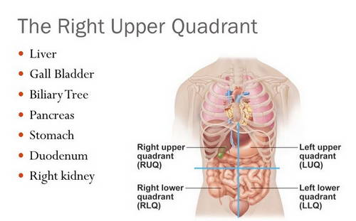
The gallbladder is located inferior (below) and posterior (behind) to the liver in the upper right quadrant (section) of the abdomen. It lies in front of the duodenum (the first section of the small intestine). The gallbladder is connected to the liver via the ducts known as the biliary tract.

What organs are in each of the 4 quadrants?
The organs in each quadrant include: RUQ: Liver, Gallbladder, Pancreas, Stomach, Small Intestine, Large Intestine, Right Kidney, Right Adrenal Gland. LUQ: Spleen, Stomach, Pancreas, Liver, Large Intestine, Small intestine, Left Kidney, Left Adrenal Gland.
Is the gallbladder in the RUQ?
The uppermost quarter on your right-hand side is your right upper quadrant (RUQ). The RUQ contains many important organs, including parts of your liver, right kidney, gallbladder, pancreas, and large and small intestine.
What is in the 4 quadrants of the abdomen?
The four quadrants of the abdomen The abdomen can also be divided into four quadrants, known as the right upper, the left upper, the right lower, and the left lower quadrants. It is common to see these quadrants abbreviated as RUQ, LUQ, RLQ, and LLQ, respectively.
Is the gallbladder in the Llq?
The right upper quadrant contains: Part of the liver. Gallbladder.
What is the most common cause of right upper quadrant pain?
Acute cholecystitis is the most common diagnosable cause for right upper quadrant abdominal (RUQ) pain in patients who present to the emergency department (ED). However, over one-third of patients initially thought to have acute cholecystitis actually have RUQ pain attributable to other causes.
What organ is right under your right rib cage?
Just under the right side of your rib cage lie several important organs, such as the pancreas, gallbladder, right kidney, and parts of your liver, and small and large intestines. Experts divide the abdomen into four quadrants, and the upper quarter on the right-hand side is the RUQ or right upper quadrant.
What quadrant is constipation?
In general, left-sided lower abdominal pain is usually caused by gastrointestinal problems such as gastroesophageal reflux disease (GERD), constipation, or irritable bowel syndrome (IBS).
What organ is in the lower left quadrant?
In the lower-left part of the abdomen, you can find the left kidney, left ureter, colon, bladder, blood vessels, and nerves. In women, you'll find the left fallopian tube and ovary.
What organs are in the lower right quadrant?
Organs found in the right lower quadrant include the appendix, the upper portion of the colon, and the right ovary and the Fallopian tube in women. The right lower quadrant may be assessed when diagnosing appendicitis, in which case, this quadrant would be tender and painful.
Where is the position of gallbladder?
Where is the gallbladder located? Your gallbladder is located in the upper right part of your abdomen (belly). It sits just under your liver.
What does left lower quadrant pain mean?
Often, left lower quadrant pain is related to conditions of the digestive tract; however, it can also be related to conditions of the body wall, skin, blood vessels, urinary tract, or reproductive organs.
What organs are on the left side of your abdomen?
The organs located in your left abdomen include your colon, left kidney, spleen, stomach, and pancreas.
What organs are found in the RUQ?
The organs within your right upper quadrant are your:Liver.Gallbladder.Part of the pancreas.Duodenum and some other parts of your large and small bowel.Right kidney (at the back behind the other organs).
What does RUQ stand for?
The right upper quadrant, or the RUQ, has many important organs that affect your health. It's important to pay attention to pain in this area.
Which of the following are organs in the right upper quadrant?
Right Upper Quadrant Organs found in this quadrant include: the liver, the gallbladder, duodenum, the upper portion of the pancreas, and the hepatic flexure of the colon.
What does an upper right quadrant ultrasound show?
A right upper quadrant ultrasound examines three organs of the digestive system: Liver. Pancreas. Gallbladder.
What are the quadrants of abdomen?
The quadrants of the abdomen refer to the four sections that the abdomen is divided into, for ease of clinical examination and communication. By di...
What organs are in each of the 4 quadrants?
The organs in each quadrant are: - RUQ - Liver, Gallbladder, Pancreas, Stomach, Small Intestine, Large Intestine, Right Kidney, Right Adrenal Glan...
How are abdominal quadrants divided?
The four abdominal quadrants are divided along the median and transverse planes, by drawing a vertical and horizontal line that perpendicularly int...
What causes gallbladder problems?
Several conditions can cause problems in your gallbladder. The most common condition is gallstones. Gallstones are typically harmless but can sometimes lead to disease states. Gallbladder issues include:
What happens to the gallbladder when you eat?
Before you start eating, your gallbladder is full of bile. When you start eating, your gallbladder receives signals to contract and squeeze the stored bile through the biliary tract. The bile eventually finds its way to your largest bile duct, the common bile duct. Bile passes through the common bile duct into the duodenum, the first part of your small intestine, where it mixes with food waiting to be digested. After you eat, your gallbladder is empty and resembles a deflated balloon, waiting to be filled up again.
What is the name of the system that carries bile from the liver?
Your gallbladder is connected to other parts of your digestive system through a series of bile ducts called the biliary tract. The biliary tract (sometimes called biliary system or biliary tree) is a pipe-like system that carries bile from your liver to your small intestine.
What is the organ that stores and releases bile?
Your gallbladder is a small, pear-shaped organ located under your liver that stores and releases bile. Bile is the fluid your liver produces that helps digest fats in the food you eat.
What is the inflammation of the gallbladder?
Cholecystitis: Cholecystitis is inflammation of your gallbladder. It can occur when a gallstone blocks bile from exiting your gallbladder. Cholecystitis causes fever and pain and usually requires surgery.
What is gallstone pancreatitis?
Gallstone pancreatitis: Gallstone pancreatitis is inflammation of your pancreas. It occurs when a gallstone travels down the common bile duct and blocks the pancreatic duct at a common point just before draining into the small intestine.
What is laparoscopic cholecystectomy?
Laparoscopic cholecystectomy: With laparoscopic surgery, your surgeon operates through a few small incisions. Laparoscopic surgery generally leads to a faster recovery, less pain and smaller scars. In most cases, cholecystectomies will be performed laparoscopically.
This organ stores and releases bile for digestion
The main function of the gallbladder is to store, concentrate, and release bile into the digestive system. It is a muscular organ that contracts when bile is needed, forcing the bile through a tube called the cystic duct.
Gallbladder Function
There are several important functions of the gallbladder, which include: 2
Anatomy of the Gallbladder
The gallbladder is a small, pear-shaped hollow organ. It is approximately an inch wide and 3 inches long, 1 and is tapered at one end where it connects to the cystic duct. It can store approximately 30 to 50 cubic centimeters (cc) of bile.
Associated Conditions
Common gallbladder conditions can involve infection, stones, inflammation or blockage of the gallbladder. 4
Symptoms
Symptoms of gallbladder problems aren't the same for everyone. Some people have no symptoms at all.
Treatment
Once a diagnosis of gallstones (or other gallbladder disorders) is made, most people with symptoms undergo removal of the gallbladder. This procedure is called a cholecystectomy . It is most often performed using laparoscopic (use of a scope with a camera, which is inserted into a very small incision) surgery.
Summary
Your gallbladder is a small organ located just below the liver on the right side of the body. Its primary function is to store and secrete bile, which helps your body digest fats.
What are Abdominal Quadrants?
The abdomen defines a region of the body located between the thorax (or chest) and the pelvis. It is separated from the thorax at the diaphragm and the pelvis at the beginning of the pelvic bones. This region is also referred to as the belly.
How to divide the abdomen into four regions?
In this system, the abdomen is divided into four regions by drawing a vertical line (dividing on the median or midsagittal plane) and a horizontal line (dividing on the transverse or transumbilical plane) that perpendicularly intersects at the umbilicus (or navel). The median (or midsagittal) plane can be visualized by drawing a vertical line from the center of the forehead to in between the two feet, this line should divide the body into two halves that are relatively equal and symmetrical. The transverse or transumbilical plane, on the other hand, divides the body into an upper half and lower half, by drawing an imaginary line that runs perpendicular to the spine, and intersects with the median plane at the navel.
How are the abdominal quadrants divided?
The four abdominal quadrants are divided along the median and transverse planes, by drawing a vertical and horizontal line that perpendicularly intersect at the navel.
What is the right lower quadrant?
The Right Lower Quadrant is the third of the abdominal quadrants. The list of organs found in the RLQ includes:
What are the organs in the abdomen?
These include the digestive organs, kidneys, and spleen. By taking a closer look at each of the four abdominal quadrants, one can learn about the different organs found in each one.
How many regions does the abdomen have?
There are two major systems by which the abdomen can be divided into smaller areas. One system involves dividing the abdomen into four regions along a vertical and horizontal axis centered at the umbilicus (navel). These four regions are known as abdominal quadrants.
What is the abdomen?
The abdomen is the region of the body between the thorax and the pelvis. It contains various important organs, such as the digestive organs, kidneys, and spleen. Because the abdomen contains so many vital organs, it is divided into abdominal quadrants for ease of study, clinical examination, and communication. An alternate system divides the abdomen into nine regions. The four quadrants are divided along the median and transverse planes by drawing a vertical and horizontal line that perpendicularly intersects at the navel. The four quadrants are:
Why is my urine dark?
Changes in urine: Patients suffering from gallbladder issues may notice darker than normal urine. Dark urine may indicate a bile duct block.
What is the term for a gallbladder that is inflamed?
Inflamed gallbladder, cholecystitis. Acute or sudden cholecystitis occurs when bile can’t leave the gallbladder. This commonly happens when a gallstone obstructs the tube that bile uses to travel into and out of the gallbladder. Chronic cholecystitis occurs if there are recurrent acute attacks.
Why is the gallbladder stiff?
This makes them stiff, limiting the gallbladder’s function and increasing the risk of gallbladder cancer. The word “porcelain” is used because the organ becomes bluish and brittle.
How long is the incision for gallbladder surgery?
During open surgery, a surgeon removes the gallbladder through a 4-6-inch-long incision in the abdomen. These surgeries often happen when the gallbladder is too inflamed or infected to remove laparoscopically or if a problem occurs during a laparoscopic procedure.
What causes chest pain?
Symptoms. Share on Pinterest. A problem with the gallbladder can cause chest pain. Symptoms of gallbladder problems include: Pain in the mid- or upper-right section of the abdomen: Most of the time, gallbladder pain comes and goes.
How to treat gallstones?
Treatment options include surgically removing the gallbladder, medications to break up gallstones, and antibiotics to treat infections.
How rare is gallbladder cancer?
Gallbladder cancer is very rare, affecting less than 4,000 Americans per year; but if it does occur, it can spread to other parts of the body.
How to scan the gallbladder?
The subcostal approach is the most commonly used initial approach to imaging the gallbladder. Begin with the patient supine, and place the transducer in the midline at the epigastrium with the transducer oriented in the sagittal plane ( Fig. 9.1A ). If there is a large left hepatic lobe, the liver may be easily seen in this position. Leaving the transducer in the midline, tilt the transducer to project the sound wave toward the patient’s right side. In some cases, this will be sufficient to localize the gallbladder. If it is not seen, move the transducer down the costal margin while projecting it cephalad under the rib margin to scan through the dome of the liver. In many patients, the liver is mostly intrathoracic. In these patients, an improved image can be obtained by placing the transducer between the ribs (intercostal approach) ( Fig. 9.1B ). Alternatively, the patient can be positioned to bring the liver down below the costal margin. Have patients sit erect or semirecumbent, and instruct them to breathe deeply with their “belly out.” As the liver descends, the liver and gallbladder may be more easily imaged. If these maneuvers fail, move the patient to a left lateral decubitus position. This position brings the liver and gallbladder more toward the midline and closer to the transducer. Scan again from the subcostal approach, then continue to move the transducer along the costal margin toward the right flank. If the anterior subcostal approach is unsuccessful, leave the patient in a left lateral decubitus position, and scan through the right flank to identify the right kidney and Morison’s pouch ( Fig. 9.1C ). Once the right kidney is in view, the transducer should be angled slightly cephalad and anterior. The fundus of the gallbladder lies in close proximity to the right kidney and will often pop into view with this maneuver. Occasionally, it is useful to roll the patient from a left lateral decubitus position into a nearly prone position and scan from a subcostal approach. This position brings the gallbladder anterior. If heavy shadowing is seen from the gallbladder fossa, moving the patient prone may help the stones fall forward and make the echogenic stones more visible themselves, helping distinguish the shadows of a gallbladder packed with stones from artifact created by bowel gas ( 12 ).
What is the portal vein on Mickey Mouse's face?
The normal portal triad looks something like Mickey Mouse, with the portal vein making up Mickey’ s face, his ears formed anteriorly by the hepatic artery on the left and the common bile duct on the right ( Fig. 9.12 inset). In the normal portal triad, the “ears” are symmetrical.
What is the path of the bile duct?
Bile ducts follow the path of the portal venous system throughout the liver. In routine scans of the liver the peripheral biliary radicles are typically not visualized unless they are enlarged. The larger common bile duct can be identified at the portal triad. If a transverse section is obtained through the portal triad, the common bile duct is seen just anterior to and to the right of the portal vein ( Fig. 9.12 ). The diameter of the common bile duct is much smaller than the main portal vein, and is often almost imperceptible. When the portal vein is viewed in its long axis, the common duct can be seen to accompany it in its course toward the duodenum. It may be difficult to see if it is not enlarged, but it can be appreciated by the irregular beaded appearance it gives just anterior to the main portal vein ( Fig. 9.10; eFigs. 9.3 and 9.4; VIDEO 9.1 ).
What is the liver acoustic window?
The liver is the major acoustic window to the abdomen. Familiarity with the liver and its surrounding structures will open up a variety of applications for abdominal scanning beyond those listed here. As clinicians become proficient in RUQ scans, they will likely become more confident in the Focused Assessment by Sonography for Trauma (FAST) exam (to assess for free fluid in Morison’s pouch) and in the scan of the proximal abdominal aorta (to detect aneurysms and dissections). Although RUQ ultrasound is primarily focused on the liver, gallbladder, and biliary tree, it may incidentally reveal pathology in adjacent areas in the right kidney, pleural space, pancreas, and aorta.
What is the right portal vein?
The right portal vein is the major landmark of the portal triad in the porta hepatis. The portal veins are characterized by bright echogenic walls that help distinguish them from other vessels in the liver. This peculiar echogenicity gives the appearance of a halo surrounding the portal venous vessels. FIGURE 9.8.
What is the best way to image the liver, gallbladder, and biliary tree?
The liver, gallbladder, and biliary tree can be imaged from a number of approaches (subcostal, intercostal, and flank) with the patient in a variety of positions (supine, semirecumbent, erect, left lateral decubitus, and prone). The optimal technique will vary based on differences in anatomy, body habitus, and bowel gas patterns. Ideally, the sonographer should begin with a few standard approaches, modified and guided by both external and internal landmarks ( Fig. 9.1 ).
Which structure is surrounded by a number of structures that can interfere with Acquiring a Quality Image?
FIGURE 9.2. The Gallbladder is Surrounded by a Number of Structures that can Interfere with Acquiring a Quality Image. The right hepatic flexure and transverse colon lie just anterior and inferior to the gallbladder. The C-loop of the duodenum often crosses just medial to the gallbladder. Air in either structure can produce shadowing and create artifact.
What is gallbladder disease?
Gallbladder disease includes inflammation, infection, stones or blockage of the gallbladder. The gallbladder is a sac located under the liver. It stores and concentrates bile produced in the liver. Bile aids in the digestion of fat and is released from the gallbladder into the upper small intestine in response to food (especially fats). Types of gallbladder disease include:
What are the symptoms of a stone in the gallbladder?
Stones lodged in the common bile duct can cause symptoms that are similar to those produced by stones that lodge in the gallbladder, but they may also cause: Jaundice. Dark urine, lighter stools or both. Rapid heartbeat and abrupt blood pressure drop.
What are the different types of gallbladder disease?
Types of gallbladder disease include: Cholecystitis (inflammation of the gallbladder) Gallstones. Chronic a calculous gallbladder disease (in which the natural movements needed to empty the gallbladder do not work well) Gangrene or abscesses. Growths of tissue in the gallbladder. Congenital defects of the gallbladder.
Where does biliary colic pain radiate?
Typically, a patient experiences a steady gripping or gnawing pain in the upper right abdomen near the rib cage, which can be severe and can radiate to the upper back. Some patients with biliary colic experience the pain behind the breastbone. Nausea or vomiting may occur.
Can gallbladder disease cause nausea?
Nausea and vomiting may occur. Chronic gallbladder disease involves gallstones and mild inflammation. In such cases, the gallbladder may become scarred and stiff. Symptoms of chronic gallbladder disease include complaints of gas, nausea and abdominal discomfort after meals and chronic diarrhea.
Can gallbladder surgery be done in outpatient?
Surgery may be warranted to remove the gallbladder if the patient has gallstones or the gallbladder is not functioning normally. Most of the time this can be performed laparoscopically (through small incisions) as an outpatient procedure.
What are the symptoms of a gallbladder infection?
When the bile builds up, it can become infected and lead to a rupture or abscess. Symptoms of a gallbladder infection may include: abdominal pain. fever.
What causes gallbladder pain?
Gallbladder inflammation pain. Inflammation of your gallbladder, a condition called cholecystitis, commonly happens when gallstones block the tube leading out of your gallbladder. This produces bile buildup, which can cause inflammation. Other conditions can also trigger inflammation of your gallbladder, including:
Why does bile hurt when you eat?
salts. water. Your liver continually makes bile until you consume food. When you eat, your stomach releases a hormone that causes muscles around your gallbladder to release the bile.
Why does my gallbladder hurt?
The most common reason for the pain is gallstones — hardened deposits of digestive fluid that can form inside your gallbladder. Inflammation or infection of your gallbladder are other possible culprits. The good news is there are treatments to effectively relieve gallbladder pain.
What causes abdominal pain, bloating, and fever?
Gallbladder cancer. Gallbladder cancer can cause abdominal pain, itching, bloating, and fever. Imaging tests may help your doctor determine if the pain you feel is due to cancer or gallstones. Appendicitis.
How do you know if you have cholecystitis?
Symptoms of cholecystitis may include: severe pain in your upper right abdomen or center of your abdomen. pain that spreads to your right shoulder or back. tenderness above your abdomen. nausea or vomiting. fe ver. These symptoms often happen after eating, especially following a large or fatty meal.
What is the stomach flu?
Gastroenteritis. Also known as the “ stomach flu ,” gastroenteritis may be mistaken for a gallbladder issue. Symptoms such as nausea, vomiting, watery diarrhea, and cramping are hallmarks of the stomach flu.
