Where does the quadriceps innervation nerve come from?
The quadriceps innervation nerve root comes from the lower back. It then branches off into the femoral nerve before it attaches to the quad muscle. Nerve roots that supply muscles in the leg branch off the spinal cord at the lower back, or lumbar spine.
What part of the brain controls the muscles of the legs?
The motor regions of the brain and spinal cord control most of the nerve impulses that stimulate muscles in this region. However, many reflex pathways are also active in the legs and foot.
What nerve roots power the quadriceps femoris?
A series of nerves that emerge between the vertebrae of the lumbar spine ultimately provide power to the quadriceps femoris. According to the Geisel School of Medicine at Dartmouth, nerve roots L2 through L4 supply the quadriceps muscle.
What is the function of the quadriceps tendon?
The tendon of quadriceps continues distally and blends with the patellar ligament which attaches to the tibial tuberosity. This significantly contributes to the stability of the patella and knee joint. The entire quadriceps femoris muscle is innervated by the femoral nerve (L2-L4). Quadriceps femoris is the most powerful extensor of the knee.

What nerve root Innervates the quadriceps?
The femoral nerve innervates the sartorius, pectineus, quadriceps femoris, and iliacus muscle of the iliopsoas. It receives nerve supply by the nerve roots L2 through L4, innervating both the hip flexor and quadriceps muscle groups. The femoral nerve is also responsible for anterior thigh and medial leg sensation.
Does the sciatic nerve affect the quadriceps?
L4: The L4 nerve root, which is the first to feed in to the sciatic nerve, supplies the quadriceps femoris muscles. These muscle help extend, or straighten, the knee. Compression at this level may cause weakness while straightening the knee, which may create the feeling of the knee buckling.
What does a pinched femoral nerve feel like?
Symptoms of Femoral Nerve pain: Pain over the front of the thigh. Loss of power of knee extension and hip flexion. Lower extremity muscle weakness. Numbness, pain, burning, or tingling sensation in the medial aspect of the lower leg and anteromedial thigh.
What spinal nerves would I have to cut to paralyze the quadriceps muscle group?
According to the Geisel School of Medicine at Dartmouth, nerve roots L2 through L4 supply the quadriceps muscle.
What are the symptoms of L5 S1 nerve damage?
Common Symptoms and Signs Stemming from L5-S1Pain, generally felt as a sharp, shooting, and/or searing feeling in the buttock, thigh, leg, foot, and/or toes.Numbness in the foot and/or toes.Weakness in the leg and/or foot muscles and an inability to lift the foot off the floor (foot drop)
Can sciatica cause quadricep weakness?
When the sciatic nerve is compressed, one or more neurological symptoms may accompany the pain. A few examples of accompanying symptoms include: Weakness in the thigh muscles. When the thigh muscles are affected, there may be a weakness felt while attempting to bring the thighs together.
How do you release a trapped femoral nerve?
Kneeling on one knee, with your foot resting on a chair behind you. Tuck your bottom under and lunge slightly forwards into hip extension. Once you feel a gentle stretch slowly curl your head an upper back down to intensify the stretch for a few seconds then repeat.
Which disc causes pain in front of thigh?
L3 or L4 impingement from a herniated disc may lead to an abnormal reflex when the area just below the knee is tapped with the rubberized reflex hammer. This is called the patellar reflex. Pain from an L3 or L4 impingement usually radiates to the quadriceps femoris muscle at the front of the thigh.
Can a herniated disc affect the femoral nerve?
The most common cause is a disc prolapse at the L2/3 or L3/4 vertebra in the spine. As the disc bulges or prolapses backwards, it can hit the branch of the femoral nerve, causing symptoms into the front of the thigh.
What does the C4 and C5 nerves control?
C4 provides sensation for parts of your neck, shoulders and upper arms. Cervical nerve 5 controls the deltoid muscles of your shoulders and your biceps. C5 provides sensation to the upper part of your upper arm down to your elbow.
What does T7 and T8 control?
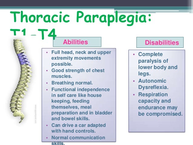
T1 and T2 (top two thoracic nerves) feed into nerves that go into the top of the chest as well as into the arm and hand. T3, T4, and T5 feed into the chest wall and aid in breathing. T6, T7, and T8 can feed into the chest and/or down into the abdomen.
What nerves are affected by T11 and T12?
They are called T11 and T12. Your lowest 2 ribs are attached to these vertebrae. The areas of the body and functions of the body controlled by the nerves exiting these vertebrae control: Kidneys, Ureters, Small Intestines, Colon, Uterus, Lymph Circulation, and the Buttocks.
Can sciatica hurt in the front of the thigh?
Common Sciatica Symptoms Usually, sciatica affects only one leg at a time and the symptoms radiate from the lower back or buttock to the thigh and down the leg. Sciatica may cause pain in the front, back, and/or sides of the thigh and leg.
What nerve causes pain in front of thigh?
The femoral nerve is located in the pelvis and goes down the front of the leg. It helps the muscles move the hip and straighten the leg. It provides feeling (sensation) to the front of the thigh and part of the lower leg.
What muscles does the sciatic nerve run through?
The sciatic nerve starts just outside the base of your spine (lumbar spine and sacral region). It runs through the top of your gluteus muscles (butt) and down the back of your thighs (hamstrings) and lower legs (calves).
What can be mistaken for sciatica?
The problem is, piriformis syndrome is often mistaken for sciatica. While both conditions interfere with sciatic nerve function, sciatica results from spinal dysfunction such as a herniated disc or spinal stenosis.
Which muscle contributes to the superficial central part of the quadriceps tendon?
The tendon of the rectus femoris muscle contributes to the superficial central part of the quadriceps tendon. The tendon of vastus medialis makes up the superficial medial part of the quadriceps tendon. The tendon of vastus lateralis comprises the superficial lateral part of the quadriceps tendon.
What is the name of the quadriceps femoris muscle?
It bears this name because it consists of four individual muscles; rectus femoris, vastus medialis, vastus lateralis, and vastus intermedius. Out of all four muscles, only the rectus femoris crosses both the hip and knee joints.
What is the lateral side of the vastus lateralis muscle?
The lateral side of the muscle is covered by the tensor fasciae latae and gluteus maximus. Its medial surface is related to vastus intermedius, from which it is separated by the lateral femoral circumflex artery and branches of the femoral nerve.
What muscle is deep to the thigh?
The proximal part of rectus femoris muscle lies deep to tensor fasciae latae, sartorius and iliacus muscles. All the contents of the anterior compartment of the thigh lie deep to rectus femoris. These include the capsule of the hip joint, vastus intermedius, anterior margins of vastus lateralis and vastus intermedius, lateral circumflex femoral artery and some branches of the femoral nerve.
What muscle is used to extend the leg at the knee joint?
These muscles differ in their origin, but share a common quadriceps femoris tendon which inserts into the patella. The function of the quadriceps femoris muscle is to extend the leg at the knee joint and to flex the thigh at the hip joint.
Where does Vastus lateralis get its blood?
Vastus lateralis receives its blood supply from three sources; The superior medial artery, which is a branch of the lateral circumflex femoral artery. The inferior medial artery, a branch of the artery of the quadriceps . The lateral artery, which is actually the first perforator of the deep femoral artery.
Which muscle is superficial to the biceps femoris muscle?
The vastus lateralis muscle lies superficial to the biceps femoris muscle, from which it is separated by the lateral intermuscular septum. The lateral side of the muscle is covered by the tensor fasciae latae and gluteus maximus. Its medial surface is related to vastus intermedius, from which it is separated by the lateral femoral circumflex artery and branches of the femoral nerve.
What nerves join to form the spinal nerve proper?
The anterior and posterior roots join to form the spinal nerve proper, containing a mixture of sensory, motor, and autonomic fibers. One of the great ways to learn anatomy effectively is to repeat as much as you can. Check out our free anatomy quizzes and guides to do this in a fun and interactive way!
What is the function of spinal nerves?
They are the structures through which the central nervous system (CNS) receives sensory information from the periphery, and through which the activity of the trunk and the limbs is regulated. Also they transmit the motor commands from the CNS to the muscles of the periphery.
How many spinal nerves are there?
Therefore, there are 12 pairs of thoracic spinal nerves, 5 pairs of lumbar spinal nerves, 5 pairs of sacral spinal nerves, and a coccygeal nerve. The cervical spinal nerves differ from this pattern.
Why do spinal nerves impinge?
These are mostly due to issues relating to the bony and cartilaginous structures surrounding the nerves as they emerge, as with the natural aging process.
How many spinal nerves are there in the cervical spine?
C1-C7 spinal nerves emerge from the vertebral canal above the corresponding vertebra, with an eighth pair of cervical spinal nerves emerging below the C7 vertebra, meaning there are a total of 8 pairs of cervical spinal nerves while there are only 7 cervical vertebrae.
How many pairs of nerves are there in the spinal cord?
They are composed of both motor and sensory fibres, as well as autonomic fibres, and exist as 31 pairs of nerves emerging intermittently from the spinal cord to exit the vertebral canal. This article will discuss the anatomy and function of the spinal nerves.
What is the structure called where the spinal cord tapers into the spinal cord?
Caudal to the level of L1/L2, the spinal cord tapers into a structure called the conus medullaris where the remaining spinal nerve rootlets exit the spinal cord at this level.
Which muscle is controlled by a branch of the spinal nerve?
Each physical movement requires one or more muscles, which is activated by a branch of a spinal nerve. For example, the biceps muscle is controlled by C6 and the triceps muscle is controlled by C7.
What are the nerves that connect the spinal cord to the body?
The spinal nerves are peripheral nerves that transmit messages between the spinal cord and the rest of the body, including muscles, skin, and internal organs. Each spinal nerve is dedicated to certain regions of the body.
What causes a pinched nerve in the spine?
A pinched nerve occurs when there is pressure or compression of a spinal nerve , and it is the most common spinal nerve disorder.
How to diagnose spinal nerve problems?
The first is a physical examination, which can identify impairment corresponding to a dermatome and/or myotome. Reflexes also correspond to spinal nerves, and they are usually diminished in these situations as well, further helping to identify which nerves are involved.
What are the major nerves in the body?
Spinal nerves are the major nerves of the body. A total of 31 pairs of spinal nerves control motor, sensory, and other functions. These nerves are located at the cervical, thoracic, lumbar, sacral, and coccygeal levels.
Why is the spinal nerve function impaired?
In these instances, the spinal nerve function is impaired because the nerve fibers in the nearby sections of the spine cease to send or receive messages to and from the spinal nerves. Treatment of spine disease depends on the cause.
Where do the spinal nerves come from?
These nerve roots emerge directly from the spinal cord—sensory nerve roots from the back of the spinal cord and the motor nerve roots from the front of the spinal cord. As they join, they form the spinal nerves on the sides of the spinal cord.
What is the function of the spinal nerve?
Therefore, once the two roots come together to form the spinal nerve, the nerve carries a combination of both sensory and motor information (i.e.
Which nerves give innervation to the lower extremities?
Lumbar Plexus – the lumbar plexus represents the continuation of lumbar spinal nerves that give innervation to the lower extremities. Sacral Plexus – the sacral plexus gives innervation to the back of the thigh, leg, bottom of the foot, as well as the pelvis.
How many nerves are in the peripheral nervous system?
The Peripheral Nervous System (PNS) consists of 12 cranial nerves, and 31 pairs of spinal nerves. The PNS acts as the system of electrical wires that allows for communication between the CNS and the body’s muscles and sensory receptors. They also control the automatic functions of the bowel, bladder, respiratory (breathing), and heart function.
How many categories of spinal nerves are there?
The spinal nerves are divided into four main categories of spinal nerves based on the location from which they branch
Where do the lumbar nerves come from?
5 lumbar (L1-L5) nerves emerge from the lumbar spine (lower back) 5 sacral (S1-S5) nerves emerge from the sacrum (the triangular bone at the base of the spine) 1 coccygeal nerve emerges from the coccyx (the tailbone) Below is a chart that outlines the main functions of each of the spine nerve roots: Spinal Nerve Root.
Which nerves carry sensory information from the skin to the brain?
There is a specific pattern to how nerves carry sensory information from our skin to our brain. Each spinal nerve carries sensory information from specific regions of our skin. These regions are called dermatomes (see below)
Where do spinal nerves exit?
they are named in accordance with the level of the spine they exit from. E.g. the C2 nerve exits between the C1-2 vertebrae, the L4 nerve exits between the L4-5 vertebrae.
What nerves are involved in the movement of the legs and feet?
Along its route through the legs, the sciatic nerve splits into the tibial and common fibular (peroneal) nerves, which in turn split into many smaller nerves in the legs and feet. The nerves of the foot help move the body and keep balance both while it’s moving and at rest.
What nerves are involved in the thigh?
The femoral, saphenous, obturator, and lateral femoral cutaneous nerves all extend from the lumbar plexus into the muscles and skin of the thigh and leg. Each of these major nerves further divides into many smaller nerve branches to stimulate individual muscles and sense touch, pain, warmth, and cold in the skin. The branches of the femoral nerve serve the quadriceps muscles and skin of the anterior and medial thigh, while its largest branch, the saphenous nerve, extends to the skin of the medial leg and foot. The obturator nerve connects to the adductor muscles and skin in the groin, while the lateral femoral cutaneous nerve connects to the skin on the anterior, posterior, and medial regions of the thigh.
What nerves are in the bottom of the foot?
The medial and lateral plantar nerves are the two largest nerves in the bottom of the foot. Working together, the plantar nerves command the many small muscles of the feet and toes to create the constant, subtle shifting of the feet that keeps us from falling down. At the same time, these nerves deliver messages to the brain ...
Which nerve connects the adductor muscles and skin in the groin?
The obturator nerve connects to the adductor muscles and skin in the groin, while the lateral femoral cutaneous nerve connects to the skin on the anterior, posterior, and medial regions of the thigh. One of the body’s largest and longest nerves is the sciatic nerve.
Where does the sciatic nerve go?
It descends from the sacral plexus through the buttocks and into the thighs to supply nerve impulses to and from the muscles and skin in the hip joints and thighs, the lower legs, feet and most of the skin below the knee. Along its route through the legs, the sciatic nerve splits into the tibial and common fibular (peroneal) nerves, ...
Which nerve connects the obturator to the adductor?
The branches of the femoral nerve serve the quadriceps muscles and skin of the anterior and medial thigh, while its largest branch, the saphenous nerve, extends to the skin of the medial leg and foot. The obturator nerve connects to the adductor muscles and skin in the groin, while the lateral femoral cutaneous nerve connects to the skin on ...
What are the functions of reflexes?
Reflexes help to maintain proper muscle tone, balance, and responsiveness of the legs and feet to stimuli such as stepping on a sharp object. The nerves of the leg and foot arise from spinal nerves connected to the spinal cord in the lower back and pelvis. As these nerves descend toward the thighs, they form two networks ...
Which region of the spinal cord is innervated by more than one spinal root?
Most muscles are innervated by more than one spinal root. Level of injury refers to the lowest area of the spinal cord where the individual exhibits normal sensory or motor functions. The spinal cord is divided into 5 regions (from top to bottom): Cervical. Thoracic.
What nerves affect the neck?
C3 – The C3-C5 nerve roots innervate the diaphragm, which is essential for breathing. Injury to the C3 nerve roots will also affect sensation at the neck as well as the ability to tilt your head right and left.
Why does motor function decrease with spinal cord injury?
This occurs because motor signals from the brain and sensory signals from the body cannot travel past damaged regions of the spinal cord. As a result, all motor functions and sensations innervated below the level of injury may be affected, depending on the severity of the injury (whether the spinal cord was completely severed or if some connections were left intact).
How many spinal nerves are there?
For each spinal cord level, there is a pair of spinal nerves (31 pairs in total), with one nerve going to the left side of the body and one going to the right. Furthermore, for each spinal nerve within the pair, there is a sensory nerve root that sends messages from the body to the brain, and a motor nerve root that sends messages from the brain to the corresponding area of the body. To give an example, at the C3 level of the spinal cord, there are nerve roots coming to (sensory) and from (motor) the spinal cord on both the right and left sides of the spine.
How many levels are there in the sacral region?
The sacral region of the spinal cord consists of 5 levels. Individuals with sacral-level spinal cord injuries have unaffected upper body functions and partial leg functions.
How many vertebrae are in the spinal cord?
The spinal cord is protected by the spine, which is composed of 33 vertebrae. Peripheral nerves (which connect the central nervous system to the rest of the body) branch out from the spinal cord in pairs and exit above or below their corresponding vertebrae (C1-C7 spinal nerves exit above the corresponding vertebrae, while the rest of the spinal nerves from C8 downward all exit below). For example, the C3 spinal nerves exit above the C3 vertebrae, while the T5 spinal nerves exit below the T5 vertebrae.
What is the spinal cord?
The spinal cord is the passageway that allows for communication between the brain and body. After a spinal cord injury, that connection is disrupted, and areas below the level of injury may no longer be able to effectively communicate with the brain. This article is going to go over the specific functions affected at each level of the spinal cord.

Anatomy
Function
- The spinal nerves have small sensory and motor branches. Each of the spinal nerves carries out functions that correspond to a certain region of the body. These are muscle movement, sensation, and autonomic functions(involuntary functions).
Associated Conditions
- Spinal nerves can be affected by a number of conditions. These situations can cause pain, sensory changes, and/or weakness. The diagnosis of a spinal nerve problem involves several steps. The first is a physical examination, which can identify impairment corresponding to a dermatome and/or myotome. Reflexes also correspond to spinal nerves, and they are usually di…
Rehabilitation
- Most of the time, spinal nerve impairment is treatable. Mild inflammation can usually be managed with anti-inflammatory medication, and pain can usually be lessened with over-the-counter pain relievers. Physical therapy and exercises can help alleviate pressure and improve posture and muscle tone, reducing pain. However, pain can be persistent and severe, requiring more aggress…
Summary
- There are 31 pairs of spinal nerves that branch out from the spinal cord. Each carries out functions that correspond to a certain region of the body, Many spine-related diseases, viral infections, and traumatic injuries can affect spinal nerves and lead to pain, weakness, and/or loss of sensation. Treatments for spinal nerve impairment depend on the cause, but a full or partial r…