What foods increase acetylcholine?
Natural Foods Containing Acetylcholine
- Eggs. Eggs are an all natural source of dietary choline, the precursor to acetylcholine. ...
- Animal Proteins. Various animal proteins are other natural foods containing choline. ...
- Seafood. Many seafood items provide a natural source of choline as well. ...
- Dairy. ...
- Plant Source of Choline. ...
How to increase acetylcholine levels naturally?
- Meditate
- Practice deep breathing
- Do Yoga
- Listen to calming music
What increases acetylcholine in the brain?
- Huperzine increases both acetylcholine & dopamine and can protect against Alzheimers by decreasing iron content in the brain ( R ).
- Betaine, folate and B12 can spare choline, through the methylation cycle.
- Jaborandi
- CRP and other markers of systemic inflammation decrease the expression of AChE, resulting in a reduced breakdown of acetylcholine.
What are the benefits of acetylcholine?
- Cognitive Enhancement
- Optimizing Cognitive Performance
- How to Get Rid of Brain Fog
- How to Improve Memory
- Best Brain Exercises for Memory
- How to Focus Better
- How to Improve Brain Function
- How to Improve Cognitive Function
- How to Improve Mental Performance
- How to Improve Mental Alertness
See more

What receptors does acetylcholine work on?
Acetylcholine also works on cholinergic muscarinic receptors in organ systems to stimulate secretions by all glands receptive to parasympathetic nerve impulses. 7
What neurotransmitter is released when your heart rate increases?
Acetylcholine is the predominant neurotransmitter in the parasympathetic nervous system. When your heart rate increases beyond what's normal, acetylcholine is released to slow your heart rate and contractions until it goes back to baseline. 6
Why should anticholinergics be avoided?
Anticholinergics have shown cognitive slowing effects and should be avoided in people over 70 due to the risk of confusion or hallucination. 10
What is the function of acetylcholine?
Acetylcholine sends messages along nerve cells through the nervous systems. All of your body’s movements depend on this communication. This means any disruption to acetylcholine functioning compromises this process and can result in illness.
What neurotransmitter is involved in muscle movement?
Acetylcholine plays an important role in muscle actions, so any drugs that influence this neurotransmitter can cause movement disruption and even paralysis.
What is the role of acetylcholine in blood pressure?
Acetylcholine plays a role in regulating blood pressure. When blood flows, it creates friction that can be seen on image signaling technology focusing on the endothelium, the cell barrier between your blood and blood vessel wall.
Why is acetylcholine important?
Acetylcholine is critical in the healthy functioning of your heart. It helps to regulate your heartbeat, blood pressure, and heart muscle contractions.
How does acetylcholine affect memory?
Acetylcholine has been implicated in learning and memory in several ways. The anticholinergic drug, scopolamine, impairs acquisition of new information in humans and animals. In animals, disruption of the supply of acetylcholine to the neocortex impairs the learning of simple discrimination tasks, comparable to the acquisition of factual information and disruption of the supply of acetylcholine to the hippocampus and adjacent cortical areas produces forgetfulness, comparable to anterograde amnesia in humans.
What enzyme breaks down acetylcholine?
After release acetylcholine is broken down by the enzyme acetylcholinesterase. Like many other biologically active substances, acetylcholine exerts its effects by binding to and activating receptors located on the surface of cells. There are two main classes of acetylcholine receptor, nicotinic and muscarinic.
How does acetylcholine affect vascular tone?
Acetylcholine in the serum exerts a direct effect on vascular tone by binding to muscarinic receptors present on vascular endothelium. These cells respond by increasing production of nitric oxide, which signals the surrounding smooth muscle to relax, leading to vasodilation.
What is the function of acetylcholine?
In the brain, acetylcholine functions as a neurotransmitter and as a neuromodulator. The brain contains a number of cholinergic areas, each with distinct functions; such as playing an important role in arousal, attention, memory and motivation.
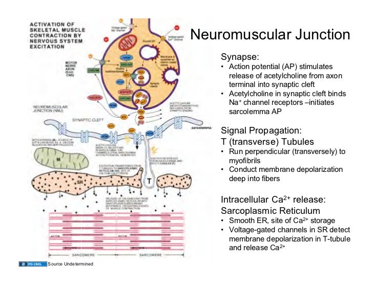
How do muscles contract?
Muscles contract when they receive signals from motor neurons. The neuromuscular junction is the site of the signal exchange. The steps of this process in vertebrates occur as follows: (1) The action potential reaches the axon terminal. (2) Calcium ions flow into the axon terminal. (3) Acetylcholine is released into the synaptic cleft. (4) Acetylcholine binds to postsynaptic receptors. (5) This binding causes ion channels to open and allows sodium ions to flow into the muscle cell. (6) The flow of sodium ions across the membrane into the muscle cell generates an action potential which induces muscle contraction. Labels: A: Motor neuron axon B: Axon terminal C: Synaptic cleft D: Muscle cell E: Part of a Myofibril
What is the name of the body system that increases or decreases the activity of acetylcholine?
Substances that increase or decrease the overall activity of the cholinergic system are called cholinergics and anticholinergics, respectively.
How is acetylcholine synthesized?
Acetylcholine is synthesized in certain neurons by the enzyme choline acetyltransferase from the compounds choline and acetyl-CoA. Cholinergic neurons are capable of producing ACh. An example of a central cholinergic area is the nucleus basalis of Meynert in the basal forebrain. The enzyme acetylcholinesterase converts acetylcholine into the inactive metabolites choline and acetate. This enzyme is abundant in the synaptic cleft, and its role in rapidly clearing free acetylcholine from the synapse is essential for proper muscle function. Certain neurotoxins work by inhibiting acetylcholinesterase, thus leading to excess acetylcholine at the neuromuscular junction, causing paralysis of the muscles needed for breathing and stopping the beating of the heart.
What is the interaction between nicotinic receptors and acetylcholine?
Acetylcholine's interaction with muscarinic receptors, as with nicotinic receptors, causes channels to open resulting in ion flow that depolarizes the muscle cell. As in skeletal muscle, the depolarization leads to muscle contraction.
What is the effect of acetylcholine on smooth muscle?
Acetylcholine’s Effect On Smooth Muscle. Acetylcholine activates a different type of receptor present in smooth muscle: the muscarinic receptor. When this receptor binds acetylcholine, one result is the release of calcium ions from internal stores. Acetylcholine's interaction with muscarinic receptors, as with nicotinic receptors, ...
What happens when acetylcholine binds to a receptor?
This means that when acetylcholine, the ligand, binds to a receptor, the receptor changes its shape in a way that lets sodium enter the muscle cell. Advertisement.
How does sodium depolarize a muscle cell?
The influx of sodium depolarizes the muscle cell in the vicinity of the motor endplate. Depolarization means the difference in charge between the inside and outside of the muscle is reduced. A different type of sodium channel, which is activated in response to depolarization, lets more sodium in and the wave of excitation spreads throughout the muscle cell. This leads to the release of calcium ions from storage sites inside the muscle cell. The calcium ions initiate a series of biochemical events involving troponin, tropomyosin and myosin that cause the muscle to contract.
What is the function of acetylcholine?
It controls the contraction of all skeletal or voluntary muscles, for instance. It also affects the contraction of smooth and cardiac muscle.
Which receptors are sensitive to acetylcholine?
The muscle cell membrane contains nicotinic receptors that are sensitive to acetylcholine. These receptor molecules, made of protein, are concentrated where acetylcholine is released. The nicotinic receptor is a ligand-gated sodium channel. This means that when acetylcholine, the ligand, binds to a receptor, the receptor changes its shape in ...
Where is acetylcholine held?
Acetylcholine is held in synaptic vesicles in nerve terminals until an electrical signal causes its release onto a specialized portion of a muscle cell membrane equipped with receptors that recognize the neurotransmitter.
What antagonists inhibit vascular relaxation?
Both the nonsubtype-specific muscarinic antagonist, atropine, and the subtype selective antagonists, pirenzepine (M1) and AFDX-116 (M2), inhibited vascular relaxation from perfusion of spinal cord tissue with acetylcholine. Because a variety of antagonist concentrations was not tested, it is not possible to determine the relative potencies of each antagonist on acetylcholine's effect. Both M1 and M2 receptors have been identified by ligand binding on spinal cord dorsal horn and intermediolateral cell column, [15] and both subtypes are involved in the analgesic and hemodynamic actions of spinally administered cholinomimetic agents. [7,16] Whereas we did not perform experiments with a wide range of antagonist concentrations, the current results are consistent with these observations of neuronal actions of acetylcholine on both muscarinic receptor subtypes in the spinal cord.
What is the release of vasorelaxant from spinal cord tissue?
These results demonstrate release of a vasorelaxant from spinal cord tissue by acetylcholine, which results from an action on muscarinic receptors and exhibits a pharmacology consistent with nitric oxide. Although precise anatomic localization of acetylcholine's action is not possible with this system, these results add to evidence that acetylcholine causes nitric oxide synthesis in the spinal cord.
What is the effect of acetylcholine perfusion?
Acetylcholine perfusion of spinal tissue caused concentration-dependent relaxations of the aortic rings, an effect blocked by each of the muscarinic antagonists, atropine, pirenzepine, and AFDX-116. Acetylcholine-induced relaxation also was antagonized by an inhibitor of nitric oxide synthase (N-methyl-L-arginine), a nitric oxide scavenger (hemoglobin) and an inhibitor of guanylate cyclase (methylene blue).
What is the role of acetylcholine in the synthesis of nitric oxide?
Acetylcholine causes synthesis of nitric oxide in vascular endothelium, and presumptive evidence in vivo suggests spinally released acetylcholine causes antinociception and increased sympathetic nervous system activity via a nitric oxide mechanism. The purpose of this study was to determine, using a recently described bioassay system, whether acetylcholine stimulates nitric oxide release from spinal cord tissue in vitro.
What solution was used to perfuse a rat spinal cord?
Rat thoracolumbar spinal cord slices were incubated in a tissue chamber and perfused with Krebs-Henseleit solution. The perfusate was then passed through endotheliumdenuded rat aortic rings and their tension was measured. Vascular rings were preconstricted with phenylephrine, then were exposed to spinal cord perfusate with increasing concentrations (10 (-12)-10 (-4)M) of acetylcholine alone or with various antagonists.
How are Sprague-Dawley rats treated?
After approval by the Animal Care and Use Committee of our institution, adult male Sprague-Dawley rats were deeply anesthetized with 50 mg/kg intraperioneal sodium pentobarbital, decapitated, and the aorta removed. The aorta was cut into 3-4-mm long rings and endothelium denuded by rubbing with stainless steel wire, then rings were mounted on transducers and tension was measured continuously with a Grass #7 polygraph (Quincy, MA). The rings were stretched to their optimum length-tension relationship by repeated exposures to 180 mM potassium chloride. Removal of endothelium was confirmed by preconstriction with phenylephrine 10 sup -6 M and lack of relaxation to 10 sup -7 M to 10 sup -6 M acetylcholine. Two rats were then killed as stated earlier and spinal cords were removed. Each spinal cord was divided into two parts, then chopped in 0.5-mm thick slices. Tissue sections from each hemispinal cord were put into an incubation chamber surrounded by a temperature-controlled water bath maintained at 26 degrees C. Tissue slices were perfused continuously with a multichannel pump (Manostat, NY) at 4 ml/min with oxygenated modified Krebs-Henseleit solution containing indomethacin 10 sup -5 M (composition in mM, 118.3 NaCl, 4.7 KCl, 2.5 CaCl 2, 1.2 MgSO sub 4, 1.2 KH 2 PO 4, 25 NaHCO 3, 0.027 EDTA, and 11 glucose), gassed with 95% Oxygen 2, 5% CO 2 at 26 degrees C. Previous studies demonstrated this tissue preparation and temperature yielded consistent responses to stimulators of nitric oxide synthase. [12] The effluent of spinal cord tissue chambers dripped onto the rings directly. Time from spinal cord tissue exposure to contact with aortic rings was < 2 s. Experiments were started after spinal cord slices had been incubated in the chamber for 60 min.
Does acetylcholine release vasorelaxant?
In summary, acetylcholine perfusion of spinal cord tissue in vitro causes concentration-dependent release of a vasorelaxant with pharmacologic properties consistent with nitric oxide. Blockade by muscarinic antagonists confirms in vivo experiments and supports the concept of nitric oxide mediation of analgesic and hemodynamic actions of spinally administered cholinomimetic agents.
Overview
- Acetylcholine has many functions in the body. It is released from cholinergic nerve synapses and acts on presynaptic (transmitter) and postsynaptic (receiver) acetylcholine receptors.1
Functions
Chemistry
Biochemistry
Diseases and disorders
Pharmacology
Acetylcholine functions in both the central nervous system (CNS) and the peripheral nervous system (PNS). In the CNS, cholinergic projections from the basal forebrain to the cerebral cortex and hippocampus support the cognitive functions of those target areas. In the PNS, acetylcholine activates muscles and is a major neurotransmitter in the autonomic nervous system.
Like many other biologically active substances, acetylcholine exerts its effects by binding to an…
Comparative biology and evolution
Acetylcholine is a choline molecule that has been acetylated at the oxygen atom. Because of the charged ammonium group, acetylcholine does not penetrate lipid membranes. Because of this, when the molecule is introduced externally, it remains in the extracellular space and does not pass through the blood–brain barrier.
History
Acetylcholine is synthesized in certain neurons by the enzyme choline acetyltransferase from the compounds choline and acetyl-CoA. Cholinergic neurons are capable of producing ACh. An example of a central cholinergic area is the nucleus basalis of Meynert in the basal forebrain. The enzyme acetylcholinesterase converts acetylcholine into the inactive metabolites choline and acetate. This enzyme is abundant in the synaptic cleft, and its role in rapidly clearing free acetyl…