Which cavity contains the toward the side?
The dorsal cavity contains the toward the side Lateral means left and right halves The midsaggital plane divides the body into medial The ____ surface of a structure is toward, or nearer the midline and away from the side
What plane separates the dorsal cavity into anterior and posterior subdivisions?
The frontal plane is the plane that divides the body or an organ into an anterior (front) portion and a posterior (rear) portion. Click to see full answer. Herein, what separates the dorsal cavity into two subdivisions?
Why do Anatomists divide the abdominal cavity into regions?
Anatomists divide the abdominopelvic cavity into smaller regions to facilitate the study of body planes. This anatomical abdominal region division is used to recognize the location of the abdomen organs and to diagnose abdominal pain.
What is a vertical cavity dividing the body into two parts?
The ventral is the larger cavity and is subdivided into two parts (thoracic and abdominopelvic cavities) by the diaphragm, a dome-shaped respiratory muscle. In this regard, what is a vertical section through the body dividing it into anterior and posterior regions called?
What is the anterior body cavity?
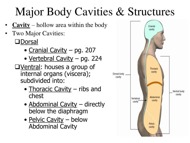
The anterior (ventral) cavity has two main subdivisions: the thoracic cavity and the abdominopelvic cavity (see Figure 4). The thoracic cavity is the more superior subdivision of the anterior cavity, and it is enclosed by the rib cage.
Which type of section would separate the anterior and posterior?
What type of cut would separate the brain into anterior and posterior parts? A frontal (coronal) section would separate the brain into anterior and posterior parts.
Which is a vertical section through the body dividing it into anterior and posterior regions called?
Coronal Plane (Frontal Plane) - A vertical plane running from side to side; divides the body or any of its parts into anterior and posterior portions.
Which structure separates the ventral body cavity into a superior and inferior portion?
The diaphragmThe diaphragm forms the floor of the thoracic cavity and separates it from the more inferior abdominopelvic cavity. The abdominopelvic cavity is the largest cavity in the body.
Which plane divides the body into anterior and posterior portions quizlet?
The coronal plane divides the body into anterior and posterior sections. Motions in the coronal plane, such as abduction and adduction, occur around an anterior-posterior axis.
What plane divides the body into two equal right and left halves?
Sagittal planePlanes: Because who said anatomy didn't require an imagination?Frontal (Coronal) planeDivides the body into anterior (front) and posterior (back) portionsSagittal planeVertical plane that divides the body into right and left sides.Midsagittal planeDivides the body at midline into equal right and left sides.2 more rows•Oct 17, 2013
What is a vertical section through the body dividing it into anterior and posterior regions called transverse median sagittal frontal?
What is a vertical section through the body, dividing it into anterior and posterior regions called? frontal. Frontal plane. A coronal plane (also known as the frontal plane) is any vertical plane that divides the body into ventral and dorsal (belly and back) sections.
What is a vertical section through the body dividing into anterior?
What is a vertical section through the body, dividing it into left and right? sagittal. What is a vertical section through the body, diving it into anterior and posterior regions called? frontal. Which of the following describes that operations of the heart and blood vessels?
Which body cavity is separated into other cavities?
thoracic cavityThe thoracic cavity is separated from the abdominopelvic cavity by the diaphragm. The thoracic cavity is further separated into the pleural cavity which contains the lungs and the superior mediastinum which includes the pericardial (heart) cavity. The organs within the ventral body cavity are called the viscera.
Which cavities are separated by the diaphragm?
The diaphragm is a thin dome-shaped muscle which separates the thoracic cavity (lungs and heart) from the abdominal cavity (intestines, stomach, liver, etc.). It is involved in respiration, drawing downward in the chest on inhalation, and pushing upward in exhalation.
Which type of plane separates the thoracic cavity from the abdominopelvic cavity?
The thoracic cavity is separated from the abdominopelvic cavity by the diaphragm. The abdominopelvic cavity is separated into the abdominal cavity and the pelvic cavity by an imaginary line parallel to the pelvis bones.
Which of these planes divides the body into frontal and back sections?
The coronal plane (frontal or Y-X plane) divides the body into dorsal and ventral (back and front) portions. It also separates the anterior and posterior portions.
What are the three divisions of the nasal cavity?
The nasal cavity is the most superior part of the respiratory tract. It extends from the vestibule of the nose to the nasopharynx, and has three divisions: Vestibule – the area surrounding the anterior external opening to the nasal cavity.
Which structure does not empty out into the lateral walls of the nasal cavity?
The only structure not to empty out onto the lateral walls of the nasal cavity is the sphenoid sinus. It drains onto the posterior roof. In addition to the paranasal sinuses, other structures open into the nasal cavity: Nasolacrimal duct – acts to drain tears from the eye. It opens into the inferior meatus.
What is the bulge in the lateral wall of the nasal cavity?
This is a bulge in the lateral wall formed by the middle ethmoidal sinus itself. The posterior ethmoidal sinuses open out at the level of the superior meatus. The only structure not to empty out onto the lateral walls of the nasal cavity is the sphenoid sinus. It drains onto the posterior roof.
What are the three conchae?
The are three conchae – inferior, middle and superior. They project into the nasal cavity, creating four pathways for the air to flow. These pathways are called meatuses: Inferior meatus – between the inferior concha and floor of the nasal cavity. Middle meatus – between the inferior and middle concha.
What is the nasal cavity?
Divisions. The nasal cavity is the most superior part of the respiratory tract. It extends from the vestibule of the nose to the nasopharynx, and has three divisions:
Which part of the brain is responsible for innervation of the nose?
The olfactory bulb, part of the brain, lies on the superior surface of the cribriform plate, above the nasal cavity. Branches of the olfactory nerve run through the cribriform plate to provide special sensory innervation to the nose.
Where is the cribriform plate located?
It contains very small perforations, allowing fibres of the olfactory nerve to enter and exit, At the level of the superior meatus, the sphenopalatine foramen is located.
Which plane divides the body into ventral and dorsal portions?
The Coronal Plane (Frontal Plane) It is also known as Y-X plane or Frontal planes; the coronal plane divides the body into ventral (front) and dorsal (back) portions. This plane also gives a clear image of the posterior and anterior portions of the body.
Where is the ventral cavity located?
A cavity which is located posteriorly to the ventral body cavity and found beneath the diaphragm and thoracic cavity . This cavity is divided into the pelvic and abdominal cavity, and it contains the organs of many systems such as renal and digestive systems.
What plane intersects the median plane?
The coronal planes intersect the median plane at a 90-degree angle and show the anatomical body parts into front and back halves. These. three are significant references plane, and all other planes are shown with. these planes, such as Parasagittal planes are parallel to the sagittal (Y-Z) plane.
How many regions are there in the abdominopelvic cavity?
The commonly abdominopelvic region is divided into four quadrants and nine regions.
How many divisions are there in the body?
The nine divisions are part of parasagittal and two transverse planes of body-centered around the navel. These divisions are important anatomically to determine the location of the organ within the abdomen and pelvic area.
What is the anatomical position?
the anatomical position is described with the help of a coordinate system, which includes three-axis (X, Y, and Z). The X-axis is going from left to. right, Z-axis from front to back, and Y-axis from up to down. In anatomical. terminology, three references plane are considered standard planes; these.
Which plane is parallel to the median plane?
A vertical plane that runs parallel to the median plane is called a Sagittal plane . This Y-Z or lateral plane separates the body right (dexter) and left (sinister) halves. The midsagittal plane (median plane) passes through the center of the body while all other sagittal planes are parallel to this midsagittal planes.
Anatomical Position
To further increase precision, anatomists standardize the way in which they view the body. Just as maps are normally oriented with north at the top, the standard body “map,” or anatomical position, is that of the body standing upright, with the feet at shoulder width and parallel, toes forward.
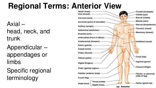
Regional Terms
The human body’s numerous regions have specific terms to help increase precision (see Figure 1.12 ).
Directional Terms
Certain directional anatomical terms appear throughout this and any other anatomy textbook ( Figure 1.13 ). These terms are essential for describing the relative locations of different body structures.
Body Planes
A section is a two-dimensional surface of a three-dimensional structure that has been cut. Modern medical imaging devices enable clinicians to obtain “virtual sections” of living bodies. We call these scans. Body sections and scans can be correctly interpreted, however, only if the viewer understands the plane along which the section was made.
Body Cavities and Serous Membranes
The body maintains its internal organization by means of membranes, sheaths, and other structures that separate compartments. The dorsal (posterior) cavity and the ventral (anterior) cavity are the largest body compartments ( Figure 1.15 ).