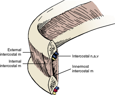
The neurovascular bundle, located in the costal groove in the undersurface of each rib, between the internal intercostal muscle and innermost intercostal muscle, supplies much of the innervation and vascular supply to the thoracic wall. The neurovascular bundle is arranged as V ein, A rtery and N erve, from the most superior to the most inferior.
What is included in the neurovascular bundle?
Neurovascular Bundle 1 Intercostal Nerves. 2 Intercostal Arteries. 3 Intercostal Veins. 4 Intercostal Lymphatics.
What is in the intercostal space?
First, let’s have a look at the contents of the intercostal space, specifically the neurovascular bundle containing the intercostal nerve, artery and vein. It is important to remember the order of these structures, with the most superior structure being the intercostal vein and below it comes the artery and below the artery, the nerve.
What are the different types of intercostal nerves?
The intercostal nerves are classified into the following 2 groups: Typical intercostal nerves (3rd, 4th, 5th, and 6th). Atypical intercostal nerves (1st, 2nd, 7th, 8th, 9th, 10th, and 11th). The normal intercostal nerves are those which stay confined to their very own intercostal spaces
What is an intercostal nerve block?
An intercostal nerve block involves injection of a short, or long-acting local anesthetic around the intercostal nerve, posterior to the region of pain. Long-term pain relief may be achieved by destruction of the nerve with phenol or a cryogenic probe.
What does the intercostal neurovascular bundle supply?
Neurovascular bundle. The neurovascular bundle, located in the costal groove in the undersurface of each rib, between the internal intercostal muscle and innermost intercostal muscle, supplies much of the innervation and vascular supply to the thoracic wall.
What structures are innervated by intercostal nerves?
The lower intercostal nerves leave their intercostal spaces anteriorly (after giving branches to the intercostal muscles, abdominal peritoneum, and skin) to innervate the anterior abdominal wall, muscles (in particular the rectus abdominis), and the overlying skin.
What is the sequence of neurovascular bundle in intercostal space?
Neurovascular bundle They are ordered vein, artery, nerve from superior to inferior (mnemonic VAN). The collateral neurovascular bundle runs at the lower edge of the space, just above the inferior rib and the order is reversed, i.e. nerve, artery, vein from superior to inferior.
What does the intercostal supply?
The posterior intercostal arteries are branches of the superior intercostal artery (upper two spaces) and the descending aorta (lower nine spaces). They supply the chest wall, parietal pleura, and, through their dorsal branches, the skin and muscles of the back and the spine and its contents.
What do the intercostal nerves control?
Unlike the nerves from the autonomic nervous system that innervate the visceral pleura of the thoracic cavity, the intercostal nerves arise from the somatic nervous system. This enables them to control the contraction of muscles, as well as provide specific sensory information regarding the skin and parietal pleura.
What is the innervation of the intercostal muscles?
External intercostal muscles They are innervated by the anterior rami of spinal nerves T1-T11, i.e. the intercostal nerves of the corresponding intercostal space. The blood supply to the external intercostals comes from the anterior and posterior intercostal arteries.
What are the intercostal nerves supplying the whole thoracic wall?
The intercostal nerves are the somatic nerves that arise from the anterior divisions of the thoracic spinal nerves from T1 to T11. These nerves in addition to supplying the thoracic wall also supply the pleura and peritoneum.
What does the anterior intercostal arteries supply?
Along with the posterior intercostal arteries, the anterior intercostal arteries supply the muscles and skin within the intercostal spaces as well as the parietal pleura.
Do the intercostal nerves innervate the pleura?
Unlike the nerves from the autonomic nervous system that innervate the visceral pleura of the thoracic cavity, the intercostal nerves arise from the somatic nervous system. This enables them to control the contraction of muscles, as well as provide specific sensory information regarding the skin and parietal pleura.
What are the intercostal nerves supplying the whole thoracic wall?
The intercostal nerves are the somatic nerves that arise from the anterior divisions of the thoracic spinal nerves from T1 to T11. These nerves in addition to supplying the thoracic wall also supply the pleura and peritoneum.
What do the thoracic nerves innervate?
These nerves mainly supply the skin over the chest and intrinsic back muscles of the thoracolumbar region. The thoracic nerves also supply some of the areas of skin over the abdomen and axilla. Spinal nerves are essential for the control of body parts by the central nervous system.
Which branch from the spinal nerves will innervate the intercostal muscles?
The intercostal nerves arise from the anterior rami of the thoracic spinal nerves from T1 to T11. The anterior division of the twelfth thoracic nerve is not technically grouped with the other intercostal nerves as it enters the abdominal wall; this nerve is instead referred to as the subcostal nerve.
What are the superficial bundles of neurovascular tissue?
Superficial bundles. Superficial neurovascular bundles do not include arteries, and consist primarily of capillaries and nerves. Because capillaries function as the sites for substance exchange between interstitial fluid and blood, they tend to have large surface area and short diffusion path.
What is neurovascular bundle?
A neurovascular bundle is a structure that binds nerves and veins (and in some cases arteries and lymphatics) with connective tissue so that they travel in tandem through the body.
How to avoid damaging neurovascular bundles?
A common anatomically informed, surgical technique to avoid damaging neurovascular bundles is to undermine anteriorly to the posterior tibial margin after reaching the fascia, in order to avoid the saphenous vein and nerve. The deep posterior compartment here is superficial and readily accessible.
Which nerve is at risk in leg surgery?
In surgeries, the principle superficial neurovascular bundles at risk are, medially, the great saphenous vein and its accompanying nerve, and, laterally, the superficial peroneal nerve.
Which bundles have smooth muscle and connective tissue?
Deep neurovascular bundles, which often include arteries, have a more complicated structure than superficial neurovascular bundles. Since arteries have high intraluminal blood pressure relative to capillaries and veins, these bundles have smooth muscle and connective tissue structures outside the endothelium.
Where is the superficial branch of the tibial?
The superficial branch then continues onto the dorsum of the foot to supply sensory fibers to the skin there. The main deep neurovascular bundle at risk is the posterior tibial. It lies on the posterior aspect of the tibialis posterior and flexor digitorum longus muscles, and medial to the belly of flexor hallucis longus.
Do arteries travel within the superficial fascia?
As arteries do not travel within the superficial fascia ( loose connective tissue under the skin ), superficial neurovascular bundles differ from deep neurovascular bundles in both composition and function.
What are the subcostal arteries?
The subcostal arteries are direct branches of the thoracic aorta. They are analogous to the posterior intercostal artery, so if there was a 12th intercostal space, they would be the 12th intercostal arteries. Like its preceding counterparts, each subcostal artery gives the anterior and posterior branches that travel along with the subcostal space below the twelfth rib. On the left side, the artery passes behind the accessory hemiazygous vein, while on the right side it passes in front of the twelfth thoracic vertebra and behind the thoracic duct and azygous vein. On either side, the arteries are posteriorly related to the sympathetic trunk, diaphragm, and adjacent pleura.
Which artery supplies the 10th and 11th intercostal spaces?
The 10th and 11th intercostal spaces are supplied by the posterior intercostal arteries only. The internal thoracic artery is a source for the upper six anterior intercostal arteries, as well as the superior epigastric and musculophrenic arteries.
What is the internal thoracic vein?
The internal thoracic veins accompany the internal thoracic arteries. They unite at about the third costal cartilage to form a single internal intercostal vein that is medial to the accompanying artery.Like most veins in the body, the internal thoracic vein has several valves along its length to promote the unidirectional flow of blood. It receives segmental tributaries at each intercostal level (similar to the points at which the corresponding arteries emerge). The pericardiophrenic vein also drains deoxygenated blood by way of the internal thoracic veins. The internal thoracic vein eventually drains directly into the ipsilateral brachiocephalic vein .
How many pairs of intercostal veins are there?
Intercostal veins. There are eleven pairs of posterior intercostal veins, ni ne pairs of anterior intercostal and one pair of subcostal veins supplying the thoracic wall. Each posterior intercostal vein forms an anastomosis with the ipsilateral anterior intercostal veins.
Why are the internal thoracic arteries called the internal mammary arteries?
The internal thoracic arteries were once referred to as the internal mammary arteries because they indirectly supply the breasts. They stem from the subclavian artery and contribute to supplying the intercostal muscles, skin and parietal pleura associated with the first six intercostal spaces.
Why is it important to know the arteries and veins of the chest wall?
Knowledge of the arteries and veins that supply the chest wall is not only important for passing exams, but also for certain emergency situations that pop up during clinical practice. This article will discuss the arteries and veins of the thoracic wall. However, a brief review of the intercostal space and the chest wall anatomy will also be included.
How many branches does the internal thoracic artery have?
The internal thoracic artery gives off five sets of branches :
Where is the neurovascular bundle palpable?
The neurovascular bundle is usually palpable with ease where it runs over the abaxial surface of the proximal sesamoid bones, making this technique the easiest regional block to perform. The site of injection is subcutaneously on the palmar aspect of the neurovascular bundle over the abaxial surface of the proximal sesamoid bones.
Which ligament is open to explore the epidural space?
The superior disk space should be explored for any bony fragments. The posterior longitudinal ligament may also be opened to explore the epidural space.
How much of the foot is affected by palmar digital block?
21 However, in clinical practice, this block desensitizes 70% to 80% of the foot. Most of the DIP joint is affected, with the exception of the proximodorsal aspect. Horses with fractures of the extensor process of the distal phalanx or injury of a collateral ligament of the DIP joint may show partial improvement after palmar digital analgesia, however. Our clinical observations have been substantiated in a recent study. Setscrews were placed near the medial and lateral aspects of the toe to simulate pain from the sole. Lameness in these horses was abolished using palmar digital analgesia performed just proximal to the heel bulbs. 22
What is a thoracic outlet syndrome?
Thoracic outlet syndrome is a disease that involves compression of the neurovascular bundle, which courses its way from the neck to the axilla and then exits the axilla. Although Hunald originally described TOS more than 200 years ago (Tyson and Kaplan 1975 ), today this condition is often associated with injuries in overhead throwing athletes ( Baker and Liu 1993, Nuber et al. 1990, Strukel and Garrick 1978, Rohrer JM et al. 1990, Safran 2004 ). Some have described two (in which venous and arterial are combined) to three categories of TOS including a neurologic compression syndrome of the brachial plexus, a vascular compression syndrome of the subclavian vein ( Toby and Korman 1989 ), and an arterial syndrome, which is caused by compression of the subclavian artery ( Freischlag and Orion 2014 ). The neurologic symptoms appear to be present in up to 90% to 97% of patients with TOS ( DiFelice GS et al. 2002, Vogel and Jensen 1985) while arterial or venous symptoms have been thought to occur much less often in only about 2% to 5% of patients ( Nemmers DW et al. 1990, Schneid K et al. 1999, Vogel and Jensen 1985 ). Overall it is estimated that around 90% are neurogenic origin, whereas less than 1% are arterial and 3% to 5% venous ( Sanders RJ et al. 2007 ). This becomes very problematic because both causes tend to create similar symptoms. Because these symptoms are inconsistent and vague some even discount TOS as an actual diagnosis ( Dale 1982 ).
What is the dye filling of axons in the mesentery?
Anterograde dye filling of axons in mesenteric nerve trunks. The only specialized axonal structures in the mesentery, revealed by biotinamide fills (apart from bundles of smooth axons of passage), were varicose branching axons on blood vessels.
How to remove the transverse process?
Next, the transverse process, lamina, and pedicle can be removed using a high-speed drill, rongeur, and curettes to identify the posterolateral thecal sac. The disk space immediately superior and inferior to the vertebrectomy site should be removed with a scalpel. The vertebral body can then be removed with either a high-speed drill or a rongeur under direct visualization ( Fig. 34.4 ).
Where is disk space cephalad?
The disk space is cephalad to the pedicle.
Where is the neurovascular bundle in the thoracotomy?
The traditional thoracotomy (postero- lateral) is performed along the 6th rib. The neurovascular bundle is shielded from injury by lifting the periosteum of the rib. Considering the position of neurovascular bundle in the intercostal space, it’s safe to add the needle, a little above the upper border of the rib below.
What nerves are used to connect the intercostal space?
Intercostal nerves is the name given to the anterior primary rami of the upper 11 thoracic spinal nerves (T1 T11) as they use the route through the intercostal spaces. The thoracic wall is supplied by the 12 pairs of the thoracic spinal nerves. Anterior and posterior rami are formed as soon as they leave the intervertebral foramina.
What is intercostal nerve block?
Intercostal nerve blockis given to make local anesthesia in 1 or more intercostal spaces by injecting the anesthetic agent around the nerve trunk near its origin, i.e., just lateral to the vertebra.
What are the features of intercostal nerves?
They may be segmental in nature unlike the anterior primary rami from some other regions of spinal cord which create nerve plexuses viz. cervical, brachial, lumbar and sacral.
Which nerves are associated with cardiac pain?
In coronary arterial disease, the cardiac pain is attributed along this nerve to the medial side of the arm. Seventh to eleventh intercostal nerves: These nerves leave the corresponding intercostal spaces to goes into the abdominal wall; for this reason they’re termed thoraco ¬ abdominal nerves.
Which nerve connects the medial cutaneous branch of the arm?
Second intercostal nerve: Its lateral cutaneous branch is named intercostobrachial nerve. It lessons around the axilla and joins the medial cutaneous branch of the arm. The intercostobrachial nerve supplies the skin of the floor of the axilla and upper part of the medial side of the arm.
Which nerve joins the ventral ramus C8 spinal nerve to create lower trunk of the brachial?
The atypical intercostal nerves are as follows: First intercostal nerve: The greater part of the nerve joins the ventral ramus C8 spinal nerve to create lower trunk of the brachial plexus. The rest of the part of thenerve is quite small and it lacks both lateral and anterior cutaneous branches.
What is intercostal nerve block?
Intercostal nerve block requires an understanding of the course of ventral primary rami of spinal nerves, their relationship to the sympathetic trunk, and the variable branching pattern of the intercostal nerves. [1] [6] [12] [13] The relationship of deep branches of the intercostal nerves to the parietal pleura allows intrapleural approaches to analgesia. [14]
Where do neurovascular structures travel?
Neurovascular structures travel in the interval between innermost and inner intercostal muscles within the intercostal space, piercing as lateral cutaneous branches near the mid-axillary line and as anterior cutaneous branches parasternally.
What are the three layers of the intercostal muscle?
The three intercostal muscle layers differentiate from hypaxial myotomes innervated by ventral primary rami of spinal nerves T1 to T11, the intercostal nerves. [3] Twelve ribs develop from segmental sclerotomes to interrupt the muscular wall and form the 11 intercostal spaces. The muscles of each intercostal space receive innervation by segmentally arranged spinal nerves denominated by the corresponding superjacent rib. Sensory innervation of the thoracic wall follows the embryonically derived pattern of overlapping dermomyotomes. The ventral primary ramus of T12 is serially homologous to an intercostal nerve but is named the subcostal nerve since it is not bounded inferiorly by a rib. This nerve, as well as the anterior branches of T7 to T11 continuing into the abdomen, are correctly termed thoracoabdominal nerves, [4] even though they are frequently referred to in the clinical literature as intercostal nerves. The three anterolateral muscle layers of the abdominal wall, as well as the rectus abdominis muscle, which they innervate, share their embryological origin and fiber directionality with the muscles of the intercostal space.
Why is the neurovascular anatomy of the thoracic wall important?
The neurovascular anatomy of the thoracic wall is clinically significant because clinicians must traverse it in such procedures as thoracentesis, thoracostomy, and thoracotomy.
How do vertebrae develop?
Vertebrae develop intersegmentally by embryonic fusion of the caudal half of the superjacent somitic sclerotome with the cranial half of the subjacent sclerotome. Ribs, myotomal derivatives, and segmental neurovasculature maintain their segmental embryonic patterning. The rib head in most ribs (2 to 9) thus articulates with two vertebrae at the costovertebral joint, but by convention, rib denomination is by reference to their subjacent vertebra. Rib 1 only articulates with the first thoracic vertebra, and ribs 10 through 12 also have only one facet at the costovertebral joint at which they articulate with the lower thoracic vertebrae.
What are the components of the thoracic wall?
The musculofascial components of the thoracic wall form a three-layered intercostal membrane between the ribs consisting of external intercostal, internal intercostal, and innermost intercostal muscles which have a primary respiratory function. [2] The external intercostal muscles act to raise the ribs in a “bucket handle” fashion, thereby increasing thoracic volume, while the internal intercostals largely depress the ribs, decreasing thoracic volume, and aiding in exhalation.
What is the lining of the coelomic cavity?
The lining of the coelomic cavity develops as a mesothelium which invests organs as visceral pleura and the internal surface of the thoracic wall as parietal pleura. The space between visceral and parietal layers comprises the pleural cavity, which normally contains only serous fluid. The lungs inhabit the two halves of the thoracic cavity, not the pleural cavity.
Which arteries accompany the intercostal nerves through the intercostal spaces?
The posterior intercostal arteries accompany the intercostal nerves through the intercostal spaces.
Where do the intercostal nerves run?
Initially, they run within the endothoracic fascia just between the parietal pleura and the internal intercostal membrane.
What is the intercostal space?
First, let’s have a look at the contents of the intercostal space, specifically the neurovascular bundle containing the intercostal nerve, artery and vein. It is important to remember the order of these structures, with the most superior structure being the intercostal vein and below it comes the artery and below the artery, the nerve.
How many pairs of spinal nerves are there in the thoracic wall?
There are 12 pairs of thoracic spinal nerves which supply the thoracic wall. They leave the intervertebral foramina as soon as they are formed, dividing into anterior and posterior rami. Now, the anterior rami of nerves T1 through T11 form the intercostal nerves that run along the intercostal spaces along the inferior borders of the ribs, ...
What is the function of the vessels and nerves in the thoracic wall?
There are numerous vessels and nerves that course throughout the bony and muscular architecture of the thoracic wall, and they provide innervation and blood supply to the structures within and around the thoracic cage.
Which nerves give rise to the dermatomal map of the trunk?
Most thoracic spinal nerves, specifically T2 through T12, give rise to the dermatomal map of the trunk through their posterior ramus and the lateral and anterior cutaneous branches of its anterior ramus.
Which nerves pass posteriorly?
The posterior rami of thoracic spinal nerves pass posteriorly, lateral to the articular processes of the vertebrae in order to supply the joints, deep back muscles and skin of the posterior thoracic region.